T. C.
SELÇUK ÜNİVERSİTESİ FEN BİLİMLERİ ENSTİTÜSÜ
Green Synthesis of Silver Nanoparticles
Sura Ahmed AL-GBURIM.Sc. THESIS
CHEMICAL ENGINEERING DEPARTMENT
2018 KONYA All Rights Reserved
III TEZ BİLDİRİMİ
Bu tezdeki bütün bilgilerın etik davranış ve akademik kurallar çerçevesinde elde edildiğini ve tez yazım kurallara uygun olarak hazırlanan bu çalışmada bana ait olmayan her türlü ifade ve bilginin kaynağına eksiksiz atıf yapıldığını bildiririm.
DECLARATION PAGE
I hereby declare that all the information in this thesis document has been obtained and presented in accordance with academic rules and ethical conduct. I also declare that, as required by these rules and conduct, I have fully cited and referenced all materials and results that are not original to this work.
Sura Ahmed Mahmood AL-GBURI 22/03/2018
IV ACKNOWLEDGEMENT
I might want to offer my genuine thanks to my supervisor Prof. Dr. Erol Pehlivan for his persistent help, support, direction, and advice all through this thesis. Without a doubt, Prof. Dr. Pehlivan’s understanding and tolerance made conceivable to finish this thesis at this every time. I might likewise want to offer my most profound thanks to Prof. Dr. Ahmet Avcı. I might likewise want to thank Prof. Dr. Masood Hussain, who has been teaching me the practical experiences in the laboratory and I benefited from his experience in my research. I might want to also thank the academic staff of Chemical Engineering Department at each of Selcuk University for their untiring help and direction all through my study. I owe an obligation of appreciation to every single one of them.
I consecrate my thesis to my dear husband Ammar Jabbar & my mother. An extraordinary sentiment appreciation to my cherishing husband, whose words always encouraged me. My mother has never cleared out my side and is very special.
At long last, I might want to thank my friends and country mates in Konya, thank you for listening, offering me counsel, and supporting me through this whole process.
Sura Ahmed AL-GBURI
V ABSTRACT
M.Sc. THESIS
Green Synthesis of Silver Nanoparticles
Sura Ahmed AL-GBURI
SELCUK UNIVERSITY
GRADUATE SCHOOL OF NATURAL AND APPLIED SCIENCE CHEMICAL ENGINEERING DEPARTMENT
Supervisor
Prof. Dr. Erol PEHLİVAN
2018, 54 pages
Jury
Prof. Dr. Erol PEHLİVAN Yrd.Doç.Dr. Serpil EDEBALİ
Yrd.Doç.Dr. Fatma KUNT
Nanotechnology includes nanoparticles (NPs) owning a size range of (1-100 nm) in at least one-dimension and have been utilized altogether concerning medicinal chemistry, nuclear material science, and all other known fields. NPs are utilized very much indeed because of its little dimension and physical properties. NPs can be obtained effortlessly by various chemical, physical, & biological methodologies. The green methodology is one of most rising production methods since this strategy is simpler than other alternate strategies, eco-friendly and minimal time exhaustion for the production. The green methodology was completed by utilizing the aqueous-phase of Teucrium chamaedrys extract, Inula helenium extract, Ocimum tenuiflorum extract together with AgNO3 solution. Silver
provides a specific contribution to this procedure in respect of its suggestive physical & chemical properties. A certain proportion of plant extract for the silver ion was prepared and the color alteration which demonstrated the development of Ag-NPs was observed during the production. The Ag-NPs were characterized by (UV-Vis) Spectrophotometer, Fourier Transform Infrared (FTIR) Spectrophotometer, Transmission Electron Microscopy (TEM), X-ray Diffraction (XRD), and Atomic Force Microscopy (AFM). We have explored the catalytic application of the Teucrium chamaedrys stabilized Ag-NPs in a reduction of the natural dye, Alizarin Red S. The UV–Vis spectrum of the dye solution was recorded before and after addition of the extract stabilized Ag-NPs as a catalyst. The importance of catalyst using the natural extract stabilized Ag-NPs for purification of the dye polluted aqueous solution was expressed in the experimental results.
Keywords: Nanoparticles, Green Synthesis, Teucrium chamaedrys, Inula helenium, Ocimum tenuiflorum leaf, Alizarin Red S.
VI ÖZET
YÜKSEK LİSANS TEZİ
GÜMÜŞ NANOPARTİKÜLLERİN YEŞİL SENTEZİ
Sura Ahmed AL-GBURI
SELÇUK ÜNİVERSİTESİ FEN BİLİMLERİ ENSTİTÜSÜ KİMYA MÜHENDİSLİĞİ ANABİLİM DALI
Danışman
Prof. Dr. Erol PEHLİVAN
2018, 54 sayfa
Jüri
Prof. Dr. Erol PEHLİVAN Yrd.Doç.Dr. Serpil EDEBALİ
Yrd.Doç.Dr. Fatma KUNT
Son yıllarda, nanoteknoloji bilim alanında büyük ilerlemeler getirmiştir. Nanoteknoloji, en az bir boyutta (1-100 nm) bir boyut aralığına sahip olan nanopartikülleri (NP'ler) kapsar ve tıbbi kimya, nükleer malzeme bilimi ve diğer tüm bilinen alanlar ile ilgili olarak kullanılmaktadır. NPler, çok küçük boyutta olması ve fiziksel özelliklerinden dolayı çok fazla kullanılmaktadır. NP’ler çeşitli kimyasal, fiziksel ve biyolojik metodolojiler ile kolayca elde edilebilir. Yeşil metodoloji en çok yükselen üretim metotlarından biridir, çünkü bu strateji diğer alternatif stratejilerden daha basit, çevre dostu ve kısa zamanda ürüne dönüşmektedir. Yeşil üretim metodu, Teucrium chamaedrys özütü, Inula helenium özütü, ve Ocimum tenuiflorum özütünün AgNO3 çözeltisi ile birlikte kullanılmasıdır.
Gümüş, fiziksel ve kimyasal özellikleri bakımından bu üretim için özel bir katkı sağlar. Gümüş iyonu için bitki özütünün belli bir oranı hazırlandı ve Ag-NP'lerin gelişimini gösteren renk değişikliği üretim sürecinde izlendi. Ag-NP'ler (UV-Vis) Spektrofotometre, Fourier Transform Infrared (FTIR) Spektrofotometre, Transmisyon Elektron Mikroskobu (TEM), X-ışını Kırınımı (XRD) ve Atomik Kuvvet Mikroskobu (AFM) ile karakterize edilmiştir. Teucrium chamaedrys eksraktı ile kararlı hale getirilmiş Ag-NP'lerin doğal boya Alizarin Red S’ nin katalitik indirgenmesi incelenmiştir. Boya çözeltisinin UV-Vis spektrumları, ekstrakt ile stabilize Ag-NP'leri çözeltiye ilave etmeden önce ve ilave ettikten sonra kaydedilmiştir. Boya ile kirlenmiş sulu çözeltinin, doğal özüt ile kararlı hale getirilmiş Ag-NP katalizörü kullanarak saflaştırılmasının önemi deneysel sonuçlarda gösterilmiştir.
Anahtar Kelimeler: Nanopartiküller, Yeşil Sentez, Teucrium chamaedrys, Inula helenium, Ocimum tenuiflorum yaprağı, Alizarin Red S.
VII CONTENTS
TEZ BİLDİRİMİ ... III DECLARATION PAGE ... III ACKNOWLEDGEMENT ... IV ABSTRACT ... V ÖZET ... VI CONTENTS ... VII LIST OF FIGURES ... IX LIST OF ABBREVIATIONS ... XI 1. INTRODUCTION ... 1
1.1. Background of the Research ... 1
1.2. Nanoparticles ... 3
1.3. Kinds of NPs ... 4
1.4. Silver nanoparticles (Ag-NPs) ... 5
2. LITERATURE REVIEW ... 6
2.2. Synthesis of Ag-NPs by bacteria ... 8
2.3. Synthesis of Ag-NPs by fungi ... 8
2.4. Synthesis of Ag-NPs by plants ... 8
2.5. Need for green synthesis ... 8
2.6. Nanosilver ... 9
2.7. Why silver? ... 9
2.8. The Activity of Ag-NPs on microbes ... 10
2.9. Application of Ag-NPs and their incorporation into other materials ... 11
3. MATERIALS AND METHODS ... 14
3.1. Preparation of plant extracts... 14
3.2. Materials ... 16
3.3. Synthesis of Ag-NPs ... 16
3.4. Characterization of Ag-NPs ... 17
4. RESULTS AND DISCUSSION ... 21
4.1. UV-Vis Spectrophotometer analysis ... 21
4.2. Transmission Electron Microscopy (TEM) ... 24
4.3. FTIR Analysis ... 29
4.4. XRD Analysis ... 32
4.5. Atomic force microscopy (AFM) Analysis... 35
4.6. The catalytic reduction of Alizarin Red S dye with prepared Ag-NPs ... 39
VIII
FUTURE WORK ... 48
IX LIST OF FIGURES
Figure 1. Engineered NPs (Elumalai et al. 2010) ... 4
Figure 2. Different methods of activity of Ag-NPs on bacteria (Morones et al 2005) ... 11
Figure 3. NPs applications in bone (Tautzenberger, Kovtun and Ignatius 2012). . 12
Figure 4. Teucrium chamaedrys flowers ... 14
Figure 5. Inula helenium rhizomes ... 15
Figure 6. Ocimum tenuiflorum leaves. ... 16
Figure 7. UV-Vis Spectrophotometer ... 17
Figure 8. UV Cuvette ... 18
Figure 9. (FTIR) Spectroscopy ... 18
Figure 10. X-ray diffraction ... 19
Figure 11. Transmission Electron Microscope ... 20
Figure 12. Atomic Force Microscopy ... 20
Figure 13. The absorbance spectrum of Ag-NPs with Teucrium chamaedrys extract showing maximum absorbance near 460 nm. A) changing volume of Ag-NPs. B) changing volume of extract. ... 22
Figure 14. The absorbance spectrum of Ag-NPs with Inula helenium extract showing maximum absorbance near 450 nm. A) changing volume of Ag-NPs. B) changing volume of extract. ... 23
Figure 15. The absorbance spectrums of Ag-NPs with Ocimum tenuiflorum extract showing maximum absorbance at 450 nm. and 425 nm. A) changing volume of Ag-NPs. B) changing volume of extract. ... 24
. ... 25
Figure 17. (A) TEM image of Ag-NPs produced with Inula helenium ... 27
Figure 17. (B) A histogram of size distribution of Inula helenium Ag-NPs ... 27
Figure 18. (A) TEM image of Ocimum tenuiflorum Ag-NPs ... 28
(B) A histogram of size distribution of Ocimum tenuiflorum Ag-NPs ... 28
Figure 19. FTIR spectra for Teucrium chamaedrys extract... 29
Figure 20. FTIR spectra for Teucrium Chamaedrys Ag-NPs ... 30
Figure 21. FTIR spectrum of Inula helenium extract (a) ... 31
Figure 22. FTIR spectra for Ocimum tenuiflorum extract (a) ... 32
Figure 23. XRD spectra for Teucrium chamaedrys Ag-NPs ... 33
Figure 24. XRD spectra for Inula helenium Ag-NPs ... 34
Figure 25. XRD spectra for Ocimum tenuiflorum Ag-NPs... 35
Figure 26. AFM for Teucrium chamaedrys extract. ... 36
Figure 27. AFM for Teucrium chamaedrys Ag-NPs. ... 36
X
Figure 30. AFM for Ocimum tenuiflorum extract ... 38 Figure 31. AFM for Ocimum tenuiflorum Ag-NPs ... 39 Figure 32. Alizarin Red S dye. ... 40 Figure 33. Alizarin Red S, NaBH4 and Ag-NPs first step (homogeneous step) .... 41
Figure 34. Glass A & B with Ag-NPs. ... 42 Figure 35. The degradation of Alizarin Red S by using NaBH4, and Ag-NPs with
(glass A), S1 ... 43
Figure 36. The degradation of Alizarin Red S by using NaBH4, and Ag-NPs with
(glass A), S2 ... 43
Figure 37. The degradation of Alizarin Red S by using NaBH4, and Ag-NPs with
(glass B), S1 second step... 44
Figure 38. The degradation of Alizarin Red S by using NaBH4, and Ag-NPs with
(glass B), S2, second step... 44
Figure. 39. Reduction of Alizarin Red S after treatment of dye with 0.5 mg of
Teucrium chamaedrys extract mediated AgNPs immobilized on a cover slip. The percent reduction efficiency up to 3 cycles (c). ... 45
XI LIST OF ABBREVIATIONS
AFM Atomic Force Microscopy Ag Silver
Ag° Silver ions AgCl Silver chloride
Ag-NPs Silver Nanoparticles AgNO3 Silver nitrate
FTIR Fourier Transform Infra-Red Spectroscopy H2O Distillated water
Hrs Hours
HRTEM High-Resolution Transmission Electron Microscopy min Minutes
NaBH4 Sodium borohydride
NPs Nanoparticles NT Nanotechnology
nm Nano-Meter
rpm Rotation per minute S1 Sample one
S2 Sample two S3 Sample three Sec Second
SiO2 Silicon dioxide
SPR Surface Plasmon Resonance TEM Transmission Electron Microscopy Temp Temperature
UV-Vis Ultra-Violet Visible Spectroscopy XRD X-Ray Diffraction
1 1. INTRODUCTION
1.1. Background of the Research
Because of quick industrialization, our planet is experienced a huge crush up, a lot of risky and unnecessary chemicals, gas or substances released to the environment and now we need to know about the secrets that exist in nature and its products that require development in the synthesis of nanoparticles.
NT applications are very reasonable for biological molecules, on account of their selective-properties (Bar et al. 2009). The synthesis of metal and semiconductor NPs is a huge region of studies because of its amplitude applications which were actualized in the improvement of new advances (Dyal et al. 2006). Ambit of NT is one of the most important extents of studies in the neoteric field of science material. NPs demonstrate totally new or enhanced properties, for example, size, and appropriation of the particles etc. The utilization of NPs and nanomaterials are developing quickly in different fields (Kaviya, Santhanalakshmi and Viswanathan 2011).
Metal NPs have highly particular surface range and a high section of surface-atoms. As a result of the interesting physicochemical qualities of NPs, involving reactant action, visual-properties, electronic-properties, anti-bacterial properties, and attractive-properties (De Gaetano et al. 2005, Zhao and Stevens 1998), they are picking up the enthusiasm of researcher for their novel strategies for the combination (Zhao and Stevens 1998). However, there is still a requirement for financial economically reasonable and additionally earth clean blend course to synthesize the Ag-NPs. Ag is notable for having a restrained impact across numerous bacterial progenies and micro-organisms ordinarily introduce in biological and industrial procedures (Jiang et al. 2004). In prescriptions, Ag and Ag-NPs have an example application involving skin balms and creams involving silver to avert contamination of consumes and open injuries (Durán et al. 2005), medicinal
2 equipment and inserts arranged with silver- fertile polymers (Becker 1999). In the textile industry, silver-embedded textures are currently utilized in sporting supplies (Klaus et al. 1999).
NPs can be inserted for utilizing different methodologies involving chemical, physical, and biological. The chemical methodology requires brief timeframe for a synthesis of the large amount of NPs; this technique requires topping operators for measure adjustment of the NPs. The chemicals utilized for NPs synthesis and adjustment are lethal and prompt non eco-friendly results. The requirement for ecological non-lethal manufactured conventions for NPs synthesis prompts the creating intense interest for biological methodologies. Therefore, there is expanding interest in "green nanotechnology" (Singhal et al. 2011). Numerous biological methodologies for extra-cellular and intra-cellular NPs have been accounted for utilizing micro-organisms involving bacteria, fungi, and plants (Mukherjee et al. 2001, Spring and Schleifer 1995).
The plants give a superior stage to NPs synthesis as they are free from lethal chemicals and additionally give normal topping operators. In addition, utilization of plant extracts additionally decreases the cost of microorganisms detachment and culture media improving the cost aggressive achievability over NPs synthesis by microorganisms (Singhal et al. 2011).
Occasionally the synthesis of NPs utilizing different plant extracts able to worthwhile over other biological methodologies which include the extremely complicated methods of keeping up microbial-societies (Sastry et al. 2003, Kannan, Selvaraj and Murty 2010). Numerous experiments have just been begun, for example, the synthesis of different metal NPs utilizing fungi like Fusarium oxysporum (Durán et al. 2005), Penicillium sp. (Hemath Naveen et al. 2010) and using some bacteria such as Bacillus subtilis etc. (Kannan et al. 2010, Elumalai et al. 2010).
3 along these lines aggravating the porousness of cell divider and cell breath (Dwivedi and Gopal 2010). The strength of the antibacterial impacts relates to the measure of the NPs. The little particles have altitude anti-bacterial exercises because of the comparable silver substance (Guidelli et al. 2011).
The development in nanotechnology is principally affecting the direction in which materials are produced and instruments are fabricated. Integration of nanoscale incorporating obstructs with practical congregations and furthermore into multifunctional gadgets is able to accomplish through a "bottom-up approach". Studies on the synthesis of nano-sized material are a great attention due to their exceptional properties such as optoelectronic, magnetic, and mechanical, which vary due to mass (Ingole et al. 2010).
1.2. Nanoparticles
The expression "nanoparticles" is preferred to define a molecule with the range of 1nm-100 nm. NPs occur in a few special morphologies, for example, circles, barrels, platelets, tubes etc (Dwivedi and Gopal 2010). Mostly, the NPs are prepared with surface modification customized to address the issues of particular applications they will be utilized for many fields. The tremendous assorted variation in which NPs as appeared in Figure 1 rising from their endless chemical nature, shape, and morphologies, the medium is available. The condition of diffusion of the particles most is imperative. The different conceivable surface changes of NPs can be subjected to make a fundamental compelling field of science in these days (Dwivedi and Gopal 2010).
4 Figure 1. Engineered NPs (Elumalai et al. 2010)
1.3. Kinds of NPs
NPs can be vastly classified into three types: (organic NPs) which involve carbon NPs (fullerenes) while, a portion of the (inorganic NPs) involves magnetic NPs, noble metal NPs (like Auo and Ago) and (semiconductor NPs) (like TiO2 and ZnO2). There is a developing attention for inorganic NPs i.e. of noble metal NPs as they furnish predominant material-properties with practical adaptability. Because of interest in accessible chemical drug specialists and drugs, inorganic NPs have been analyzed as effort devices for medical evidence and additionally to treat diseases. Inorganic nanomaterial has been generally utilized for cell conveyance because of their flexible features like wide accessibility, rich usefulness, great similarity, and the ability of focused medication conveyance and controlled arrival of drugs (Xu et al. 2006).
5 1.4. Silver nanoparticles (Ag-NPs)
Ag-NPs are so interesting because of their individual properties (size and shape be based on visual, electrical and magnetic properties). A few physical and chemical procedures have been utilized for synthesizing Ag-NPs (Klaus et al. 1999, Senapati 2005). The most famous chemical approaches, involving chemical reduction utilizing an assortment of organic and inorganic reducing agents, electrochemical systems, physicochemical reduction, and radiolysis are broadly utilized for the synthesis of Ag-NPs. As of late, NPs synthesis is among the most motivating logical zones of the request, and there is developing thoughtfulness regarding the production of NPs utilizing ecologically cordial techniques (Green-chemistry) (Ramya and Subapriya 2012).
This section points a view of Ag-NPs arrangement by physical, chemical, and green synthesis approaches. We described a current state and future prospects, particularly the efforts and limitations of the above-mentioned approaches for industries. In addition, we look at the applications of Ag-NPs and they are synthesized into different materials, the mechanistic parts of the antimicrobial impacts of Ag-NPs (Ramya and Subapriya 2012).
6 2. LITERATURE REVIEW
2.1. Methods for nanoparticle synthesis
2.1.1. Physical approaches
In general, essential physical procedures involve evaporation, condensation and laser application. Different metal NPs for example (silver, gold, nickel, and iron) have already been synthesized utilizing the evaporation-condensation procedure. The nonattendance of solvent smearing in the arranged thin films and the consistency of NPs apportionment are the merits of physical methodologies in differentiation with the chemical procedure (Kruis, Fissan and Rellinghaus 2000, Magnusson et al. 1999). It was shown that Ag-NPs could be synthesized through a little ceramic heater with a local heating source (Jung et al. 2006). The evaporated vapor can cool at the reasonable quick rate since the temperature gradient in the region of the heater surface is exceptionally steep in comparison with that of a tube furnace. This makes conceivable the arrangement of small NPs in high concentration. This technique is useful as NPs production for long-term tests for the inhalation toxicity research, and as a calibration device for NPs measurement equipment (Jung et al. 2006).
Ag-NPs can be produced by laser removal of metallic mass materials in solution (Mafuné et al. 2001, Mafune et al. 2000, Kabashin and Meunier 2003, Dolgaev et al. 2002, Sylvestre et al. 2004). One imperative preferred standpoint of laser ablation procedure contrasted with different strategies for the creation of metal colloids is the nonattendance of chemical reagents in aqueous phase. Consequently, unadulterated and uncontaminated metal colloids for assist applications can be set up by using this type technique (Tsuji et al. 2002).
7 2.1.2. Chemical approaches
The most widely recognized approach for the synthesis of Ag-NPs is the application of reducing organic and inorganic agents for a chemical reduction. It is imperative to utilize defensive materials to stabilize dispersive NPs during the course of metal NPs planning and ensure the NPs that can be stayed onto NPs surfaces, preventing their agglomeration (Oliveira et al. 2005). The existence of some surfactants involving some functional groups (e.g., thiols, amines, acids, and alcohols) for relation with the surfaces of a particle can stabilize particle development, and conserve particles from precipitation, flocculation, sedimentation, agglomeration, or losing some surface properties.
The straightforward one-step process, Tollens strategy, has been utilized for the synthesis of Ag-NPs with a planned size. In the adjusted Tollens technique, Ag+ is reduced by saccharides in the presence of ammonia, yielding Ag-NPs films (50-200 nm), silver hydrosols (20-50 nm) and Ag-NPs of various shapes (Yin et al. 2002).
2.1.3. Biological approaches
In recent years, the advancement of proficient green chemistry strategies utilizing natural reducing, capping, and stabilizing agents to make Ag-NPs with required morphology and size has turned into a main focus of researchers. The biological techniques can be utilized to synthesize Ag-NPs without the utilization of any toxic, dangerous and costly chemical substances, furthermore rapid synthesis, controlled toxicity, controlling on size characteristics, economical and eco-friendly approach (Ahmad et al. 2003, Ankamwar et al. 2005). Many investigations have revealed a fruitful synthesis of Ag-NPs utilizing organisms (micro-organisms and biological procedures) (Korbekandi, Iravani and Abbasi 2009, Iravani 2011).
8 2.2. Synthesis of Ag-NPs by bacteria
The major proof of bacteria synthesizing Ag-NPs was set up utilizing the Pseudomonas stutzeri AG259 strain that was disconnected from silver mine (Haefeli, Franklin and Hardy 1984). The techniques associated with the opposition are efflux frameworks, modification of solubility and lethality through reduction or oxidation or, on the other hand, precipitation of metals, and absence of particular metal transport frameworks (Husseiny et al. 2007).
2.3. Synthesis of Ag-NPs by fungi
At the point when in comparison with bacteria, fungi can create great quantities of NPs in light of the fact that they can discharge bigger measures of proteins which specifically mean higher efficiency of NPs (Mohanpuria, Rana and Yadav 2008). The mechanism of Ag-NPs generation by accompanying steps: catching of Ag+ ions at the roof of the fungal-cells and the resulting reduction of the Ag+ (Mukherjee et al. 2001). Although the exact mechanism associated with Ag-NPs creation by fungi isn't completely decoded, it is trusted the previous case is in charge of the procedure. Noteworthy, the obstacle of utilizing microorganisms for Ag-NPs is a very weak procedure in differentiation with plant extracts.
2.4. Synthesis of Ag-NPs by plants
The main feature of utilizing plant extracts for Ag-NPs synthesis is that they are effortlessly accessible, safe, and nontoxic have an expansive assortment of metabolites that can help in the diminishment of Ag+, and are faster than microorganisms in the synthesis (Kaviya et al. 2011).
2.5. Need for green synthesis
Biosynthesis of NPs is a sort of bottom-up approach where the principle reaction happening is reduction/oxidation. The requirement for biosynthesis of NPs
9 ascended as physical and chemical procedures were expensive. Regularly, chemical synthesis strategy causes the existence of a portion of the toxic material absorbed on the matrix of the material that can have an unfavorable impact in the medicinal applications (Parashar, Saxena and Srivastava 2009). This isn't an issue when it comes to biosynthesized NPs by means of green synthesis way (Begum et al. 2009). They are normally in charge of the diminishment of metal compounds into their relevant NPs. The development of efficient green chemistry methods for the production of Ag-NPs gives headway over chemical and physical strategy as it is low cost, effortlessly scaled up for synthesis and in this technique, there is no compelling reason to use high driving force, energy consumption, temperature and poisonous chemicals (Begum et al. 2009).
2.6. Nanosilver
One of the materials utilized as a part of silver is (nanosilver). Because of its anti-microbial characteristics, nanosilver particles are smaller than 100 nm & comprise of around 20-15,000 silver-atoms (Chen and Schluesener 2008). At the nanoscale, Ag-NPs display some physicochemical properties (like pH subordinate parceling to solid and dissolved particulate matters) and biological activities compared with the regular metal (Lok et al. 2006). This is because of the higher surface area per mass, allowing great quantities of atoms to interact with their parameter. Because of the characteristics of Ag at the nanoscale, nanosilver in these days is utilized as a part of an expanding number of a purchaser and medicinal products. Since Ag is a delicate white glossy component; an imperative utilization of Ag-NPs is to give products an Ag wraps up (Ramya and Subapriya 2012).
2.7. Why silver?
Ag is a major element that found in the earth. It is unusual, but naturally occurring element, a little harder than gold and very adaptable and elastic. The pure Ag has the most elevated electrical and warm accessibility of all metals and has the
10 least contact protection. Ag can be available in three states: Ag°, Ag2+, and Ag3+. The former two are the most plenteous ones, the last is unstable in the aquatic environment (Ramya and Subapriya 2012). Ag is insoluble in water, however metallic salts, for example, AgNO3 and AgCl are soluble in water (Shahverdi et al. 2007). Dissolvable Ag compounds, for example, silver salts, have been utilized as a part of treating irresistible sicknesses (Ramya and Subapriya 2012). Although the acute poisonous quality of Ag in nature is upon to the availability of free Ag+, researchers have proven that small quantity of Ag+ in the solution phase is too little to prompt toxicity (Shahverdi et al. 2007). Metallic Ag seems to posture negligible hazard to health, while dissolvable Ag compounds are all the more promptly ingested and can possibly deliver inverse impacts (Drake and Hazelwood 2005). The wide assortment of employment of Ag permits introduction through different ways of the body. Since Ag in any frame isn't believed to be poisonous to the invulnerable, apprehensive and it isn't thought to be cancer-causing (Furst and Schlauder 1978), in this manner, Ag is generally non-dangerous metal (Chen and Schluesener 2008).
2.8. The Activity of Ag-NPs on microbes
The forming of free radicals by the Ag-NPs might be thought to be another system by which the cells die. There has been electron turn reverberation spectroscopy considers that there is an arrangement of free radicals by the Ag-NPs when in contact with the microbes, and these free radicals can harm the cell layer and make it permeable which can at last prompt cell death (Danilczuk et al. 2006, Kim et al. 2007). It has been suggested that there can be an arrival of Ag+ by the NPs (Feng et al. 2000), and these ions can associate with the thiol collection of numerous indispensable enzymes and inactivate them (Matsumura et al. 2003). It has additionally been discovered that the NPs can tweak the signal transduction in microscopic organisms. It is a well-established actuality that phosphorylation of protein substrates in microscopic organisms impacts bacterial signal transduction. Dephosphorylation is noted just in the tyrosine deposits of gram-negative microscopic organisms. The phosphotyrosine profile of bacterial peptides is adjusted by the NPs. It was discovered that the NPs dephosphorylate the peptide substrates on
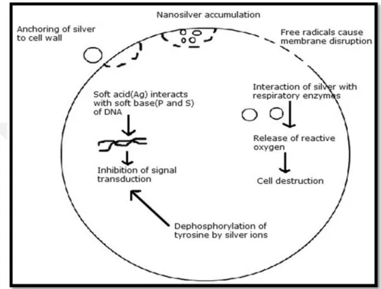
11 tyrosine deposits, which prompts signal transduction hindrance and in this manner the stoppage of growth. It is, however, necessary to understand that additional research is required on the topic to thoroughly establish the claims (Hatchett and White 1996) (Figure 2).
Figure 2. Different methods of activity of Ag-NPs on bacteria (Morones et al. 2005)
2.9. Application of Ag-NPs and their incorporation into other materials
NPs are of incredible attention because of their small size & a large surface to volume ratio, which prompt to both chemical & physical variances in their properties contrast with the bulk of main part of a similar chemical composition, like mechanical, biological properties, catalytic action, thermal and electrical conductivity, optical absorption and liquefying point (Daniel and Astruc 2004). Thus, the production of novel materials with modern methods can be performed by checking the particle shape and size at the nanometer scale. NPs show size and shape-dependent properties which are of enthusiasm for applications going from bio-sensing and catalysts to optics, antimicrobial activity, and electronic transistors. These particles likewise have numerous applications in various areas, for example,
12 medical imaging, nano-composite materials, filter medium, and medicine conveyance (Lee et al. 2008, Tan et al. 2006). Ag-NPs have drawn the consideration of scientists in light of their broad applications in territories, for example, incorporated circuits (Kasthuri, Kathiravan and Rajendiran 2009, Kotthaus et al. 1997), sensors (Kotthaus et al. 1997), bio-labelling, filters, antimicrobial antiperspirant filaments (Wenzheng and Guangwen 2003), cell electrodes (Klaus-Joerger et al. 2001), low-cost paper batteries (silver nano-wires) (Hong et al. 2006) and antimicrobials (Cho et al. 2005, Durán et al. 2007). Ag-NPs have been utilized broadly as antimicrobial agents in the health industry, food storage, texture coatings and various ecological applications (Durán et al. 2007, Cho et al. 2005).
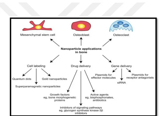
Figure 3. NPs applications in bone (Tautzenberger, Kovtun and Ignatius 2012).
In Figure 3, a medicinal NPs application in bone was presented. In general, therapeutic impacts of silver particles (in suspension form) depend on important aspects, involving particle size (surface region and power), particle shape (catalytic movement), particle concentration (therapeutic list) and particle charge (oligo dynamic quality) (Yoon et al. 2007). It was discovered that Ag-NPs attached to sulfur-containing proteins of bacteria, and caused death. In addition, fluorescent
13 estimations of cell-free supernatants demonstrated the impact of Ag-NPs on recombination of microbes. The connection of Ag+ or NPs to the cell divider caused the gathering of envelope protein precursors bringing about the prompt dispersal of the proton motive force (Liu and Zhao 2009). The catalytic technicality of Ag-NPs composites and their harm to the cell by collaboration with phosphorous and sulfur-containing compounds, for example, DNA, has been additionally investigated (Sharma, Yngard and Lin 2009). Besides, Ag-NPs displayed destabilization of the external film and break of the plasma layer, along these lines causing consumption of intracellular (Lok et al. 2006).
14 3. MATERIALS AND METHODS
3.1. Preparation of plant extracts
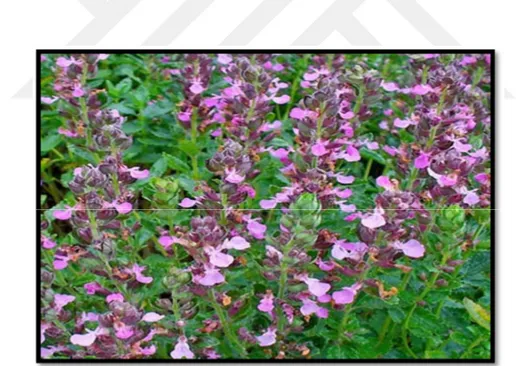
3.1.1. Plant extracts from Teucrium chamaedrys flowers
Dry flowers of Teucrium chamaedrys, (Figure 4) were washed many times with H2O to clean the dirtiness and dust particles. Then, that all of the flowers were powdered into uniform particles in a mortar by rubbing them with a porcelain bar. After that, the plant extract was prepared by adding 15 g of flower powder into pure water medium in a 500 ml of the conical flask. Then, the solution was held for 24 hr in an incubator. After that, the supernatant was isolated and filtered with a filter-paper with the assistance of vacuum filter. Following filtering step, the solution was utilized for converting silver ions (Ag+) into Ag-NPs (Ago) form.
15 3.1.2. Plant extracts from Inula helenium rhizomes
Dry rhizomes of Inula helenium (Figure 5) were washed many times with H2O to clean the dirtiness and dust particles. Then, that all of the rhizomes were grinded into uniform powder in a mortar by rubbing them with a porcelain bar. After that, plant extract was prepared by adding 10 g of rhizomes powder into pure water medium in a 500 ml of conical flask and the solution was incubated for 2 days. Then the supernatant was isolated and filtered with a filter-paper with the assistance of vacuum filter. After that, the solution was utilized for converting silver ions (Ag+) into Ag-NPs (Ago) form.
Figure 5. Inula helenium rhizomes
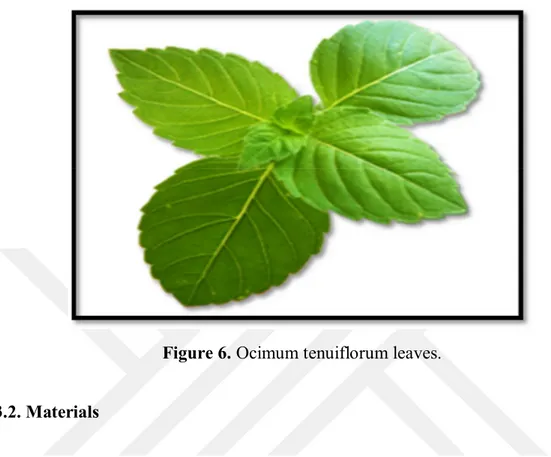
3.1.3. Plant extracts from Ocimum tenuiflorum leaves
Dry leaves of Ocimum tenuiflorum, (Figure 4) were washed many times with H2O to remove the dirtiness and dust particles. Then, that all of the leaves were grinded into uniform powder in a mortar by rubbing them with a porcelain bar. After that, the plant extract was prepared by adding 20 g of leaves powder into pure water medium in a 500 ml of conical flask. The solution was held for 24 hr in an incubator. And the supernatant was isolated and filtered with a filter-paper with the assistance
16 of vacuum filter. After that, the solution was utilized for converting silver ions (Ag+) into Ag-NPs (Ago) form.
Figure 6. Ocimum tenuiflorum leaves.
3.2. Materials
In experiments, there are many chemical and reagents were used directly in the green synthesis, characterization, and application of Ag-NPs. NaOH (98%) and HNO3 (65%) were purchased from Sigma-Aldrich. NaBH4 were taken from Merck KGaA Germany. Alizarin Red S dye was supplied from Sigma-Aldrich. AgNO3 with 97% purity (Panreac Quimica S.A.U) was utilized in the undertaken study and all standards and solutions have been prepared in deionized water by dissolving suitable quantity. All the experiments were conducted at room temperature.
3.3. Synthesis of Ag-NPs
Five different concentration ratios of plant extracts and silver nitrate solution were prepared (1:1, 2:1, 3:1, 4:1 and 5:1) by increasing the concentration of plant extracts in the solution phase. 5 ml AgNO3 solution was added to the prepared plant extract. After that, the reduced Ag-NPs were utilized for measuring UV-Vis spectra of the solution.
17 3.4. Characterization of Ag-NPs
Many methods are applied for the characterizing different Ag-NPs. Here we have discussed the essential techniques that have been utilized for the characterization of the Ag-NPs in this thesis. These methods are Ultraviolet-Visible spectrophotometer (UV-Vis), X-Ray Diffraction (XRD), Transmission Electron Microscopy (TEM), Atomic Force Microscopy (AFM) and Fourier Transforms Infrared spectroscopy (FTIR) (Umamaheswari, Lakshmanan and Nagarajan 2017).
3.4.1. UV-Vis Analysis
A visual property of Ag-NPs was analyzed by using UV-Vis spectrophotometer (HITACHI U-3900) (Umamaheswari et al. 2017). After the addition of AgNO3 to the plant extract, the spectrum of the solution was taken in various time periods (0 to 24 hr) between 400 nm to 470 nm wavelength.
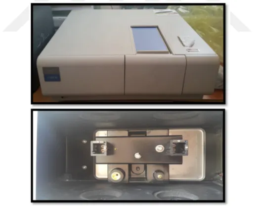
Figure 7. UV-Vis Spectrophotometer
Figure 7 shows UV-Vis spectrometer which applied for the measurement of absorbance of prepared solutions.
18 Figure 8. UV Cuvette
Figure 8. presents the UV cuvette used in UV-Vis analyses.
3.4.2. FTIR Analysis
A chemical structure of the produced Ag-NPs was studied by utilizing FTIR spectrometer (Vertex 70) and the spectrum was given in Figure 9. The implementation of this instrument was described in a detailed way in the reference (Durán et al. 2005).
Figure 9. (FTIR) Spectroscopy
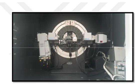
3.4.3. XRD Analysis
In XRD instrument, a large fraction of the X-rays are not easily absorbed or transmitted by the sample but, they are scattered quickly. At the point when an X-beam hits an atom, the electron clouds around the atom begin to sway with an indistinguishable recurrence from the incoming beam creating an electric field. All
19 directions have damaging involvement, that is, the joining waves are out of the stage and there is not any resultant energy leaving the solid specimen (Bruker AXS).
The atoms arranged like regular pattern in a crystal and later on the constructive interference can be obtained. The waves will be on stage and there will be very much characterized X-beams leaving the specimen under different headings. Thus, a diffracted ray might be portrayed as a ray made out of a substantial number of scattered beams commonly strengthening each other. X-ray diffraction provides a useful tool to study the construction and creation of the materials which is a key requirement for understanding materials properties as shown in (Figure 10) (Singhal et al. 2011).
Figure 10. X-ray diffraction
3.4.4. TEM Analysis
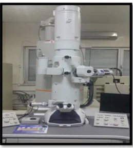
Morphology and size of the Ag-NPs were researched by TEM pictures utilizing (JEOL, JEM-2100 ELECTRON MICROSCOPE) instrument. A thin film of the specimen was set up on a carbon covered copper matrix by simply dropping a little amount of the specimen on the grid and drying under the bulb as shown Figure 11 (Singhal et al. 2011).
20
Figure 11. Transmission Electron Microscope
3.4.5. AFM Analysis
Atomic Force Microscopy (NT-MDT Molecular Devices and Tools for (Nano Technology)) was given in Figure 12. This device is the most adaptable and intense microscopic innovation for studying specimens at the nano-scale. The atomic power microscope can not only picture in three-dimensional topography, as well as gives different sorts of surface measurements to the requirements of researchers and engineers. It is effective on the grounds that an AFM can produce pictures at nuclear determination with angstrom scale determination elevation data with least specimen planning (Umamaheswari et al. 2017).
21 4. RESULTS AND DISCUSSION
4.1. UV-Vis Spectrophotometer analysis
After addition of Teucrium chamaedrys extract, Inula helenium extract and Ocimum tenuiflorum extract to the aqueous phase of AgNO3 of different concentrations, the mixture showed an imperceptible change in color at room temperature with time from yellowish to wine-red in 48 hours. The control experimental sets showed no variation in color under the same experimental conditions at the end of the reaction. The reduction of silver ion to Ag-NPs was reflected in spectral data acquired by utilizing UV-Vis spectrophotometer. It showed an absorbance peak around 460 nm for all five samples of Teucrium chamaedrys as shown in (Figure 13), 450 nm for all five samples of Inula helenium as shown in (Figure 14), and 450 nm /425 nm for all five samples of Ocimum tenuiflorum as shown in (Figure 15 A&B).
22 Figure 13. The absorbance spectrum of Ag-NPs with Teucrium chamaedrys extract
showing maximum absorbance near 460 nm. A) changing volume of Ag-NPs. B) changing volume of extract.
23 Figure 14. The absorbance spectrum of Ag-NPs with Inula helenium extract showing maximum absorbance near 450 nm. A) changing volume of Ag-NPs.
B) changing volume of extract.
24 Figure 15. The absorbance spectrums of Ag-NPs with Ocimum tenuiflorum extract
showing maximum absorbance at 450 nm. and 425 nm. A) changing volume of Ag-NPs. B) changing volume of extract.
In all experiments pH of the solution phase was adjusted to 7 because this value is optimum value to obtain a better spectrum. When compared to the three plant extracts after UV-Vis Spectrophotometer analysis we found that Teucrium chamaedrys extract was better than the others. The spectrum recorded at 460 nm and this value was more convenient compared to the other extracts. The magnitude, peak wavelength and spectral bandwidth of the spectrum of nanoparticles are dependent on particle size, shape, and material composition. The speed of interaction of Teucrium chamaedrys extract with Ag-NPs was more than the other extracts. We confirmed that Teucrium chamaedrys extract has more possibility to reduce Ag+ into Ago compared to the other extracts.
4.2. Transmission Electron Microscopy (TEM)
The size, shape, and crystallinity of the arranged Ag-NPs were investigated B
25 by utilizing TEM. The TEM works on basic criteria that cannot be distinguished the essential standards from the light magnifying microscope and it uses electrons rather than light. The wavelength of electrons is much smaller than that of light and the ideal determination achievable for TEM pictures was obtained from a light microscope. Therefore, TEM can detect the most interesting points of internal structure and again as little as individual atoms.
4.2.1. TEM analysis of Teucrium chamaedrys Ag-NPs
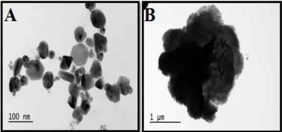
TEM analysis of a sample containing 10 mL of Teucrium chamaedrys Ag-NPs was realized. The analysis was done by the reaction of 4 ml of leaf extract, 1 ml silver nitrate solution and 5 ml H2O and these results were represented in Figure 16. TEM image showed a large portion of the particles are almost spherical, while several of them are slightly elliptical in shape with sizes of up to 500 nm in Figure 16. (A). Figure 16. (B) is a histogram of size distribution of Ag-NPs shows the obtained nanoparticles sizes are in the range of 20–84 nm and a few of them are agglomerated. It is evident that there is variation in particle sizes and the average size estimated at 64.8 nm.
Figure 16. (A) TEM image of Ag-NPs produced with Teucrium chamaedrys .
26 Figure 16. (B) A histogram of size distribution of Teucrium chamaedrys
Ag-NPs
4.2.2. TEM analysis of Inula helenium Ag-NPs
The sample contains 10 mL of Inula helenium Ag-NPs. The sample for the measurement was prepared by the reaction of 4 ml of leaf extract, 2 ml silver nitrate solution and 4 ml H2O. The final situation after the reaction was represented in Figure 17. TEM image (A) shows the mixture of plates (triangles, pentagons, and hexagons) and spheres. The most spherical shapes were prevalent. It’s clear that the triangles, pentagons, and hexagons structures are plate structures with sizes of up to 500 nm. Figure 16. (B) is a histogram of size distribution of Ag-NPs. This figure shows the obtained nanoparticles and they are in the range of sizes 15–80 nm and a few of them are agglomerated. It is evident that there is variation in particle sizes and the average size estimated at 47.5 nm.
27 Figure 17. (A) TEM image of Ag-NPs produced with Inula helenium
Figure 17. (B) A histogram of size distribution of Inula helenium Ag-NPs
4.2.3. TEM analysis of Ocimum tenuiflorum Ag-NPs
TEM analysis of a sample containing 10 mL of Ocimum tenuiflorum Ag-NPs was completed for this aim. 3 ml of leaf extract, 2 ml silver nitrate solution and 5 ml H2O were replaced in a container. After the reaction completed, TEM image was taken. Figure 18. (A) shows the analysis. When the morphology of the
28 nanoparticles was considered, it can be assumed in spherical form. Under cautious perception, it is apparent that the Ag-NPs encompassed by a dim thin layer of different materials, which we assume are capping organic material from Ocimum tenuiflorum leaf broth while several of them are slightly elliptical in shape with sizes of up to 500 nm. Figure 18 (B) is a histogram of size distribution of silver nanoparticles. It shows the obtained nanoparticles sizes they are in the range of sizes 3–20 nm and a few them are agglomerated in the solution. It is evident that there is variation in particle sizes and the average size was estimated at 9.5 nm.
Figure 18. (A) TEM image of Ocimum tenuiflorum Ag-NPs
29 The three plant extracts were compared with each other after TEM analysis. We found that Teucrium chamaedrys extract was more homogeny than the others. We can see that the distances between the particles are very small. For that reason, the molecules are coherent to each other.
4.3. FTIR Analysis
Fourier Transform Infrared Spectroscopy estimations were completed to analyze the peaks at bio-molecules for capping silver ion from the solution phase.
4.3.1. FTIR Analysis for Teucrium chamaedrys extract
50 ml of Teucrium chamaedrys extract were used for the FT-IR analysis and results were displayed in Figure 19. The FT-IR analysis revealed the strong bands at 1391.25 cm−1 corresponds to C-N stretching of the aromatic amine group and peak at 1009.28 cm−1 corresponds to N-H group. The bands between 2500-3500 cm-1 correspond to O-H stretching H-bonded alcohols and phenols.Furthermore, a stretch was found around 688.36 cm−1.
30 4.3.2. FTIR Analysis for Teucrium chamaedrys Ag-NPs
In this section, we used 50 ml from Teucrium chamaedrys Ag-NPs as shown in Figure 20. The FT-IR analysis revealed that the band for Ag-NPs was found around 500 cm-1 disappeared after reaction between the plant extract and Ag-NPs while the band appeared at 1371.78 cm-1 corresponds to C-N stretching of the aromatic amine group. The stretch for Ag-NPs was found around 823.84 cm-1 corresponds to Ag-NPs formation.
Figure 20. FTIR spectra for Teucrium Chamaedrys Ag-NPs
4.3.3. FTIR Analysis for Inula helenium extract
In this part of the experiment, we used 50 ml Inula helenium extract. After drying of the extract, FTIR spectrum was taken and given in Figure 21(a). The FT-IR analysis revealed that the strong bands at 3415.31 cm-1 correspond to O-H stretching H-bonded alcohols and phenols, while 2358.52 cm−1 and 2928.38 cm−1 corresponds to C-H group. The bands at 1618.95 cm−1, 1445.39 cm−1, and 104137 cm−1 show the stretching, C=C corresponds to aromatic amino groups.
31 4.3.4. FTIR Analysis for Inula helenium Ag-NPs
In this part of the experiment, 50 ml of solution from Inula helenium Ag-NPs was used for the production of Ag-NPs. Figure 21(b). The FT-IR analysis revealed that the band at 1000 cm-1 corresponds to Ag-NPs and reduced to 1038.48 cm-1 after reaction between the plant extract and Ag-NPs. The band appeared at 1623.77 cm-1 corresponds to C=C aromatic amino groups. Also, we can see other small peaks which disappeared after the interaction of extract with Ag-NPs.
Figure 21. FTIR spectrum of Inula helenium extract (a) FTIR spectrum of Inula helenium Ag-NPs (b)
4.3.5. FTIR Analysis for Ocimum tenuiflorum extract
The pellets of the solid powder Ocimum tenuiflorum extract and the extract capped with Ag-NPs was prepared for FT-IR analysis as shown was taken in Figure 22. The FT-IR spectrum belongs to Ocimum tenuiflorum extract was given in Figure 22 (a).
32 4.3.6. FTIR Analysis for Ocimum tenuiflorum Ag-NPs
The solid powder Ocimum tenuiflorum and the extract capped Ag-NPs was prepared for FT-IR analysis. As shown in Figure 22 (b), the curve represents plant extract with Ag-NPs. We observed here that some of the peaks were reduced at the band 1500 cm-1 corresponds to Ag-NPs after the interaction between the plant extract and Ag-NPs. Also, we can see the band 2400 cm-1 corresponds to C-H group the peaks disappeared, while the band appeared at 1391 cm-1 corresponds to N-H group. Also, we can see other bands at 2923 cm-1 corresponds to C-H group and it is transferred and disappeared after the interaction of extract with Ag-NPs.
Figure 22. FTIR spectra for Ocimum tenuiflorum extract (a) FTIR spectra for Ocimum tenuiflorum Ag-NPs (b)
4.4. XRD Analysis
4.4.1. XRD Analysis for Teucrium chamaedrys Ag-NPs
XRD spectrum shown in Figure 23 indicated the diffraction peaks around 22o, 30o, 36o, 42o.Which are indexed the (111), (200), (220) and (311) of the cubic
33 face-centered silver. These sharp Bragg pinnacles may have performed because of capping agent balancing out the nanoparticles. XRD pattern shows sharp peaks, therefore Bragg repercussion proposes that strong X-ray dispersing centers in the crystalline stage because of capping agents. The extract has capping agents and they influenced the synthesized of nanoparticles overgrowth and aggregation as well as to control the structural characteristics of the resulted nanoparticles and for their stability in a precise manner. XRD conclusions likewise proposed that the crystallization of the bio-organic phase happened on the surface of Ag-NPs. The expanding of crests in the XRD paradigms of solids is due to particle size effects. More extensive pinnacles imply littler particle size and reflect the impacts because of experiential conditions on the nucleation and development of the crystal nuclei. XRD pattern of crystalline materials shows sharp peaks while that of amorphous shows single broad diffused peaks in the crystalline stage of XRD pattern.
Figure 23. XRD spectra for Teucrium chamaedrys Ag-NPs
4.4.2. XRD Analysis for Inula helenium Ag-NPs
XRD spectrum of synthesized Inula helenium Ag-NPs as shown in Figure 24 displayed discrete diffraction crests about 38°, 45°, 65° and 78°. ° representing Bragg’s peaks with (111), (200), (220) and (311) planes respectively explain the
face-34 centered cubic (fcc). These sharp Bragg pinnacles may have come about because of crystalline nature of Ag-NPs. XRD pattern of crystalline materials shows sharp peaks while that of amorphous shows single broad widespread peak in the crystalline stage of XRD.
Figure 24. XRD spectra for Inula helenium Ag-NPs
4.4.3. XRD Analysis for Ocimum tenuiflorum Ag-NPs
The crystalline impression of Ocimum tenuiflorum Ag-NPs was confirmed by XRD analysis and showed in Figure 25 displayed four discrete diffraction peaks at 37.6°, 44.7°, 65° and 76.3°. The expanding of X-ray crests were basically because of the small particle size dimension. XRD study indicated that the synthesized plant extract capped silver nanoparticles were crystalline in nature. Four different bands having 2θ values of 37.6°, 44.7°, 65° and 76.3° representing Bragg’s peaks with (111), (200), (220) and (311) planes respectively explain the face-centered cubic (fcc) structure of Ocimum tenuiflorum capped silver nanoparticles as shown in Figure 25. Similarly, the XRD pattern of Ocimum tenuiflorum extract capped silver nanoparticles were pure crystalline in nature showing peaks with different values of 2Ѳ.
35 Figure 25. XRD spectra for Ocimum tenuiflorum Ag-NPs
4.5. Atomic force microscopy (AFM) Analysis
4.5.1. AFM Analysis for Teucrium chamaedrys extract
AFM results display the surface morphology of the monodisperse Ag-NPs utilizing Teucrium chamaedrys extract. The particle size of the Ag-NPs that ranges from 10 µm to 50 µm was observed. The topographical picture of Ag-NPs indicated that they are agglomerated and formed distinct nanoparticles as shown in Figure 26 & Figure 27. The shining spots on the micrograph showed that the Ag-NPs are in ball-shaped.
36 Figure 26. AFM for Teucrium chamaedrys extract.
Figure 27. AFM for Teucrium chamaedrys Ag-NPs.
4.5.2. AFM Analysis for Inula helenium extract.
AFM results display the surface morphology of the monodisperse silver nanoparticles obtained by using Inula helenium extract. The morphology of synthesized Ag-NPs was observed to be very changeful, and the greater part of them exhibited in ball-shaped. The particle size of the Ag-NPs that ranges from 10 µm to 50 µm was observed. As shown in Figure 28 and Figure 29, the Ag-NPs
37 manufactured by Inula helenium extract were bigger in size and less monodisperse in differentiation with those produced by the other examined species.
Figure 28. AFM for Inula helenium extract
Figure 29. AFM for Inula helenium Ag-NPs
38 4.5.3. AFM Analysis for Ocimum tenuiflorum extract
AFM results displayed the surface morphology of the monodisperse Ag-NPs obtained with Ocimum tenuiflorum extract as shown in Figure 30 and Figure 31. The roughness of the coating can be seen on the surface of the individual crystals of Ag-NPs. The particle size of Ag-NPs varied in the ranges from 10 µm to 50 µm. The images suggested that there was obviously a variation in crystal heights, but even within a larger context, there were over 1µm variations in topography between regions of crystalline development. It indicates that each crystal has a special characterized triangular cross-section.
39 Figure 31. AFM for Ocimum tenuiflorum Ag-NPs
When the three plant extracts were compared with each other after AFM analysis, we found that Teucrium chamaedrys extract was better than others because Teucrium chamaedrys extract is monodisperse (particles have the same shape) in comparison with others which are heterodisperse (particles have the different shape). Monodisperse Ag-NPs is better for the catalytic degradation of dyes.
4.6. The catalytic reduction of Alizarin Red S dye with prepared Ag-NPs
4.6.1. Alizarin Red S dye
A notable utilization of alizarin in modern times is a smearing operator in biological research in order to it smears free calcium and certain calcium compounds a red or light purple color. Alizarin keeps on being utilized commercially as a red textile dye.
40 The properties of Alizarin Red S dye:
-Solubility in water : slightly to sparingly soluble.
-Chemical formula : C14H8O4
-Acidity (pKa) : 6.94
Figure 32. Alizarin Red S dye.
In the research, the catalytic reduction of Alizarin Red S dye with Ag-NPs obtained from Teucrium chamaedrys extract was completed. The catalytic reduction of Alizarin Red S dye was performed (a quartz cuvette 4 cm high and with a 1cm optical path) in two steps. The first step is a presentation of a homogeneous solution of 1.5 ml Alizarin Red S dye and 0.5 ml deionized water (H2O) was prepared and then UV-Vis spectrophotometer analyze, after the addition of 0.1 ml sodium borohydride (NaBH4) solution and measured it again. Then, 0.1 ml of Ag-NPs was added to the mixture and measured at different times by taking pure water as a reference in another quartz cuvette. The distance between the light source and the cuvette containing the mixture was kept constant at all times during the measurements. The same procedures were tried again in a quartz cuvette by adding 0.1 ml of Ag-NPs. This sample was numbered as sample1 (S1). Then, the UV-visible spectra were
41 observed at a different time for samples S2 and S3. The same volume of Ag-NPs (0.1 ml) solution and the same concentrations of Alizarin Red S and NaBH4 were added to the sample container and S2 and S3 spectrums were measured at different times. The reduction periods started and it was completed after a short time, the shape and the size of Ag-NPs were changed. Then the reduction of Ag-NPs reached to zero. The overall time for the reduction reached to zero was 30 min. and was showed in Figure 33.
Figure 33. Alizarin Red S, NaBH4 and Ag-NPs first step (homogeneous step)
In the second step, we added the same quantity from an aqueous solution of 1.5 ml from Alizarin Red S dye with 0.5 ml deionized water (H2O) the solution was measured by UV-Vis spectrophotometer. After that, 0.1 ml NaBH4 was added to the solution and UV-Vis measurement of the solution was completed. The difference in this step labeled as glass (A) and glass (B) was showed in Figure 34.
42 Figure 34. Glass A & B with Ag-NPs.
The glass A & glass B were weighted and after that Ag-NPs was added into the glasses and they were dried by a heater at 200 °C. Then, they were weighted again. The variation between the weights of them was presented as the value of Ag-NPs as shown in Figure 34. After that, these glasses were used to analyze with UV-Vis spectrophotometer.
The nanoparticles in glass (A) and glass (B) were replaced in the quartz cuvette. Then, the dye solution in the glass (A) was reduced by Ag-NPs. UV-visible spectrum was taken at a certain time for the sample S1. S2 was prepared by taking the same volume Ag-NPs in the glass (A) and the same concentrations of Alizarin Red S and NaBH4. The same procedure was completed for the glass (B), and then the reduction of Alizarin Red S was completed after a short time. The total time for the reduction of dye using Ag-NPs in glass (A) for S1 reached to zero was in 40 min. The overall time for the reduction using Ag-NPs in glass (A) for S2 was 35 min. as shown in Figure 35 and Figure 36.
43 Figure 35. The degradation of Alizarin Red S by using NaBH4, and Ag-NPs with
(glass A), S1
Figure 36. The degradation of Alizarin Red S by using NaBH4, and Ag-NPs with (glass A), S2
44 The overall time for the reduction of the dye by using glass (B) for S1 until reached to zero was 33 min. The overall time for the reduction of the dye by using glass (B) for S2 was 32 min. as shown in Figure 37 and Figure 38.
Figure 37. The degradation of Alizarin Red S by using NaBH4, and Ag-NPs with (glass B), S1 second step
Figure 38. The degradation of Alizarin Red S by using NaBH4, and Ag-NPs with (glass B), S2, second step
45 The first step was a homogeneous phase with mixing (using drops wise addition of Ag-NPs) and the second step heterogeneous phase using Ag-NPs without mixing (using glass with Ag-NPs). They were compared each other by observing that first step was better because the spending time for the degradation was short and this explained that when applying Ag-NPs as drop wise faster than using Ag-NPs on the surface of the glass. It was proved that when using Ag-NPs covered on the surface of the glass, the reaction became slow because the density of the solution contains glass is higher than the Ag-NPs in the solution phase.
Figure. 39. Reduction of Alizarin Red S after treatment of dye with 0.5 mg of Teucrium chamaedrys extract mediated AgNPs immobilized on a cover slip. The
46 5. CONCLUSIONS
A fast, eco-friendly, and convenient green method was developed for the synthesis of Ag-NPs from Ag-NPs nitrate using plant extracts. It is green, high yield, fast, and low-cost approach for producing Ag-NPs. The color changes of plant extract occurred in the production phases due to the surface reaction during the interaction of the components present in the extracts. The formation of Ag-NPs were resulted and this formation was confirmed by AFM, XRD, FT-IR, UV–Vis spectroscopy, and TEM.
There are many ways to synthesize Ag-NPs that include the physical, chemical, and biological approaches. For the production of Ag-NPs physicochemical approach is very expensive and also using of a toxic chemical is not an effective process. Ag-NPs have antimicrobial, anti-inflammatory, antioxidant and anti-cancer properties. The green chemistry synthetic path was applied to obtain Ag-NPs in our research. The field of nanotechnology is still in its infancy, and further research should focus on the mechanism of Ag-NPs formation that may lead to accurate monitoring of the process that eventually leads to the synthesis of nanoparticles with strict control over the size and shape. In this research, Ag-NPs were successfully synthesized by a biological method from the plant extracts. This approach to green chemistry towards the synthesis of Ag-NPs has many advantages. The process is easy and it is economic. The quick biological synthesis of Ag-NPs using plant extracts appears environmental friendly and simple. It is an effective way for a synthesis of Ag-NPs. Most importantly, the reaction was simple and convenient to handle and believed to have advantages over other biological synthesis.
During the study optimization of various reaction, the parameters were carried out such as precursor salt concentration, the volume of extracts. pH is an important factor which can play a vital role in the fabrication of nanoparticles. In our study pH was found to be 7 to obtain small size nanoparticles by controlling size at nm level.
47 When three plant extracts were compared after UV-Vis Spectrophotometer analysis we found that Teucrium chamaedrys extract was better than the others. The maximum peak was recorded at 460 nm. This value was bigger than the others extracts. The magnitude of the spectrum, peak wavelength and spectral bandwidth of the nanoparticles spectrums are dependent on particle size, shape, and plant extract composition. The speed of interaction for Teucrium chamaedrys extract with Ag-NPs was more than the others plant extracts. It was concluded that Teucrium chamaedrys extract has more ability to reduce Ag+ into Ago than others plant extracts.
When three plant extracts were compared after TEM analysis we found that Teucrium chamaedrys extract was more homogeneous than the other plant extracts. It can be seen that from the distances between the nanoparticles are very small. For this reason, the molecules look more coherent to each other.
When compared to the three plant extracts after AFM analysis, we found that Ag-NPs produced with Teucrium chamaedrys extract was better than other extracts because monodisperse particles (the same shape) were produced with this extract compared to other extracts. They were in heterodisperse (particles with a different shape). Monodisperse Ag-NPs is better for the catalytic degradation of Alizarin Red S.
For the degradation of Alizarin Red S dye, various sizes Ag-NPs were synthesized by using Teucrium chamadryes extract. The UV-VIS results showed that peaks rely on the size of the Ag-NPs obtained by using the extracts. Ag-NPs is suitable catalyst of Alizarin Red S. In the solution phase, the small size of Ag-NPs had the most effective catalytic action, and the reaction rate decreased with increasing size of the Ag-NPs.
In the reduction of Alizarin Red S, first, dropwise addition of Ag-NPs homogenous solution was employed in the solution phase including the dye. Then, in the second step, Alizarin Red S solution was inserted in the heterogenic solid
Ag-48 NPs phase on the glass surface. This stage is without mixing of Ag-NPs with Alizarin Red S. That means direct contacting of the dye with Ag-NPs on the surface of the glass.
When comparing two methods, we observed that the first method was better than the second method because the spending time for the degradation of the dye solution was short. This explained that the application of Ag-NPs in the solution was faster than Ag-NPs applied on the glass surface. When using the Ag-NPs on the surface of the glass was less effective than the dropwise addition of Ag-NPs. The reaction became in a slow way when Ag-NPs applied on the stationary surface because the amount of Ag-NPs in the solution phase is higher than Ag-NPs on the glass surface. The applied Ag-NPs were recovered easily and applied in multiple cycles without losing in catalytic efficiency.
FUTURE WORK
In future plans, I will use Ag-NPs generated from other plant extracts and their applications in different dyes. The importance of Ag-NPs application in pharmaceutical, medicinal, dentistry, and industrial areas will be considered. We will find the different implementation of Ag-NPs in many different technologies.