Aim: This study evaluated the effect of different irrigation protocols on elastic modulus and biomechanics of single‑rooted premolar tooth using with nano‑indentation and finite element analysis (FEA). Materials and Methods: Root canals of single‑rooted human teeth were prepared, divided into eight groups, and irrigated with (1) 2.5% NaOCl + 17% EDTA; (2) 2.5% NaOCl + 17% EDTA + 2.5% NaOCl; (3) 2.5% NaOCl + SmearClear; (4) 2.5% NaOCl + 2% chlorhexidine; (5) 1.3% NaOCl + MTAD; (6) 5.25% NaOCl; (7) 17% EDTA; and (8) saline. The roots were vertically sectioned, and elastic modulus of the root dentine was measured using nano‑indenter device at coronal, middle, and apical third. Data were recorded as megapascal and statistically analyzed (one‑way analysis of variance, Tukey tests). Three‑dimensional FEA model of a premolar tooth was created, and the inner root dentine was modified to simulate the effect of irrigation protocols on root dentine. The elastic properties of inner root dentine layer in the FEA models were modified for each group according to the data obtained with nano‑indentation. A 300‑N load was applied at the buccal cusp and central fossa of the models with a 45° angle. The stresses were calculated using von Mises stress criteria. Results: All irrigation protocols affected the elastic modulus of root dentine. Groups 2 and 3 showed similar elastic modulus values (P > 0.05), whereas the lowest values were obtained in group 7 (P < 0.05). No statistically significant differences were found between groups 4, 5, and 8 (P > 0.05). Conclusion: Despite the effect of different clinically used irrigation protocols on elastic modulus of the inner dentine, this does not affect the biomechanics of the roots.
Keywords: Dentine, elastic modulus, finite element analysis, irrigants, nano-indentation
The Effect of Different Irrigation Protocols on Elastic Modulus
of Dentine and Biomechanics of Single‑Rooted Premolar Tooth:
A Nano‑Indentation and Finite Element Analysis Study
B Durmuş, AA Hale1, E Oğuz2, B Sema1
of tetracycline, citric acid, and detergent) are other widely used solutions.[4]
Studies have indicated that irrigation solutions affect the mechanical properties of dentine.[5,6] There are several techniques and tools for analyzing the mechanical
Introduction
C
leaning with root canal irrigants is themost accepted method for the removal of tissue remnants and dentine debris during
instrumentation.[1] The most commonly used
irrigating solutions are sodium hypochlorite (NaOCl), ethylenediaminetetraacetic acid (EDTA), and chlorhexidine (CHX);[2] SmearClear (SybronEndo, Orange, CA, USA) (contains 17% EDTA, cetrimide, and a specific surfactant)[3] and MTAD (Dentsply, Tulsa, OK, USA) (an irrigant consisting Department of Endodontics, Faculty of Dentistry, University of Necmettin Erbakan, Departments of 1Endodontic and 2Prosthodontics, Faculty of Dentistry, University of Selçuk, Konya, Turkey
Abstract
Address for correspondence: Dr. B Durmuş, Department of Endodontics, Faculty of Dentistry, University of Necmettin Erbakan, Konya, Turkey. E‑mail: [email protected]
This is an open access journal, and articles are distributed under the terms of the Creative Commons Attribution‑NonCommercial‑ShareAlike 4.0 License, which allows others to remix, tweak, and build upon the work non‑commercially, as long as appropriate credit is given and the new creations are licensed under the identical terms.
For reprints contact: [email protected]
How to cite this article: Durmuş B, Hale AA, Oğuz E, Sema B. The effect of different irrigation protocols on elastic modulus of dentine and biomechanics of single-rooted premolar tooth: A nano-indentation and finite element analysis study. Niger J Clin Pract 2019;22:101-7.
Date of Acceptance: 19-Oct-2018
Access this article online
Quick Response Code:
Website: www.njcponline.com
DOI: 10.4103/njcp.njcp_85_18
properties of materials. Nano‑indentation is one of the widely used one among these. The advantage of nano‑indentation test over the others is that it is capable of probing the mechanical properties and behavior of small volumes of dental structures.[7]
The biomechanics of the tooth is an important parameter for its survival.[8] Structure loss because of several reasons is a major problem in endodontically treated teeth because changes in the elastic properties of dentine may affect the biomechanics of the tooth, since there is limited dentine when compared with sound teeth.[8] Finite element analysis (FEA) is a well‑known technique that is widely used in all fields of science and dentistry. Some dental and medical research that cannot be conducted on live tissues or simulated in a laboratory can be carried out using FEA.[9]
The effect of irrigation protocols on the elastic modulus of dentine was studied by several researchers in the past.[5,6] To our knowledge, none of those studies conducted a detailed measurement from coronal to apical and from canal lumen to the inner root dentine. Furthermore, clinically accepted and widely used irrigation protocols were not compared in detail. The effect of irrigation solutions on the elastic modulus of dentine was simulated in a previous FEA study;[8] however, the elastic properties of the structures were provided from the manufacturers or from previously published articles. The effect of changes in the elastic properties of dentine because of widely used clinical irrigation protocols was also not simulated in the past. Thus, this study was planned to evaluate the effect of different clinically used irrigation protocols on the elastic modulus of root dentine from coronal to apical and from canal lumen to inner root dentine, and to evaluate the effect of these changes on stresses within the tooth under loading using FEA. The first hypothesis to be tested in the study was that different irrigation protocols do not change the elastic modulus of root dentine from coronal to apical and from canal lumen toward the inner root dentine. The second hypothesis of the study was that changes in the elastic modulus of dentine because of different clinically used irrigation protocols do not affect the stress distributions within the endodontically treated tooth.
Materials and Methods
This study protocol was approved by the Ethical Committee of Medical School, Selçuk University, Konya, Turkey (protocol number 2012/10). Forty single‑rooted extracted premolar teeth were selected with similar dimensions, and without caries, coronal or root filling, or cracks. The soft tissue remnants covering the
root surfaces were removed using 0.01% NaOCl with gauze and a fine brush. The teeth were then stored until use in distilled water at 4°C for a maximum of 1 month. The crowns were removed with a high‑speed bur under water cooling, and root lengths were standardized to 15 ± 1 mm. The roots were randomly divided into five groups according to the irrigation protocol (n = 5) and three control groups (n = 5). Root canals were then prepared using ProTaper rotary files (Dentsply, Maillefer, Ballaigues, Switzerland) to size F4 of the ProTaper instrument. The sequence followed was SX, S1, S2, F1, F2, F3, and F4.
The irrigation protocols were done with a 30‑gauge slot‑tipped needle, and the study groups were as follows: Group 1: 2 mL 2.5% NaOCl (Çağlayan Kimya, Konya, Turkey) after each file change and final 5 mL 17% EDTA (Werax, Spot Diş Deposu, İzmir, Turkey) for 5 min.
Group 2: 2 mL 2.5% NaOCl after each file change, final 5 mL 17% EDTA for 5 min, and then 5 mL 2.5% NaOCl. Group 3: 2 mL 2.5% NaOCl after each file change and final 5 mL SmearClear for 5 min.
Group 4: 2 mL 2.5% NaOCl after each file change and final 5 mL 2% CHX (Klorhex, Drogsan, Ankara, Turkey) for 5 min.
Group 5: 2 mL 1.3% NaOCl (Çağlayan Kimya) after each file change and final 5 mL Biopure MTAD for 5 min.[6]
Group 6: 2 mL 5.25% NaOCl (Wizard, Rehber Kimya, İstanbul, Turkey) after each file change and final 5 mL 5.25% NaOCl for 5 min.
Group 7: 2 mL 17% EDTA after each file change and final 5 mL 17% EDTA for 5 min.
Group 8: 2 mL saline after each file change and final 5 mL saline for 5 min.
Before CHX and Biopure MTAD irrigations, root canals were irrigated with 2 mL of distilled water to avoid colored precipitate. All roots were irrigated with 10 mL of distilled water finally to remove the remaining irrigants.
Nano‑indentation testing
The roots were split in the buccolingual direction with a low‑speed diamond saw (Accutom‑50; Struers, Copenhagen, Denmark) under constant water irrigation. Samples were embedded in epoxy resin (Epon 815; Nissin, Tokyo, Japan). After setting, exposed surfaces were polished using carbide paper with a grit size of 600–2000. This was followed by polishing with 3 and 1
μm polishing diamond paste in sequence. Samples were stored in distilled water at 4°C until nano‑indentation testing (CSM Instruments, SA, Switzerland). The indenter used was a pyramidal‑triangular‑shaped Berkovich indenter, and the maximum force applied was 20 mN. Each indent was accomplished at a 40‑mN/min loading ratio and a 20‑s delay of the maximum load at the intertubular dentine. The maximum penetration depth was 1400 nm. The nano‑indentation test was performed in each sample from apical to the cervical region at 2.5, 5.5, and 8.5 mm for the measurement of the elastic modulus. Three measurements were done in each test region starting from the root canal lumen toward the cement [at 10, 50, and 90 μm; Figure 1]. Each group had a total of 45 indentations (3 regions × 3 deepness × 5 samples).
FEA
A three‑dimensional FEA model of a mandibular premolar tooth was allocated based on the anatomical data described by Nelson.[10] The FEA model included enamel, dentine, cementum, pulp, and periodontal ligament and consisted of 105,443 nodes and 68,036 elements. Assuming that the root canal was prepared using a rotary file, it was enlarged from 1 mm less than the apex to the coronal with a 6% taper. The FEA study models were modified based on this initial model. In these models, a 90‑μm‑thick additional layer associated with the root canal lumen was created within the root dentine. All the materials and structures used in this study were assumed to be homogeneous, isotropic, and linear elastic except for this 90‑μm‑thick dentine layer associated with the root canal lumen. Data, which were obtained from the nano‑indentation test for each irrigation protocol for each region and deepness (total of 45 indentations [3 regions × 3 deepness {10, 50, and 90 μm} × 5 samples]), were averaged and used as the elastic modulus of this dentine layer to simulate the affected dentine. The elastic modulus of the remaining root dentine structure was achieved from the measurements, which was obtained from the saline group. The elastic modulus and Poisson’s ratios of all the other materials and structures used were obtained from the literature[8,9] or from the manufacturers [Table 1]. Initially, the root canal models were assumed to be prepared with irrigation protocols and unfilled, and then the same models were assumed to be root‑filled and coronally restored [Figure 2].
A 300‑N load was applied to the functional buccal cusp and central fossa at a 45° angle to the long axis of the tooth to calculate the stress patterns.[9] Nodes at the outer surface of the roots were assumed as fixed in all directions to calculate the stress distributions.
To simulate adhesion between the structures, all interfaces were considered as completely bonded. The finite elements on the x, y, and z‑axes for each model were assumed as fixed for the boundary conditions. FE modeling was accomplished using the SolidWorks software program (SolidWorks 2009; SolidWorks Corp., Concord, MA, USA), and analyses were run using the COSMOSWorks structural‑analysis program (SolidWorks Corp., Waltham, MA, USA).
The stresses were recorded using von Mises criteria. A standard view of a mid‑sagittal section from each model was provided. The stress distribution scale range was limited to 0–10 MPa. To provide better visualization, calculated numeric data were transformed into color images.
Statistical analysis
Elastic modulus values were statistically analyzed using SPSS 15.0 (SPSS, Chicago, IL, USA) for Windows, using one‑way analysis of variance with a subsequent Tukey’s multiple comparison test (P < 0.05).
Results
Nano‑indentation test
Table 2 shows the mean elastic modulus values after treatment with different irrigation protocols. The results of the statistical analysis according to the regions were as follows:
Coronal region: No difference was found among the elastic modulus of dentine in groups 4, 5, and 8 (P > 0.05). These three groups were found to be significantly different from the others (P < 0.05). No difference was found among groups 1, 2, 3, and 6 (P > 0.05). Elastic modulus of the dentine was found to be lowest in Group 7 (Cosmos <0.05).
Figure 1: Nano‑indentaion measurement points at three regions from root canal lument to cementum
Midcoronal region: No difference was found among groups 1, 4, 5, 6, and 8 (P > 0.05). The elastic modulus values of these groups were significantly higher than the other test groups (P < 0.05). Groups 2 and 3 showed similar elastic modulus values (P > 0.05), while the
lowest values were obtained in group 7 with 17% EDTA (P < 0.05).
Apical region: Similar to the coronal region, the elastic modulus values were higher in groups 4, 5, and 8 (P < 0.05). Groups 1, 2, 3, and 6 were also found to be similar to each other (P > 0.05) and significantly different from the other groups (P < 0.05). The lowest values were found with 17% EDTA (P < 0.05).
FEA
Table 3 shows the maximum and total von Mises stress values for each group. The maximum stress values in root dentine were found to be similar in groups 4 (2.5% NaOCl + 2% CHX), 5 (1.3% NaOCl + MTAD), and 8 (Saline), whereas affected dentine showed higher maximum stress values in the models that were assumed to be prepared but kept unfilled. The models that were assumed to be root‑filled showed almost identical maximum stress values in dentine. The affected dentine showed higher stress values in groups 4, 5, and 8. The affected dentine showed lower values in groups 2 (2.5% NaOCl + 17% EDTA + 2.5% NaOCl) and 7 (17% EDTA) in the unfilled models and higher values in groups 4, 5, and 8. Nevertheless, total stress values were similar in all groups.
Figures 3 and 4 show the stress distributions and accumulations within the models, which represent teeth treated with different irrigation protocols. von Mises stress values and domains in the unfilled models did not show any differences among the groups. It was observed
Figure 3: Von Mises stress patterns calculated in the corresponding FEA models within the prepared models. Stress values were specified in the color scale. (1) 2.5% NaOCl + 17% EDTA, (2) 2.5% NaOCl + 17% EDTA + 2.5% NaOCl, (3) 2.5% NaOCl + SmearClear, (4) 2.5% NaOCl + 2% chlorhexidine, (5) 1.3% NaOCl + MTAD, (6) 5.25% NaOCl, (7) 17% EDTA, (8) saline, (9) natural tooth
Table 1: Mechanical properties of the isotropic and unisotropic materials used
Elastic modulus (GPa) Poisson’s ratio (ν) Dentine Group 1a 17.25 0.31 Group 2a 15.56 0.31 Group 3a 16.48 0.31 Group 4a 18.52 0,31 Group 5a 18.31 0.31 Group 6a 17.11 0.31 Group 7a 14.01 0.31 Group 8a 19.64 0.31 Enamel[9] 41 0.31 Pulp[9] 0.003 0.45 Periodontal ligament[9] 0.0000689 0.45 Composite resin[8] 12 0.30 Gutta‑percha[9] 0.14 0.45
aAcquired from the manufacturer
Table 2: Mean elastic modulus values of root dentine obtained at totally 90 µm (mean±SE)
Groups Elastic Modulus (GPa)
Region Mean
Apical Middle Cervical
1 17.5±0.81b 17.13±0.57a 17.12±0.73b 17.25±0.4 2 14.88±0.68b 16.65±0.63b 15.16±0.48b 15.56±0.35 3 17.71±0.82b 16.52±0.7b 15.2±0.5b 16.48±0.41 4 19.17±0.66a 17.92±0.86a 18.46±0.67a 18.52±0.42 5 18.6±0.53a 18.5±0.64a 17.82±0.59a 18.31±0.33 6 16.76±0.78b 17.21±0.69a 17.34±0.76b 17.11±0.42 7 14.42±0.75c 13.89±0.63c 13.71±0.66c 14.01±0,38 8 19.8±0.87a 20.12±0.9a 18.99±0.99a 19.64±0.52 The common letters on the columns are not statistically significant (a > b > c > d; P<0.05). SE=Standard error
Figure 2: The structures, materials, and the loading conditions of the FEA models. (a) The unfilled model and (b) the model assumed to be filled with gutta‑percha and restored using composite resin
b a
that the stress intensity of the input cavity enamel dentine junction consisted of, but was dispersed in, the natural tooth models that stress coronal dentine and enamel [Figure 3].
von Mises stress values and domains in the models that were considered filled were almost identical [Figure 4]. High stress accumulations were observed along the root canal dentine walls and access cavity.
Discussion
The penetration of different irrigation solutions into dentine has shown different results in previous studies because distinct testing protocols were used. Kuga et al.[11] found the depth to be 107 μm when dentine bars were placed in the irrigation solution for 20 min, while Zou et al.[12] recorded a depth of 77–123 μm when using different concentrations for 2 min. In this study, the measurements were performed at 10, 50, and 90 μm, starting from the root canal lumen and considering that the maximum penetration depth of irrigation protocols is around 100 μm.[11] Three measurements were carried out for each region (coronal, median, and apical) and a distance of 40 μm was preserved between the measurement points to avoid residual stresses, as mentioned by He et al.[13] The results indicated that different irrigation protocols affected the elastic modulus of dentine; thus, the first hypothesis of the study that different irrigation protocols do not change the elastic modulus of root dentine from coronal to apical and from canal lumen toward the inner root dentine should be rejected.
In this study, irrigation protocols were applied at room temperature; thus, the effect of temperature was eliminated. In contrast to previous studies, which used high concentrations of irrigation solutions for longer periods,[5,6] this study’s irrigation protocols were conducted in accordance with clinical protocols to get more clinically relevant results. For this reason, lower concentrations of NaOCl, Biopure MTAD, SmearClear, and CHX were used as final irrigating solutions and then compared with control groups of EDTA, 5.25% NaOCl, and saline.
The effect of NaOCl on the elastic modulus of root dentine has previously been published.[14] This effect may change depending on the concentration of the solution.[5] In this study, dentine bars[6] or extended application times for the irrigation solutions[12] were not used; instead, clinical protocols were followed. The results indicated that the elastic modulus of dentine decreased with increased concentration of NaOCl [5.25% vs. 1.3%; Table 2]. Conversely, Grigoratos et al.[5] found that NaOCl concentrations Figure 4: Von Mises stress patterns calculated in the corresponding
FEA models within the filled/coronally restored. Stress values were specified in the color scale. (1) 2.5% NaOCl + 17% EDTA, (2) 2.5% NaOCl + 17% EDTA + 2.5% NaOCl, (3) 2.5% NaOCl + SmearClear, (4) 2.5% NaOCl + 2% chlorhexidine, (5) 1.3% NaOCl + MTAD, (6) 5.25% NaOCl, (7) 17% EDTA, (8) saline, (9) natural tooth
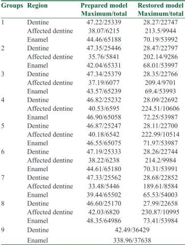
Table 3: Maximum and total von Mises stress values obtained within the FEA models representing prepared
but unfilled and root‑filled teeth
Groups Region Prepared model
Maximum/total Restored model Maximum/total
1 Dentine 47.22/25339 28.27/22747 Affected dentine 38.07/6215 213.5/9944 Enamel 44.46/65188 70.19/53992 2 Dentine 47.35/25446 28.47/22797 Affected dentine 35.76/5841 202.14/9286 Enamel 42.04/65331 68.01/53997 3 Dentine 47.34/25379 28.35/22766 Affected dentine 37.19/6077 209.4/9701 Enamel 43.57/65239 69.4/53993 4 Dentine 46.82/25232 28.09/22692 Affected dentine 40.53/6595 224.51/10606 Enamel 46.90/65058 72.25/53987 5 Dentine 46.87/25247 28.11/22700 Affected dentine 40.18/6542 222.99/10514 Enamel 46.55/65075 71.97/53987 6 Dentine 47.19/25333 28.26/22744 Affected dentine 38.22/6238 214.2/9984 Enamel 44.61/65180 70.31/53991 7 Dentine 47.33/25562 28.68/22852 Affected dentine 33.48/5446 189.61/8584 Enamel 39.44/65502 65.53/54003 8 Dentine 46.60/25170 27.99/22658 Affected dentine 42.03/6820 230.87/10995 Enamel 48.35/64986 73.41/53984 9 Dentine Enamel 42.49/36429 338.96/37638
A natural tooth was added as ninth group for comparison; values are given in MPa
did not significantly decrease dentine’s elastic modulus. These different results might be because of the conditions mentioned above.
The common concentrations used for EDTA are 15%–17%.[15] When the elastic modulus of dentine was evaluated in this study according to the regions, the effect of 17% EDTA on root dentine from coronal to the apical was found to be similar [Table 2]. The lowest values were achieved with 17% EDTA [Group 7; Table 2], while its effect on elastic modulus of root dentine was changed when used in combination with NaOCl [groups 1 and 2; Table 2]. NaOCl irrigation followed by 17% EDTA opens the dentinal tubular orifices, erodes the intertubular dentine, and reduces dentine microhardness.[16‑18] Several in vitro studies have shown that this combination removed the inorganic and organic phases of the dentine.[19‑21]
The toughness of dehydrated dentine is significantly lower than when in the hydrated state.[22,23] When the dentinal tubules lose water, the “water‑induced effects” such as inherent plasticity and distinct strain response in the directions parallel and perpendicular to the dentinal tubules are lost.[19,24] Kinney et al.[25] reported different values when the elastic modulus of dentine was measured under dry or wet conditions (23.9 vs. 20 GPa, respectively). This water loss was reported to be fully restorable by rehydration. In this study, the dentine samples were kept in wet conditions between the measurements; however, the testing procedure was applied under dry conditions with dynamic loading. Considering the rapidity of the testing procedures, the possible effect of dehydration on the elastic modulus of dentine was ignored.
FEA is a widely used technique; however, many details are idealized, simplified, or ignored.[9] Three‑dimensional modeling is therefore necessary. However, it is not possible to simulate all of the structures because they are not homogeneous; this is the case with dentine. Therefore, in this study, all the structures except dentine were assumed to be homogeneous.[8] A perfect simulation still could not be carried out for dentine because an affected dentine layer was modeled and the elastic modulus of the affected dentine layer (average of coronal, middle, and apical values) was achieved from the first part of the study. While doing this, the effects of dentinal tubules, intrapulpal hydrostatic pressure, and the elastic modulus gradient on the mechanical properties of dentine were ignored, as in the previous FEA studies.[8] This is one other important limitation of this study. The stress distributions or accumulations within the FEA models were found to be similar in this study, showing
that the outcome of the elastic modulus change does not affect the biomechanics of the roots. Therefore, the second hypothesis that changes in the elastic modulus of dentine because of different irrigation protocols do not affect the stresses within an endodontically treated tooth must be accepted.
Belli et al.[8] reported that both MTAD and 17% EDTA changed the stress dynamics within the root dentine when the clinical protocol was not simulated. As a result, these authors concluded that MTAD should be used according to the clinical protocol as advised by Machnick et al.[6] Therefore, in this study, a clinical protocol of 1.3% NaOCl with MTAD was applied. MTAD showed similar results with physiological saline, confirming the results of this previously published article. In addition, this result should be related to the concentration of 1.3% NaOCl.
The maximum stress was noticeably reduced toward the apical region of the root [Figure 4]. This decreased stress distribution in the middle/apical third of the root was attributed to the shape of the tooth and its interaction with the supporting bone.[19] Also, a number of studies have suggested that there are no major differences in the mechanical properties of dentine from teeth with vital pulp and root‑filled teeth.[26,27]
The maximum and total stress values were similar in CHX, MTAD, and saline [Table 3]. In accordance with previous studies, the results of this study showed that using the clinical protocol for MTAD and CHX causes no adverse effects on the mechanical properties of root canal dentine.[6,14] Considering the other advantages of CHX such as remineralizing the demineralized dentine[28] or other positive properties of MTAD,[4,6] the results of this study suggest that CHX and MTAD have beneficial qualities as root canal irrigants.
Conclusion
Within the limitations of this study it was concluded that (1) Despite the effect of different clinically used
irrigation protocols on elastic modulus of the inner dentine, this does not affect the biomechanics of the roots
(2) CHX and MTAD showed a similar effect with saline on the elastic properties of root dentine when used according to the clinical protocol.
Acknowledgements
This study was based on the work partially performed by Durmuş Alperen Bozkurt for fulfillment of the degree of Doctor of Philosophy, University of Selçuk, Turkey.
Financial support and sponsorship
Supported, in part, by Scientific Research Projects Coordination Center (BAP) of Selçuk University (12202015), Konya, Turkey and Selçuk University.
Conflicts of interest
There are no conflicts of interest.
References
1. Cruz‑Filho AM, Sousa‑Neto MD, Savioli RN, Silva RG, Vansan LP, Pecora JD. Effect of chelating solutions on the microhardness of root canal lumen dentin. J Endod 2011;37:358‑62.
2. Dutner J, Mines P, Anderson A. Irrigation trends among American Association of Endodontists members: A web‑based survey. J Endod 2012;38:37‑40.
3. Sadegh M, Sohrabi H, Kharazifard M, Afkhami F. Effect of smear clear and some other commonly used irrigants on dislodgement resistance of mineral trioxide aggregate to root dentin. J Clin Exp Dent 2017;9:e617‑21.
4. Zehnder M. Root canal irrigants. J Endod 2006;32:389‑98. 5. Grigoratos D, Knowles J, Ng YL, Gulabivala K. Effect of
exposing dentine to sodium hypochlorite and calcium hydroxide on its flexural strength and elastic modulus. Int Endod J 2001;34:113‑9.
6. Machnick TK, Torabinejad M, Munoz CA, Shabahang S. Effect of MTAD on flexural strength and modulus of elasticity of dentin. J Endod 2003;29:747‑50.
7. Marshall GW, Habelitz S, Gallagher R, Balooch M, Balooch G, Marshall SJ. Nanomechanical properties of hydrated carious human dentin. J Dent Res 2001;80:1768‑71.
8. Belli S, Eraslan O, Eraslan O, Eskitascioglu M, Eskitascioglu G. Effects of NaOCl, EDTA and MTAD when applied to dentine on stress distribution in post‑restored roots with flared canals. Int Endod J 2014;47:1123‑32.
9. Belli S, Eraslan O, Eskitaşcıoğlu G. Effect of different treatment options on biomechanics of immature teeth: A finite element stress analysis study. J Endod 2018;44:475‑79.
10. Nelson SJ. The permanent mandibular premolars. In: Wheeler’s Dental Anatomy, Physiology and Occlusion. 10th ed. St Louis, MO: Saunders; 2015. p. 159‑63.
11. Kuga MC, Gouveia‑Jorge E, Tanomaru‑Filho M,
Guerreiro‑Tanomaru JM, Bonetti‑Filho I, Faria G. Penetration into dentin of sodium hypochlorite associated with acid solutions. Oral Surg Oral Med Oral Pathol Oral Radiol Endod 2011;112:e155‑9.
12. Zou L, Shen Y, Li W, Haapasalo M. Penetration of sodium hypochlorite into dentin. J Endod 2010;36:793‑6.
13. He LH, Fujisawa N, Swain MV. Elastic modulus and stress‑strain response of human enamel by nano‑indentation. Biomaterials 2006;27:4388‑98.
14. Ari H, Erdemir A. Effects of endodontic irrigation solutions on mineral content of root canal dentin using ICP‑AES technique. J Endod 2005;31:187‑9.
15. Perez F, Rouqueyrol‑Pourcel N. Effect of a low‑concentration EDTA solution on root canal walls: A scanning electron microscopic study. Oral Surg Oral Med Oral Pathol Oral Radiol Endod 2005;99:383‑7.
16. Çalt S, Serper A. Smear layer removal by EGTA. J Endod 2000;26:459‑61.
17. Niu W, Yoshioka T, Kobayashi C, Suda H. A scanning electron microscopic study of dentinal erosion by final irrigation with EDTA and NaOCl solutions. Int Endod J 2002;35:934‑9. 18. Saleh A, Ettman W. Effect of endodontic irrigation solutions on
microhardness of root canal dentine. J Dent 1999;27:43‑6. 19. Kishen A. Biomechanics of fractures in endodontically treated
teeth. Endodont Topics 2015;33:3‑13.
20. Sayin TC, Serper A, Cehreli ZC, Otlu HG. The effect of EDTA, EGTA, EDTAC, and tetracycline‑HCl with and without subsequent NaOCl treatment on the microhardness of root canal dentin. Oral Surg Oral Med Oral Pathol Oral Radiol Endod 2007;104:418‑24.
21. Zhang K, Kim YK, Cadenaro M, Bryan TE, Sidow SJ, Loushine RJ, et al. Effects of different exposure times and concentrations of sodium hypochlorite/ethylenediaminetetraacetic acid on the structural integrity of mineralized dentin. J Endod 2010;36:105‑9.
22. Habelitz S, Marshall GW Jr, Balooch M, Marshall SJ. Nanoindentation and storage of teeth. J Biomech 2002;35:995‑8.
23. Kruzic JJ, Nalla RK, Kinney JH, Ritchie RO. Crack blunting, crack bridging and resistance‑curve fracture mechanics in dentin: Effect of hydration. Biomaterials 2003;24:5209‑21.
24. Nalla R, Kinney J, Ritchie R. On the fracture of human dentin: Is it stress‑or strain‑controlled? J Biomed Mater Res A 2003;67:484‑95.
25. Kinney J, Habelitz S, Marshall S, Marshall G. The importance of intrafibrillar mineralization of collagen on the mechanical properties of dentin. J Dent Res 2003;82:957‑61.
26. Huang TJ, Schilder H, Nathanson D. Effects of moisture content and endodontic treatment on some mechanical properties of human dentin. J Endod 1992;18:209‑15.
27. Sedgley CM, Messer HH. Are endodontically treated teeth more brittle? J Endod 1992;18:332‑5.
28. Kim DS, Kim J, Choi KK, Kim SY. The influence of chlorhexidine on the remineralization of demineralized dentine. J Dent 2011;39:855‑62.
![Figure 1]. Each group had a total of 45 indentations (3 regions × 3 deepness × 5 samples).](https://thumb-eu.123doks.com/thumbv2/9libnet/4976237.100843/3.918.478.833.801.1060/figure-group-total-indentations-regions-deepness-samples.webp)