1
Differential expression of full length and N-terminally truncated
FAM134B isoforms in normal physiology and cancer
Umur Keles1,2, Evin Iscan1,2, Huriye Erbak Yilmaz2, Gokhan Karakulah1,2, Aslı Suner3, Erhan Bal1,
Nilgun Tasdemir4, Ayse Derya Cavga4, Umut Ekin1,2, Zeynep Mutlu1, Sila Kahyaoglu1, Muhittin A.
Serdar5, Nese Atabey1, Mehmet Ozturk1*
1Izmir Biomedicine and Genome Center, Izmir, Turkey; 2Izmir International Biomedicine and Genome
Institute, Dokuz Eylul University, Izmir, Turkey; 3Department of Biostatistics and Medical Informatics,
Ege University, Izmir, Turkey; 4Department of Molecular Biology and Genetics, Bilkent University,
Ankara, Turkey; 5 Department of Biochemistry, Acıbadem University, İstanbul, Turkey.
Running head: FAM134B isoforms in normal physiology and cancer
*Corresponding Author: Mehmet Ozturk: Izmir Biomedicine and Genome Center Dokuz Eylul University
Health Campus, 35320 Balcova, Izmir, Turkey; Tel. 00 90 232 299 41 00; E-mail:
[email protected]
Abstract
Selective autophagy of the endoplasmic reticulum, namely phagy, is mediated by
localized receptors, which are recognized and sequestered by GABARAP/LC3B-decorated phagophores,
and transferred to lysosomes for degradation. Being one such receptor, FAM134B plays critical roles in
cellular processes such as protein quality control and neuronal survival. FAM134B has also been
associated with different cancers, although its exact role remains elusive. We report here that the
FAM134B gene encodes not one but at least two different protein isoforms; the full length, and the
terminally truncated forms. Their relative expression shows extreme variation, both within normal tissues
2
and among cancer types. Expression of full length FAM134B is restricted to the brain, testis, spleen and
prostate. In contrast, N-terminally truncated FAM134B is dominant in the heart, skeletal muscle, kidney,
pancreas and liver. We compared wild-type and knockout mice to study the role of the Fam134b gene in
starvation. N-terminally truncated FAM134B-2 was induced in the liver, skeletal muscle and heart, but not
in the pancreas and stomach following starvation. Upon starvation, Fam134b-/- mice differed from
type mice by less weight loss, less hyperaminoacidemic and hypocalcemic response, but increased levels
of serum albumin, total serum proteins and α-amylase. Interestingly, either N-terminally truncated
FAM134B or both isoforms were down-regulated in liver, lung and colon cancers. In contrast,
upregulation was observed in stomach and chromophobe kidney cancers.
New & noteworthy
We reported tissues expressing FAM134B-2 such as the kidney, muscle, heart and pancreas,
some of which exhibit stimulated expression upon nutrient starvation. We also demonstrated the
effect of Fam134b deletion during ad libitum and starvation conditions. Resistance to weight loss
and hypocalcemia, accompanied by an increase in serum albumin and α-amylase levels, indicate
critical roles of Fam134b in physiology. Furthermore, the differential expression of FAM134B
isoforms was shown to be significantly dysregulated in human cancers.
Keywords: Endoplasmic reticulum (ER), autophagy, reticulophagy, ER-phagy, gene expression,
hepatocellular carcinoma, gene knockout
Supplemental material available at:
URL:
https://figshare.com/s/289c471e4ae7749290a3
DOI:
https://doi.org/10.6084/m9.figshare.12047520
3
Introduction
FAM134B (also called RETREG1) is a critical gene for neuronal survival. FAM134B
function mutations cause hereditary sensory and autonomic neuropathy type II, and its knockdown results
in structural alterations of the cis-Golgi compartment, inducing apoptosis in some primary dorsal root
ganglion neurons (25). This pathology is associated with a major role of FAM134B in selective ER
degradation by autophagy, or ER-phagy. Mutant FAM134B proteins that cause sensory neuropathy in
humans (25) are also unable to act as ER-phagy receptors (22). The autophagy receptor FAM134B is
tethered to the ER membrane by its reticulon-homology domain (RHD), which is composed of two large
transmembrane domains (TM12 and TM34), and binds to phagophore-associated LC3/GABARAB
proteins via its C-terminally located LIR (LC3-interacting region) motif (6, 22).
FAM134B was shown to mediate lysosomal degradation of bulk ER fragments during starvation
(22) or during ER stress response caused by misfolded luminal proteins (13, 14). On the other hand,
FAM134B overexpression was found to induce ER stress, followed by cell death in HeLa cells (27). Early
studies also reported that FAM134B expression is enhanced in fatty pig adipocytes, and that its
experimental overexpression induced lipid deposition (43). Recently, the overexpression of FAM134B in
mouse white adipose tissue was reported to increase obesity by promoting adipogenesis via enhanced
mitophagy (8).
There are also several reports linking FAM134B to different cancer types as both a tumor
suppressor and promoter. Initially, FAM134B was described as an oncogene that is able to transform the
mouse NIH 3T3 cell line and found to be overexpressed in human esophageal cancer (38). Additional
studies showed that its experimental downregulation induces significant reductions in cell proliferation,
colony formation, wound healing, migration and invasion capacities of esophageal cancer cells (19). On
the other hand, FAM134B displayed features of a tumor suppressor in colon cancer, since lower levels of
the FAM134B protein expression are associated with larger tumor size, advanced cancer stages and higher
recurrence rates (21). In breast cancer, the FAM134B expression is relatively higher in estrogen
positive or non-triple negative subtypes and positively correlated with patient relapse-free survival, which
4
is suggestive of a tumor suppressor role (11). In further support of a tumor suppressive role, we also
identified FAM134B as a hepatocellular senescence-associated gene, serving as a robust biomarker for the
differentiation of hepatocellular carcinoma (HCC) from cirrhosis (42). In contrast, Zhang et al. recently
reported a highly elevated expression of FAM134B in HCC compared to normal liver tissues (45). In
addition, HCC patients with a higher expression of FAM134B have shorter overall survival and
free survival. Experimentally, the knockdown of FAM134B with shRNA inhibits cell growth and motility,
as well as tumor formation and metastasis in nude mice, and its overexpression leads to increased cell
proliferation and motility, as well as increased tumor formation and metastasis (45).
Given the discordant and controversial reports on the biological roles of FAM134B in literature,
herein we performed a detailed exploration of this interesting gene by in silico, in vitro and in vivo
approaches in terms of its structure, expression and implications in both normal physiology and cancer.
Methods
Genome and transcriptome data
The illustration of human and mouse FAM134B transcript variants was recreated using the NCBI
reference sequence (RefSeq) database with the following accession codes: human FAM134B-1,
NM_001034850.2; human FAM134B-2, NM_019000.4; mouse isoform-1, NM_001034851.2; mouse
isoform-2, NM_025459.3; mouse isoform-3, NM_001277315.1; mouse isoform-4, NM_001277316.1;
mouse isoform-5, NM_001277317.1; mouse isoform-6, NM_001277318.1. The exon annotation was
based on the UCSC genome browser.
The expression data of FAM134B variants in human healthy tissues were retrieved from the
Genotype Tissue Expression (GTEx) database, version 3 as RPKM (Reads Per Kilobase Million) values
(3). Bar graphs were generated using Log2 (RPKM+1) excluding the standard deviation.
Cancer and normal tissue expression data of human FAM134B isoforms were downloaded directly
from The Cancer Genome Atlas Splicing Variant Database (TSVdb) as RSEM values (RNA-Seq by
5
Expectation Maximization) (36). This database provides isoform level expression value of any human
gene across 33 tumor types, and researchers can retrieve and interpret the isoform expression variations
between or across clinical subgroups. Statistical analyses of tumor and non-tumor groups were performed
using the nonparametric Mann-Whitney U test, and p<0.01 was accepted as significant.
Protein alignment and modeling
Human, mouse and bovine amino acid sequences of FAM134B isoforms were obtained from the
University of California Santa Cruz (UCSC) Genome Browser. The sequences were aligned utilizing the
alignment function of UniProt (5), and the reticulon domains of FAM134B were predicted through the
PSIPRED workbench (7), using MEMSAT transmembrane topology prediction method (32). The LC3B
binding LIR motif had previously been identified, and the homolog LIR amino acid sequence was used for
illustration.
Reverse Transcriptase Polymerase Chain Reaction
Total RNA from tissues were extracted using the NucleoSpin RNA isolation kit (Macherey-Nagel)
and converted to cDNA using the RevertAid First Strand cDNA Synthesis Kit (ThermoFisher) as per
manufacturer’s instructions. The PCR was set up in a 20µl reaction mixture with total 20ng of cDNA per
reaction by using the MyTaq DNA polymerase kit (Bioline). The oligonucleotide sequences for human
FAM134B isoforms are as follows: hFAM134B-1-F, 5’- GAGAAGCCTCAGTGAAAGCTG-3’;
hFAM134B-2-F, 5’-TTTGGACCAGGCAAAAGCTGG-3’, hFAM134B-Common-R,
GCAACCGTGAGGCTAATCTTAGGA-3’. The oligonucleotides used for mouse Fam134b mRNA
isoforms are as follows: isoform1-F, 5'- TTCTGGTTCCTTGCCTTGAC-3’; isoform2-F, 5’-
TGCTGGAGTGAGAGAGCCTGT-3’; isoform3-F, 5’- CATAATAGTCCACTCCTCGGC-3’;
F, 5’- TCACGGTGCTGGAGTGAGAGC-3’; isoform5-F, 5’- CTCTGAGGTAATAGGCTCCTG-3’;
isoform6-F, 5’- AGTGTTATGAAATGGGTCACAG-3’; common reverse, 5’-
CAGAAGGTAGCTGAGTATGAC-3’; isoformX common-F, 5’-TCCTGTGCGTGCTTCTTGTGAG-3’;
6
isoformX1-R, 5’- AAGCGCTCCTCCTCTCTC-3’; isoformX2-R, 5’-AAAGCGCTCCTCCTCTAC-3’;
HPRT-F, 5’- CACAGGACTAGAACACCTGC-3’; HPRT-R, 5’- GCTGGTGAAAAGGACCTCT-3’.
The RT-PCR was performed under the following conditions: 3 minutes at 94°C (initial denaturation), 1
cycle; 30 seconds at 94°C (denaturation), 30 seconds at 58 °C (annealing), 30 seconds at 72°C (extension),
35 cycles; 3minutes at 72°C (final extension), 1 cycle. Samples were run at 1.5 % agarose gel for 45
minutes at 90V using Tris Acetic acid EDTA (TAE) in both agarose gel and running buffer. The DNA
was stained with SafeView (ABM Inc.) by adding into both the agarose gel and the running buffer.
Ethical Statement
All animal experiments were approved by the IBG Animal Experiments Local Ethics Committee
(IBG-AELEC). Human samples used in the RT-PCR were approved by the Ethics Committee of Dokuz
Eylul University. Written consent was obtained from all patients prior to liver transplantation.
Animal studies
The Fam134b
mice (22) were a kind gift from Christian Hübner. Male and female mutant mice,
along with their wild type counterparts, were housed and bred in individually ventilated cages (IVC) under
a 12-hour light/dark cycle at 22±2 °C ambient temperature and 55±10 % humidity. All wild-type
(Fam134b+/+) and mutant (Fam134b-/-) animals used in this study were derived by crossing founder
Fam134b+/- mouse colony. Unless used for starvation experiments, all animals were fed ad libitum with
standard sterile chow and water. For genotyping, tail DNA samples were extracted and subjected to PCR
analysis using the following primers: Fam134b_wt_forward, 5’-ACCCCATAGTTCATACTAGGC-3’;
Fam134b_mut_forward, 5’-CATGGCAATGACATTTCTCC -3’; and Fam134b_reverse,
CGTAACAGAGGTTGGTGAGG-3. 280bp and 420bp product size represent the wild type and mutant
alleles respectively.
Prior to starvation, the animals were synchronized by a pre-starvation for 24 hours, which is
followed by free access to food for 2 hours (12). After 2 hours of feeding, the chow was removed from
7
cages of starvation groups (WT-ST and KO-ST), but ad libitum (WT-AL, KO-AL) groups were allowed
to access chow. Access to drinking water was free at all times. All animals were weighed at the beginning
and the end of the starvation procedure. Animal age and numbers are given in the related figure legends.
For the termination of experiments, the animals were sacrificed using cardiac puncture-mediated
exsanguination, followed by cervical dislocation under deep terminal anesthesia. Immediately after
euthanasia, liver, spleen, kidney, heart and lung tissues were collected, weighed and snap frozen in liquid
nitrogen. The whole heart tissue was used for further experimentation without compartmental dissection.
The skeletal muscle tissues of mice were collected from gastrocnemius muscles. Animal and organ weight
values were analyzed by the one-tailed nonparametric Mann-Whitney U test using the GraphPad Prism 8
software.
Bovine tissues were collected freshly from a local slaughterhouse, snap frozen in liquid nitrogen
and transferred to the laboratory under the same conditions. Bovine kidney samples were taken from
kidney cortex.
Serum Analyses
Whole blood was collected by cardiac puncture and immediately transferred to sterile
microcentrifuge tubes with extra care to prevent hemolysis. Samples were left at room temperature for 20
minutes until the blood clot was formed and centrifuged for 10 minutes at 2000xg at 4°C. Mouse serum
biochemistry analyses were performed using photometric methods by an automated analyzer, Architect
c16000 (Abbott) as previously shown (24, 40). Total cholesterol, HDL cholesterol and triglyceride levels
were directly measured, whereas the VLDL cholesterol levels were calculated by the Triglyceride/5
formula. LDL cholesterol levels were also calculated by the Friedewald equation: LDL=(Total
Cholesterol-(VLDL+HDL))(15). Serum amino acids and catabolites were analyzed by the TSQ Endura
Triple Quadrupole Mass Spectrometer (Thermo Fisher Scientific, USA)(18). Due to insufficient serum
materials, some parameters have not been tested in all experimental subjects, as indicated in related
figures.
8
All parameters were analyzed by the one-tailed nonparametric Mann-Whitney U test, using the GraphPad
Prism 8 software.
Plasmids
Full length FAM134B-1 and FAM134B-2 coding sequences were obtained using PCR and cloned
into p3xFLAG-CMV-10 and p3xFLAG-CMV-14 vectors using HindIII and BamHI restriction enzyme
digestion sites. Using these vectors, coding sequences were sub-cloned into Tet-inducible pcDNA4TO,
pEGFP-C3 and pmCherry-C2 vectors. p3xFLAG-CMV-14 and pcDNA4TO vectors contain neomycin and
zeocin antibiotic selection markers, respectively.
Cell culture and generation of stable cell lines
Cells were maintained in RPMI medium supplemented with 10% Fetal Bovine Serum, 100 IU/ml
penicillin, 100µg/ml streptomycin and 1x non-essential amino acid (ThermoFisher). For
immunofluorescence experiments, Huh7 cells were chemically transfected with mCherry-FAM134B-1-
and EGFP-FAM134B-2. Cells were fixed after 24 hours without further antibiotic selection. Cell lines
expressing inducible FAM134B variants were generated by transfecting pcDNA4TO-FAM134B-1 and
pcDNA4TO-FAM134B-2 vectors into Hep3B-TRex, a Tet repressor (pcDNA6TR) expressing cell line.
FAM134B-1 and FAM134B-2 containing p3xFLAG-CMV-14 vectors were transfected into Hep3B cells.
The chemical transfection of plasmids was performed using the FuGENE HD (Promega) transfection
reagent. After transfection, cells were maintained under Zeocin (pcDNA4TO, 10ug/ml) or Neomycin
(p3xFLAG-CMV, 300ug/ml) selection for 3-4 weeks, and single cell derived clones were picked,
expanded and screened by western blot.
Antibodies
The antibodies used in western blot are as follows: rabbit anti-FAM134B, (Sigma, HPA012077),
mouse anti-FLAG (Sigma, F1804), rabbit anti-LC3B (Cell signaling, 2775S), rabbit anti-GOPC (Cell
9
Signaling, 8576), rabbit anti-Syntaxin 6 (Cell Signaling, #2869), rabbit anti-GM130 (Abcam,
ab31561
),mouse anti-58K (Abcam, ab27043) and mouse-anti Calnexin (Santa Cruz, sc23954).
Cell fractionation experiments
A fractionation experiment was performed using a slightly modified version of a previously
established protocol (17). Briefly, equal amounts (5x10^6 from each) of FAM134B-1 and FAM134B-2
expressing Hep3B cell pellets were homogenized in 5 ml 0.35M sucrose containing homogenization
buffer with 50mM Tris-HCL pH 7.6, 25mM KCL, 10mM MgCl2, 2mM DTT using a motor homogenizer
(Stuart). After centrifugation at 14.000xg, 4 °C for 10 minutes, the supernatant was carefully loaded on
top of the discontinuous sucrose gradient, which is made up of 10ml 1,5M sucrose (top) and 10ml 2M
sucrose (bottom). Samples were centrifuged at 70.000xg, 4°C for 16h using SW32 Ti rotor (Beckman
Coulter). By puncturing the bottom of the tubes, 500ul fractions were collected. Fraction samples were
mixed with 4x Laemmli loading buffer (with 2-mercaptoethanol), making the total loading buffer
concentration to 1x. Samples were directly loaded on polyacrylamide gel.
Western Blot
Protein extraction from animal tissues and cell pellets were performed using RIPA lysis buffer
containing 150mM NaCl, 1% Nonidet P-40, 0.5% Sodium deoxycholate, %0.1 SDS, 25mM pH7.4 Tris,
1x protease inhibitor cocktail (cOmplete, ROCHE), 1mM NaOV3 and 1mM NaF. Cell pellets were
directly lysed in RIPA buffer, while mouse liver, pancreas, muscle, heart, testis, fat, lung and stomach
tissues were dipped in liquid nitrogen and immediately grounded into fine tissue powder with the help of a
mortar and pestle. RIPA was directly added on the finely grounded tissue by taking extra care to prevent
the tissue from thawing. Samples were kept on ice and vortexed every five minutes for 30 minutes, which
was followed by centrifugation at 13.300xg, 4°C for 30 minutes. They were treated with Laemmli loading
buffer containing 2-mercaptoethanol and boiled at 95°C for 3 minutes, and were then loaded on
polyacrylamide gel and run at 90V for protein stacking and 110V for protein resolving. Proteins were wet
10
transferred to PVDF membrane at 400mA for 2 hours. Membranes were blocked with 5% Non-Fat Milk
Powder dissolved in TBS-T (50mM Tris-HCl, pH 7.5, 150mM NaCl, 0.5% Tween-20) for 1 hour at room
temperature. After blocking, the membranes were incubated with the primary antibody diluted in blocking
solution overnight at 4 °C. Membranes were treated with HRP conjugated secondary antibodies diluted in
blocking buffer for 1 hour at room temperature. Following the incubation step of each antibody,
membranes were washed three times with TBS-T. Protein bands were viewed using SuperSignal West
Dura (ThermoFisher), and membrane images were taken via western blot imaging system (Vilber
Lourmat).
Immunofluorescence
Cells were seeded on coverslips before transfection. One day after transfection, they were fixed
either with ice cold methanol (FAM134B-FLAG and 58K staining) for 20 minutes or 4%
paraformaldehyde (EGFP and mCherry conjugated FAM134B) for ten minutes at room temperature.
Fixed cells transfected with FLAG tagged FAM134B were blocked with 5% BSA in PBS-T (137 mM
NaCl, 2.7 mM KCl, 8 mM Na2HPO4, and 2 mM KH2PO4, pH 7.4, 0.1% Tween-20) for 1 hour at room
temperature. Cells were then incubated with the primary antibody in 2% BSA overnight at 4°C. They were
then incubated with anti-mouse and anti-rabbit secondary antibodies, which are conjugated with 488nm
and 594nm fluorophores respectively, for 1 hour at room temperature. EGFP-FAM134B-2 and
FAM134B-1 transfected cells were not incubated with an antibody. Cells were stained with 100ng/ml
DAPI solution for 1 minute. They were washed three times with PBS-T in between all staining
procedures. Coverslips were mounted on glass slides using mounting medium (ProLong Gold Antifade
Mountant, Invitrogen). Confocal images were taken using Zeiss LSM880 and processed using the ImageJ
software.
Results
11
Genomic organization of FAM134B and its protein isoforms
The human FAM134B gene is located on chromosome 5p, and spans a 144 kb region. The
structural organization of the human and mouse full-length orthologs are quite similar, with 9 exons and 8
introns. Two transcript isoforms, namely FAM134B-1 and FAM134B-2, are generated from the human
(Fig. 1A-top) and bovine (not shown) genes, while the mouse gene serves as a template to generate six
isoforms (Fig. 1A-bottom). The FAM134B-1 isoform is generated by transcriptional initiation from the
DNA sequence 5’ of exon 1, whereas other isoforms have alternative start sites all located in intron 3 in
both human and mouse (Fig. 1A). The translation of these transcripts generates a full length FAM134B-1
protein isoform and an N-terminally truncated FAM134B-2 protein isoform in both human and bovine
species. The mouse also generates a FAM134B-1 isoform, as well as several N-terminally truncated
isoforms (all short forms are referred to as FAM134B-2 in this report for simplicity).
The domain organization of the full length FAM134B-1 is characterized by a hydrophilic
terminal domain, followed by the hydrophobic RHD that spans the cytoplasmic leaflet of the ER
membrane, and a hydrophilic C-terminal domain. The RHD of FAM134B is composed of two
transmembrane domains, namely TM12 and TM34, which are separated by a hydrophilic loop facing the
cytoplasm (6, 22). On the other hand, N-terminally truncated FAM134B-2 protein isoforms are composed
of a short hydrophilic N-terminal domain, followed by the single hydrophobic TM34 domain of the RHD,
and completed by an intact hydrophilic C-terminal domain (Fig. 1B). Both short and long isoforms harbor
a LIR motif located near the end of the hydrophilic C-terminal domain (Fig 1B).
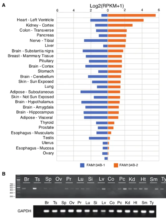
FAM134B isoforms are differentially expressed in normal adult tissues
The localization of transcription start sites of all short isoforms within intron 3 of the FAM134B
gene strongly suggested that the expression of long and short FAM134B transcript isoforms are controlled
by different factors and/or mechanisms. This led us to compare the tissue-specific expression of
length and truncated FAM134B isoforms. First, we analyzed normal tissue expression data on human
isoforms available at the GTEx database (3). We observed striking differences between tissues in terms of
12
FAM134B gene expression (Fig. 2A).FAM134B-1 is strongly expressed in the brain, which also expresses FAM134B-2 at lower levels. A
stronger expression of FAM134B-1 was also seen in the tibial nerve, adipose tissue and testis. Many other
tissues including the skeletal muscle, heart, kidney cortex, colon, pancreas, liver and stomach displayed a
strong expression of FAM134B-2 compared to FAM134B-1. A few tissues such as the uterus and ovary
were observed to have a weak expression of both FAM134B isoforms (Fig 2A). We strived to confirm the
differential expression of FAM134B isoforms by an RT-PCR analysis of a panel of 14 human tissues. As
shown in Fig. 2B, experimental data confirmed the differential expression of 1 and
2 transcripts in these tissues. For example, the brain, testis, spleen and prostate were rich in FAM134B-1
expression. In contrast, the skeletal muscle, pancreas and liver displayed a stronger expression of
FAM134B-2. As expected, no significant signal was observed in the ovarian tissue.
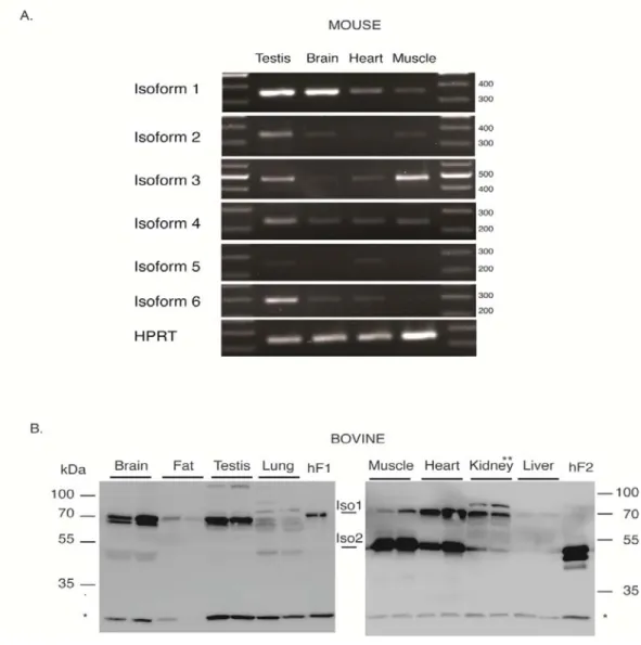
We also analyzed the expression patterns of Fam134b transcripts in several mouse tissues. As
shown in Fig. 3A, mouse testis strongly expressed all isoforms except for isoform-5. The mouse brain
showed a strong expression of isoform-1 with a weak expression of isoform-2, -4 and -6. The heart was
weakly positive for 1, -3, -4, -5 and -6. The muscle stood apart by a strong expression of
3, together with a weak expression of isoform-1, -2, and-4.
Differential expression of FAM134B protein isoforms in normal tissues
A further verification of FAM134B isoform expression in different tissues was performed at
protein level by western blotting. We initially applied western blotting experiments to bovine species,
which express only two transcript isoforms, similar to human, as stated above. As shown in Fig. 3B,
bovine FAM134B-1 and FAM134B-2 proteins migrated with apparent molecular weights of 70 kDa and
50 kDa, respectively, similar to human isoforms. The dominant expression of FAM134B-1 was observed
in the brain, adipose, testis and kidney, while FAM134B-2 was dominant in the heart and muscle. The
weak expression of both isoforms was detected in the lung tissue. The bovine liver stood apart by little to
no expression of both FAM134B protein isoforms.
13
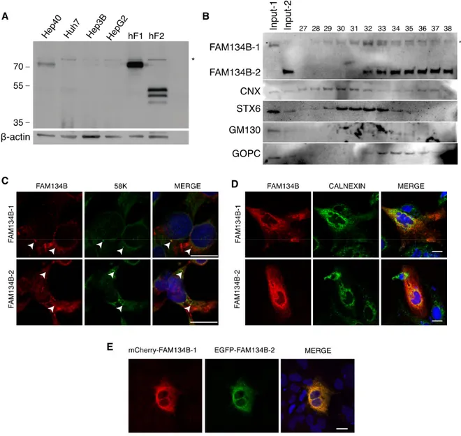
FAM134B isoforms differentially localize in sub-cellular compartments
FAM134B has previously been identified as an ER and Golgi-associated protein (22, 25). As the
studied isoform(s) has not been specified in these studies, we went on to compare the two isoforms for
their sub-cellular localization. Initial analysis of four well-differentiated (16, 44) liver cancer cell lines
failed to detect FAM134B isoforms in these cell lines, except for the Hep40 displaying a weak
1 expression (Fig. 4A). We constructed Hep3B-derived cells stably expressing FLAG-tagged FAM134B-1
or FAM134B-2.
In order to investigate the subcellular localization of both isoforms in the same experiment, we
performed cell fractionation studies followed by western blot analysis. We first prepared cell extracts
(Fig.4B, lines input-1 and input-2 respectively) prior to a sucrose gradient. Sucrose fractions were then
collected from the bottom of the gradient making tube and subjected to western blotting using antibodies
for FAM134B, as well as Golgi markers GOPC (9), GM130 (30) and STX6 (34) and CNX (ER, but leaky;
ref. (33)).
FAM134B-2 co-localized with GOPC and cis-Golgi marker GM130, but not with trans-Golgi
marker STX6 fractions, while calnexin was expanded to many fractions and co-localized with both
isoforms. On the other hand, both FAM134B isoforms partially co-localized with Golgi marker 58K (4),
demonstrated by immunofluorescent staining (Fig. 4C). Immunofluorescence analysis of FAM134B-1 and
FAM134B-2 indicated that when ectopically expressed, they mostly co-localize with each other, and they
also co-localize with Calnexin (Fig. 4D and E), suggesting ER-localization.
Starvation increases FAM134B-2 RNA and protein in the liver
As FAM134B is involved in ER-phagy during starvation (22), we studied its expressional changes
in mice following starvation. Wild-type and Fam134b-/- mice were either fed ad libitum or starved up to
48h, followed by an expression analysis of FAM134B isoforms. Tissues obtained from Fam134b-/- mice
14
were used as negative controls. Our FAM134B antibody cross-reacted with several non-specific proteins
in mouse tissues, thus limiting our analysis to only the FAM134B-2 isoform (Fig. 5A, C, D).
We first analyzed LC3B and FAM134B-2 expression in the liver after 48h of starvation. Autophagy
induction in the liver as a response to starvation was shown by a decrease in p62 protein levels and
induced levels of autophagosome-associated LC3B-II and LC3B-I (Fig. 5A). We also noticed that LC3B-I
levels were more pronounced in Fam134b-/- mice compared to wild-type mice (Supplemental Fig. S1).
FAM134B-2 was not detected in Fam134b-/- mice, as expected. Fam134b+/+ and Fam134b+/- mice showed
an induced expression after starvation, at 50 kDa (Fig. 5A and Supplemental Fig. 1). In addition to the
50kDa FAM134B-2, a specific smaller 35kDa protein was also induced in wild type animals (Fig. 5A,
marked with a single asterisk). Additional starvation experiments performed for 24h have exhibited an
even stronger stimulation than of 48h (Supplemental Fig. S2).
To test whether FAM134B protein accumulation in starved animal liver was associated with
transcriptional upregulation, we performed an RT-PCR analysis of six mouse isoforms along with two
additional putative protein coding transcripts, X1 and X2. The starvation induced the expression of
isoform-3, -4 and -6 strongly and isoform-2 weakly, while the level of isoform-5 was not changed. There
was little to no expression of isoform-1 in both fed and starved conditions (Fig. 5B). There was also no
induced expression of putative transcripts X1 and X2.
Starvation increases FAM134B-2 in the muscle and heart
In order to observe possible starvation induced FAM134B-2 response in other organs, we expanded
our analysis with mouse heart, muscle, pancreas and stomach. Similar to the liver, FAM134B-2 protein
levels as well as shorter protein fragments near 35kDa in the muscle and heart were elevated following
starvation (Fig. 5C). Stimulation of autophagy was confirmed by increased shift from LC3B-I to LC3B-II
(Supplemental Fig. 3). Under the same conditions, there was no change in FAM134B-2 levels in the
pancreas or stomach (Fig. 5D).
15
Upon starvation, Fam134b-/- mice display less weight loss and increased serum albumin levels along
with disrupted amino acid release
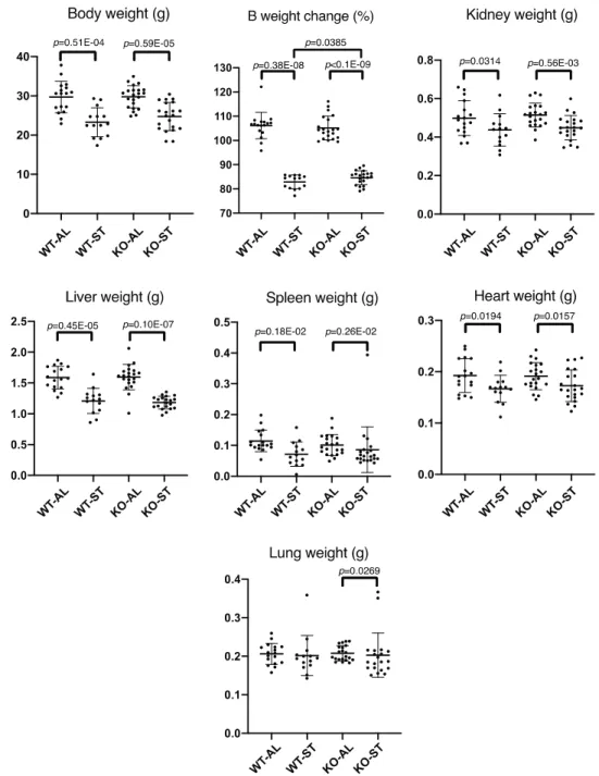
In order to analyze the in vivo response of Fam134b-deficient tissues to starvation, we compared
Fam134b+/+ and Fam134b-/- mice for their response to nutrient starvation for 36h. The initial body weights
of tested animals were similar with no significant differences (n=73) (Supplemental Fig. S4). Starvation
induced significant whole body and tissue-specific weight loss in both wild-type and Fam134b-/- mice
(Fig. 6). Among the tissues analyzed, there was significant weight loss in the liver, kidney, spleen and
heart for both wild type and mutant animals, and significant loss of lung weight was observed only in
mutant animals. Interestingly, starved mutant animals slightly resisted body weight loss compared to wild
type mice, as shown by body weight change as percentage (Fig 6, Body weight change).
Autophagic proteolysis in the liver following starvation makes a significant contribution to the
maintenance of blood glucose levels via conversion of amino acids to glucose, and accordingly, many
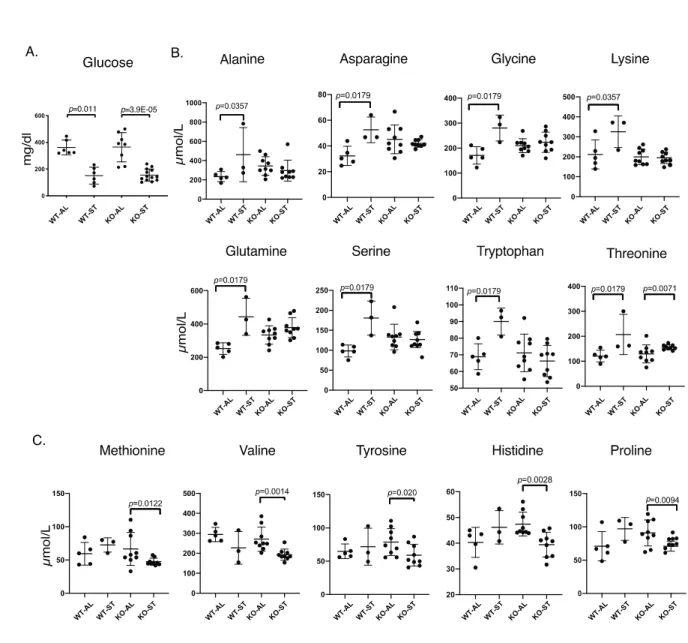
amino acids are released into the blood by 24h of starvation (12). Therefore, we measured serum glucose
and amino acid levels in both wild-type and Fam134b-/- mice after 36h of starvation. There was a
significant decrease in serum glucose in both wild type and mutant mice, the fall in Fam134b-/- being more
significant (Fig 7A). Wild-type animals displayed a significant increase in serum levels of amino acids
glutamine, asparagine, alanine, serine, glycine, tryptophan, threonine and lysine. Fam134b-/- mice failed to
increase the levels of these amino acids with the exception of threonine (Fig. 7B). There was no change in
the levels of histidine, proline, valine, methionine and tyrosine in wild-type mice, but interestingly, all
these five amino acids underwent a significant drop in Fam134b-/- mice (Fig. 7C).
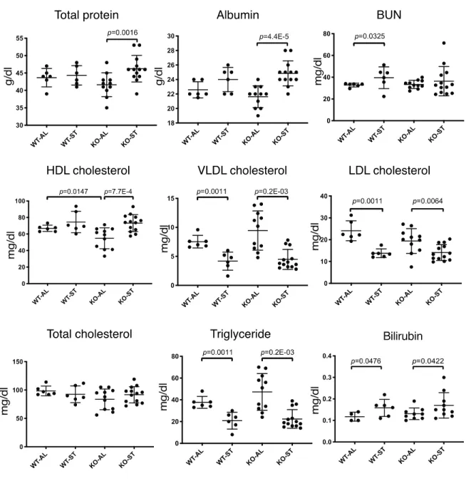
Other changes in starved Fam134b-/- mice
We further investigated the Fam134b associated phenotype by analyzing serum levels of proteins,
nutrients, electrolytes and enzymes. Interestingly, baseline High-density lipoprotein (HDL) cholesterol
levels were significantly lower in mutant animals. Starvation induced a decrease in serum levels of
16
triglycerides, Very-low density lipoprotein (VLDL) and Low-density lipoprotein (LDL) cholesterol, but
not in total cholesterol in either group of animals (Fig. 8).
Starvation also induced serum levels of albumin and total protein in Fam134b-/-, but not in
type mice (Fig. 8). Wild type animals showed elevated levels of blood urea nitrogen (BUN), as reported
previously (41), but no such significant change was observed in mutant mice.
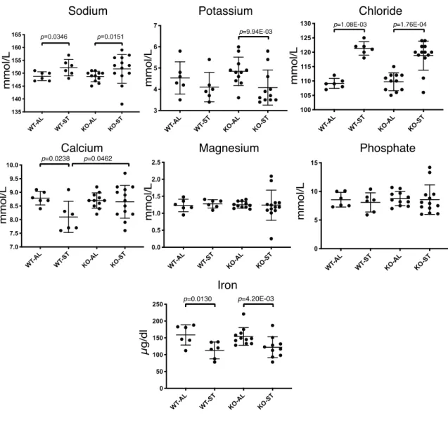
Changes in serum ions such as sodium, potassium, chloride, calcium and iron were also observed
(Fig. 9). Upon starvation, sodium, potassium and chloride displayed significant increases in both wild-type
and Fam134b-/- mice. In contrast, calcium levels in wild-type animals and iron levels in both groups were
decreased. The lack of starvation-induced hypocalcemia in Fam134b-/- mice was noteworthy.
Finally, starvation resulted in liver injury as evidenced by increased levels of serum AST and ALT
in both groups of animals. Of particular interest, serum α-amylase activity was significantly upregulated in
Fam134b-/-, but not in wild-type mice (Fig. 10), suggesting starvation-induced pancreas injury.
Expression of FAM134B isoforms is suppressed or stimulated in different cancers
As stated earlier, the expression changes of FAM134B have been described in esophageal, colon,
breast and liver cancers (9-14). Our observations described so far show that the human FAM134B gene
encodes at least two different protein isoforms: one full length with an intact RHD domain (i.e.
FAM134B-1) and an N-terminally truncated form with a disrupted RHD domain (i.e. FAM134B-2).
Previously published data on FAM134B in cancers did not specify what type of FAM134B protein is
implicated in different cancers. In order to clarify this issue, we collected following data sets from the
TSVdb database: hepatocellular carcinoma (371 tumors and 50 non-tumor liver), lung adenocarcinoma
(515 tumors and 59 normal), lung squamous carcinoma (501 tumors and 51 normal), colon
adenocarcinoma (285 tumors and 41 normal) stomach adenocarcinoma (415 tumors and 35 normal) and
kidney chromophobe cancer (66 tumors and 25 normal), and performed an isoform level differential
expression analysis on these publicly available transcriptome data.
17
As shown in Fig. 11, both FAM134B-1 and FAM134B-2 isoforms were positive in non-tumor colon,
lung, (Fig. 11A), stomach and kidney (Fig. 11B) tissues. In contrast, only FAM134B-2 was positive with a
marginal expression of FAM134B-1 in non-tumor liver samples (Fig. 11A). HCC samples displayed a
significant down-regulation of FAM134B-2 (Fig. 11A). We confirmed this finding by performing an
PCR analysis on HCC tumors (n=11) and non-tumor liver tissues (n=16), which showed a loss of the
FAM134B-2 signal in most HCC tumors analyzed (Fig. 11C).
A significant down-regulation of both FAM134B-1 and FAM134B-2 was observed in colon and
lung cancers (Fig. 11A). In contrast, FAM134B-2 was significantly upregulated in stomach
adenocarcinoma. Finally, both FAM134B-1 and FAM134B-2 were significantly upregulated in
chromophobe renal cell carcinomas (KICH). Other tumors including breast and prostate cancer did not
display significant changes in the FAM134B isoform expression (not shown).
Discussion
We performed a comprehensive analysis of the FAM134B gene, its transcripts and encoded protein
isoforms together with its implications in nutrient starvation and cancer. Our main findings indicate that
human and bovine species express at least two different transcript isoforms encoding a full-length and an
N-terminally truncated protein isoform. In contrast, mice encode at least six different transcript isoforms,
giving rise to one full-length and five N-terminally truncated proteins. During the preparation of this
report for publication, we became aware of a publication by Kohno et al. (23) on the identification of a
novel FAM134B-2 transcript isoform capable of encoding an N-terminally truncated protein in the livers
of fasted mice. This transcript may represent either isoform-2 or isoform-3 described in our report (Fig. 1).
In addition to isoform-2 and -3, mouse tissues exhibited the expression of isoform -4, -5 and -6, all
capable of encoding four different N-terminally truncated proteins that differ from each other at the first
45 amino acids of the N-terminal region (Fig. 1B).
18
The FAM134B-2 protein lacks the N-terminal tail and the TM12 reticulon structure of the
length protein, but retains the TM34 reticulon along with the C-terminal region, which harbors the LC3B
binding LIR motif. It was previously shown that the remaining reticulon structure, namely TM34, is
critical for membrane shaping (6), and that the LIR motif of FAM134B-2 still mediates ER-phagy (23).
However, FAM134B-2 is unlikely to be a mere compensatory protein for FAM134B-1, since FAM134B-1
cleavage by the zika virus yields a truncated protein very similar to FAM134B-2 that is unable to promote
reticulophagy (26).
Our subcellular localization studies with human FAM134B-1 and human FAM134B-2 indicated
that loss of integrity of the RHD in the shorter isoform did not abolish the ability of FAM134B-2 to
localize to the ER (Fig. 4B and D). However, the two isoforms of FAM134B did not completely
localize, as shown in Fig. 4B. Co-localization of FAM134B-2 with the Golgi marker GOPC, GM130 (Fig.
4B) and 58K (Fig. 4C) but not with STX6 strongly suggested that this form may be preferentially
localized at the ER membrane close to the Golgi apparatus. Indeed, mouse liver FAM134B-2 was shown
to be localized at both the rough and smooth ER (23).
In order to detail the physiological relevance of FAM134B variants, we analyzed their distribution
in human, bovine and mouse tissues. One of our most striking findings is the identification of the
FAM134B-2 transcript as the dominantly expressed form in the skeletal muscle, heart, kidney cortex,
colon, pancreas, liver and stomach. This contrasts sharply with the dominant expression of FAM134B-1 in
the brain, tibial nerve, adipose tissue and testis. Our western blot analyses with bovine and mouse tissues
have further shown that the tissue-specific expression of FAM134B is conserved during evolution (Figs.
3, 5). This may indicate that FAM134B-1 and FAM134B-2 are involved in some tissue-restricted
functions. For example, FAM134B-2 may be involved in the lysosomal degradation of a subgroup of ER
components rather than bulk ER degradation (or turnover), which was previously elaborated for the liver
(23). As the FAM134B-2 protein is distinctly co-localized to ER and Golgi, it may have a role during the
transfer of protein and lipid molecules from ER to Golgi, or in autophagy-related changes during the
trafficking, processing, and sorting of the newly synthesized membrane and secretory proteins and lipids.
19
Reticulon Homology Domain (RHD) containing proteins are well known as ER-shaping elements,
and their deficiency results in a disturbed ER network (10). Furthermore, proteins harboring a single
transmembrane structure (e.g. TM12 or TM34 alone) were previously shown to be involved in shaping ER
tubules (46). Despite carrying a truncated RHD, a pronounced expression of FAM134B-2 in the muscle
and heart might indicate a similar structural role in ER shaping and organization. However, besides the
previously identified sensory neuropathy, no major phenotype has been observed even in aged animals,
indicating that Fam134b deficiency is well-tolerated, possibly due to a compensatory mechanism which is
yet to be understood.
From simple prokaryotes to humans, all living organisms are equipped with complex survival
mechanisms to cope with nutrient scarcity. Starvation initiates a cascade of events including autophagy,
thereby mobilizing energy sources to meet the energy demands of vital organs. In this complex
mechanism, the liver, heart, skeletal muscle and kidney play critical roles in regulating the release and
uptake of major energy sources such as amino acids, lipids, glucose and ketone bodies (37). Considering
the critical role of starvation in autophagy, we performed comparative analyses of wild-type and
Fam134b-/- mice (29). Starvation for 48h resulted in the accumulation of LC3B-I, as well as the
autophagy-associated LC3B-II form in the livers of wild-type mice. A similar accumulation was observed
in Fam134b-/- animals even in the absence of starvation (Supplemental Fig S1), which implies a possibly
disturbed autophagic flux. The stimulated expression of FAM134B-2 in the muscle and heart but not in
the pancreas and stomach strongly indicates a possible involvement of this protein in the physiological
response against nutrient starvation.
The FAM134B-2 protein, which was weakly positive in the liver of fed animals, was induced
strongly by starvation, as reported previously (23). However, besides the verification of the previously
reported FAM134B-2 increase at 50kDa, we surprisingly observed another specific band at 35kDa, which
was also increased upon nutrient starvation, not only in the liver but also in the muscle and heart. Indeed,
this shorter isoform could be a post translational modification of FAM134B-2, as well as a protein product
of short isoform transcripts.
20
The starvation-induced FAM134B accumulation in different tissues urged us to carry out a series of
analyses to compare wild-type and Fam134b-/- mice under fed and starved conditions. Fam134b-/- mice
under fed conditions did not differ from wild-type animals, with the exception of decreased serum HDL
cholesterol levels. Since HDL production and clearance from blood are primarily performed by the liver
(39), hepatic FAM134B-2 might be involved in the regulation and/or maturation of HDL particles.
Following 36h of starvation, both wild type and Fam134b-/- mice displayed significant weight loss
in total body as well as in liver. Although we found some significant differences in other tissues, the
weight loss was less pronounced. There were also significant changes in blood parameters, in line with the
literature (20, 31, 41).
When we compared starvation responses of wild-type and mutant mice, we identified several
important aberrations associated with Fam134b deficiency. Firstly, mutant mice were significantly more
resistant to body weight loss, as opposed to wild-type mice. Secondly, mutant mice showed profound
aberrations in serum amino acid dynamics. As reported previously, starvation-induced hypoglycemia and
a decrease in insulin levels with maintained glucagon levels triggers an autophagic response in the liver,
resulting in a surge of amino acids released into the blood at around 24h of fasting (12). Accordingly, we
observed an increase in the serum levels of alanine, asparagine, glycine, lysine, glutamine, serine,
tryptophan and threonine with wild-type mice. In contrast, mutant mice failed to elevate the levels of all
these amino acids with only one exception. This observation strongly suggests that in the absence of
FAM134B-2, liver tissue cannot undergo sufficient autophagic proteolysis to produce these amino acids.
Another interesting observation with Fam134b-/- mice was a decrease in the serum levels of methionine,
valine, tyrosine, histidine and proline. Thus, it appears that these normal constituents of serum are
depleted during starvation, probably because they are converted to glucose. Such a compensation may be
necessary when autophagic degradation of proteins is defective.
Glucagon in circulation is one of the key factors for energy mobilization upon nutrient starvation
(12). Glucagon was previously shown to downregulate serum calcium levels by altering the
gastrointestinal uptake (1). The steady state calcium levels in mutant mice during starvation strongly
21
suggest a function in calcium homeostasis, likely through intestinal absorption. Finally, Fam134b-/-, but
not wild-type mice, displayed abnormal increases in blood α-amylase levels upon starvation, which
indicates pancreatic injury, salivary gland malfunction or other intraabdominal inflammations (28). As
demonstrated in western blot experiments, FAM134B-2 is predominantly expressed in the pancreas, in
which basal autophagy is necessary to maintain α-amylase secreting acinar cells (2). Furthermore, another
ER-phagy receptor, CCPG-1, was shown to be critically involved in secretory acinar cells, and its absence
results in the accumulation of insoluble α-amylase particles and unfolded protein response (UPR) (35).
Therefore, the α-amylase increase in Fam134b-/- could be partially attributed to a defective ER-phagy,
which may lead to a discharge or leak of excessive α-amylase into the bloodstream.
Our comprehensive analysis of FAM134B-1 and FAM134B-2 expression in the TCGA tumor
collection has clearly established that FAM134B-2, and to a lesser degree FAM134B-1, are closely
involved in cancer. The loss of FAM134B-2 expression was observed in HCC, colon and lung cancers. In
contrast, stomach and chromophobe renal cell carcinomas displayed a significant upregulation of the same
transcript. FAM134B-1 levels were generally low in non-tumor tissues, but a significant loss was observed
in colon and lung cancers, as well as a significant increase in chromophobe renal cell carcinomas. Thus,
cancers can be grouped into three classes depending on FAM134B-2 gene expression; those with
significant downregulation such as HCC, colon and lung cancers, those with significant upregulation such
as stomach and a subtype of kidney cancers, and those with the FAM134-2 expression likely not changing
significantly in the third group of remaining cancers.
The past few years have been fruitful for the understanding of the molecular roles of FAM134B,
but without defining its isoforms and their relevance in the whole organism. To our knowledge, this is the
first paper to define the differential expression of FAM134B isoforms in various tissues under normal and
starved conditions, as well as in cancer. These findings greatly help us further understand the functional
roles of FAM134B isoforms in diseases such as cancer.
22
Funding: This study is supported by Dokuz Eylul University, Department of Scientific Research
Projects and IBG’s institutional funds.
Acknowledgements: We would like to thank the IBG-Vivarium Rodent Facility and the optical
imaging core facility for their great support in the experimental procedures. We are also deeply
grateful to Deniz Donmez for her efforts in improving the manuscript’s language. We also thank
Christian Hübner for his kind donation of Fam134b-/- animals.
The Genotype-Tissue Expression (GTEx) Project was supported by the Common Fund of the Office
of the Director of the National Institutes of Health, and by NCI, NHGRI, NHLBI, NIDA, NIMH and
NINDS. The data used for the analyses described in this manuscript were obtained from: the GTEx
Portal on 11/14/2018 and/or dbGaP accession number phs000424.v3.p1.
Statement of contribution:
• UK: Designed and performed in vivo and in vitro experiments
• HEY, UE, SK, ZM: Carried out in vivo experiments and serum analyses
• AS, GK: performed statistical analyses
• EI, EB: performed and reported in vitro experiments
• ADC, NT: designed and cloned FAM134B plasmids, and carried out preliminary experiments
• NA: Contributed to the study by providing valuable HCC patient cDNA and by providing
guidance on its usage
• MAS: Contributed to the study by providing results and interpretation of serum amino acid
analyses.
• MO: Supervised, designing, planning, performing and reporting of all experimentation.
Competing Interests:The authors have declared that no competing interest exists.
References
1. Aliapoulios MA, Morain WD, Kacoyanis GP. Glucagon as a hypocalcemic and
23
hypophosphatemic agent in the rat. Gastroenterology 65: 912–918, 1973.
Antonucci L, Fagman JB, Kim JY, Todoric J, Gukovsky I, Mackey M, Ellisman MH,
Karin M. Basal autophagy maintains pancreatic acinar cell homeostasis and protein synthesis
and prevents ER stress. Proc Natl Acad Sci U S A 112: E6166–E6174, 2015.
Ardlie KG, DeLuca DS, Segrè A V., Sullivan TJ, Young TR, Gelfand ET, Trowbridge
CA, Maller JB, Tukiainen T, Lek M, Ward LD, Kheradpour P, Iriarte B, Meng Y,
Palmer CD, Esko T, Winckler W, Hirschhorn JN, Kellis M, MacArthur DG, Getz G,
Shabalin AA, Li G, Zhou YH, Nobel AB, Rusyn I, Wright FA, Lappalainen T, Ferreira
PG, Ongen H, Rivas MA, Battle A, Mostafavi S, Monlong J, Sammeth M, Melé M,
Reverter F, Goldmann JM, Koller D, Guigó R, McCarthy MI, Dermitzakis ET, Gamazon
ER, Im HK, Konkashbaev A, Nicolae DL, Cox NJ, Flutre T, Wen X, Stephens M,
Pritchard JK, Tu Z, Zhang B, Huang T, Long Q, Lin L, Yang J, Zhu J, Liu J, Brown A,
Mestichelli B, Tidwell D, Lo E, Salvatore M, Shad S, Thomas JA, Lonsdale JT, Moser
MT, Gillard BM, Karasik E, Ramsey K, Choi C, Foster BA, Syron J, Fleming J,
Magazine H, Hasz R, Walters GD, Bridge JP, Miklos M, Sullivan S, Barker LK, Traino
HM, Mosavel M, Siminoff LA, Valley DR, Rohrer DC, Jewell SD, Branton PA, Sobin LH,
Barcus M, Qi L, McLean J, Hariharan P, Um KS, Wu S, Tabor D, Shive C, Smith AM,
Buia SA, Undale AH, Robinson KL, Roche N, Valentino KM, Britton A, Burges R,
Bradbury D, Hambright KW, Seleski J, Korzeniewski GE, Erickson K, Marcus Y,
Tejada J, Taherian M, Lu C, Basile M, Mash DC, Volpi S, Struewing JP, Temple GF,
Boyer J, Colantuoni D, Little R, Koester S, Carithers LJ, Moore HM, Guan P, Compton
C, Sawyer SJ, Demchok JP, Vaught JB, Rabiner CA, Lockhart. The Genotype-Tissue
Expression (GTEx) pilot analysis: Multitissue gene regulation in humans. Science (80- ) 348:
648–660, 2015.
Bashour AM, Bloom GS. 58K, a microtubule-binding Golgi protein, is a
formiminotransferase cyclodeaminase. J. Biol. Chem. (1998). doi: 10.1074/jbc.273.31.19612.
24
Bateman A. UniProt: A worldwide hub of protein knowledge. Nucleic Acids Res 47: D506–
D515, 2019.
Bhaskara RM, Grumati P, Garcia-Pardo J, Kalayil S, Covarrubias-Pinto A, Chen W,
Kudryashev M, Dikic I, Hummer G. Curvature induction and membrane remodeling by
FAM134B reticulon homology domain assist selective ER-phagy. Nat Commun 10, 2019.
Buchan DWA, Jones DT. The PSIPRED Protein Analysis Workbench: 20 years on. Nucleic
Acids Res. (2019). doi: 10.1093/nar/gkz297.
Cai M, Zhao J, Liu Q, Wang X, Wang Y. FAM134B improves preadipocytes differentiation
by enhancing mitophagy. Biochim Biophys Acta - Mol Cell Biol Lipids 1864, 2019.
Charest A, Lane K, McMahon K, Housman DE. Association of a Novel PDZ
containing Peripheral Golgi Protein with the Q-SNARE (Q-soluble
sensitive Fusion Protein (NSF) Attachment Protein Receptor) Protein Syntaxin 6. J Biol Chem
276: 29456–29465, 2001.
Chiurchiù V, Maccarrone M, Orlacchio A. The role of reticulons in neurodegenerative
diseases. NeuroMolecular Med 16: 3–15, 2014.
Dai X, Hua T, Hong T. Integrated diagnostic network construction reveals a 4-gene panel and
5 cancer hallmarks driving breast cancer heterogeneity. Sci Rep 7: 1–15, 2017.
Ezaki J, Matsumoto N, Takeda-Ezaki M, Komatsu M, Takahashi K, Hiraoka Y, Taka H,
Fujimura T, Takehana K, Yoshida M, Iwata J, Tanida I, Furuya N, Zheng DM, Tada N,
Tanaka K, Kominami E, Ueno T. Liver autophagy contributes to the maintenance of blood
glucose and amino acid levels. Autophagy 7: 727–736, 2011.
Forrester A, De Leonibus C, Grumati P, Fasana E, Piemontese M, Staiano L, Fregno I,
Raimondi A, Marazza A, Bruno G, Iavazzo M, Intartaglia D, Seczynska M, Anken E,
Conte I, De Matteis MA, Dikic I, Molinari M, Settembre C. A selective ER ‐phagy exerts
25
procollagen quality control via a Calnexin‐ FAM 134B complex . EMBO J 38: 1–16, 2019.
Fregno I, Molinari M. Endoplasmic reticulum turnover: ER-phagy and other flavors in
selective and non-selective ER clearance. F1000Research 7: 454, 2018.
Friedewald WT, Levy RI, Fredrickson DS. Estimation of the concentration of low-density
lipoprotein cholesterol in plasma, without use of the preparative ultracentrifuge. Clin. Chem.
(1972). doi: 10.1093/clinchem/18.6.499.
Fuchs BC, Fujii T, Dorfman JD, Goodwin JM, Zhu AX, Lanuti M, Tanabe KK.
Epithelial-to-mesenchymal transition and integrin-linked kinase mediate sensitivity to
epidermal growth factor receptor inhibition in human hepatoma cells. Cancer Res 68: 2391–
2399, 2008.
Gangalum RK, Horwitz J, Kohan SA, Bhat SP. αA-crystallin and αB-crystallin reside in
separate subcellular compartments in the developing ocular lens. J Biol Chem 287: 42407–
42416, 2012.
Hautem J, Saintier A. Direct quantification of amino acids in plasma by liquid
chromatography-tandem mass spectrometry for clinical research. ThermoScientific Tech Note
65382, [date unknown].
Islam F, Gopalan V, Law S, Tang JC on, Lam AK yin. FAM134B promotes esophageal
squamous cell carcinoma in vitro and its correlations with clinicopathologic features. Hum
Pathol 87: 1–10, 2019.
Jensen TL, Kiersgaard MK, Sørensen DB, Mikkelsen LF. Fasting of mice: A review. Lab
Anim 47: 225–240, 2013.
Kasem K, Gopalan V, Salajegheh A, Lu CT, Smith RA, Lam AKY. The roles of JK-1
(FAM134B) expressions in colorectal cancer. Exp Cell Res 326: 166–173, 2014.