ii
ELICITING IMMUNE RESPONSE BY USING NANOSTRUCTURES
A THESIS SUBMITTED TO
GRADUATE SCHOOL OF ENGINEERING AND SCIENCE OF BILKENT UNIVERSITY
IN PARTIAL FULFILLMENT OF THE REQUIREMENTS FOR THE DEGREE OF
MASTER OF SCIENCE IN NEUROSCIENCE By GÖKHAN GÜNAY June, 2017
iii
ELICITING IMMUNE RESPONSE BY USING NANOSTRUCTURES By Gökhan Günay,
June, 2017
We certify that we have read this thesis and that in our opinion it is fully adequate, in scope and in quality, as a thesis for the degree of Master of Science.
Ayşe Begüm Tekinay (Advisor)
Michelle Marie Adams
Reyhan Neslihan Gürsoy
Approved for the Graduate School of Engineering and Science:
Ezhan Karaşan
i
ABSTRACT
ELICITING IMMUNE RESPONSE BY USING NANOSTRUCTURES
Gökhan GÜNAY M.Sc. in Neuroscience
Advisor: Ayşe Begüm Tekinay, PhD June, 2017
The ability of dendritic cells to coordinate innate and adaptive immune responses makes them essential targets for vaccination strategies. Presentation of specific antigens by dendritic cells is required for the activation of the immune system against many pathogens and cancer, and nanoscale materials can be functionalized for active targeting of dendritic cells. In this work, we integrated an immunogenic, carbohydrate melanoma-associated antigen-mimetic GM3-lactone molecule into mannosylated peptide amphiphile nanofibers to target dendritic cells through DC-SIGN receptor. Based on morphological and functional analyses, when dendritic cells were treated with peptide nanofiber carriers, they showed significant increase in antigen internalization and a corresponding increase in the surface expression of the activation and maturation markers CD86, CD83 and HLA-DR, in addition to exhibiting a general morphology consistent with dendritic cell maturation. These results indicate that mannosylated peptide amphiphile nanofiber carriers are promising candidates to target dendritic cells for antigen delivery. Overall these structures are proven to be effective in terms of dendritic cell activation and maturation and hold high potential to be used with a variety of antigens for different immunotherapy purposes.
ii
KEYWORDS: Cancer immunotherapy, cancer antigens, dendritic cells, carbohydrate melanoma associated antigen, peptide amphiphiles
iii
ÖZET
NANOYAPILAR KULLANILARAK İMMÜN YANIT TETİKLENMESİ
Gökhan GÜNAY Nörobilim, Yüksek Lisans Tez Danışmanı: Ayşe Begüm Tekinay
Haziran, 2017
Dendritik hücrelerin doğuştan gelen ve adaptif bağışıklık tepkilerini koordine etme kabiliyeti onları aşı stratejileri için önemli hedefler haline getirmektedir. Dendritik hücrelerin spesifik antijenler sunması, birçok patojen ve kansere karşı bağışıklık sisteminin aktivasyonu için gereklidir. Nano ölçekli materyaller, dendritik hücrelerin aktif hedeflenmesi için işlevselleştirilebilir. Bu çalışmada, DC-SIGN reseptörü vasıtasıyla dendritik hücreleri hedeflemek için immünojenik bir karbonhidrat olan melanoma ile ilişkili antijen-taklidi GM3-lakton molekülünü mannozile peptid amfifil nanofiberlere integre edilmiştir. Dendritik hücreler peptid nanofiber taşıyıcılarla muamele edildiğinde, antijenin hücre içine alımında önemli anlamlı bir artış gözlenmiştir ve aktiflik ve olgunlaşma belirteçleri olan CD86, CD83 ve HLA-DR'nin yüzey ekspresyonunda da buna eşdeğer bir artış görülmüştür. Ayrıca dendritik hücre olgunlaşması ile tutarlı genel bir hücre morfolojisi de gözlenmiştir. Bu sonuçlar, mannosillenmiş peptit amfifil nanofiber taşıyıcılarının, antijen sunumu için dendritik hücreleri hedeflemeye yönelik umut vaadeden adayları olduğunu göstermektedir. Genel olarak, bu yapıların dendritik hücre aktivasyonu ve
iv
olgunlaşması açısından etkili olduğu kanıtlanmıştır ve farklı immünoterapi amaçları için çeşitli antijenler ile kullanılmak için yüksek bir potansiyel bulundurmaktadır
ANAHTAR SÖZCÜKLER: Kanser immünoterapisi, kanser antijenleri, dendritik hücreler, karbonhidrat melanoma ile ilişkili antijen, peptit amfifiller
v
ACKNOWLEDGEMENTS
First, I would like to express my gratitude to my advisor Prof. Ayşe Begüm Tekinay. I am grateful for the opportunity to have a part in her laboratory, additionally I am grateful for her extreme patience and guidance throughout my master education and additionally she provided me independence during my studiers which helped me improve myself even more. I also would like to thank Prof. Mustafa Özgür Güler for his support during my master education.
I would like to acknowledge the graduate scholarship from BIDEB 2210-C TÜBİTAK (The Scientific and Research Council of Turkey) for financial support.
First and for most I would like to thank Dr. Melis Şardan Ekiz and Melike Sever for guiding me throughout the projects and believing in the projects as much as I did, more importantly for being such good collaborators and wonderful friends.
I would like to thank my dearest friends from İYTE; İdil UYAN for supporting me and being there for me no matter what, her friendship, support and positivity, Merve ŞEN for her support and being wonderfully weird like Pheobe Buffay, Nurcan Haştar for her warm friendship, turkish coffes and for always pointing out the right answer, Burak ARIÖZ and Fırat AŞIR for being amazing brothers and mentors throughout all these years, together we have been through much and we always help each other no matter what.
I would like to thank my lab members starting from Canelif YILMAZ, Zeynep ORHAN and Fatih YERGÖZ for their support, Özge UYSAL and Mustafa BETER for our many many discussions about anything, Nuray GÜNDÜZ, Elif ARSLAN,
vi
Gülistan TANSIK, Ahmet Emin TOPAL for their support. I specially thank here to Alper Devrim ÖZKAN for being helpful to anyone.
I would like to express my thanks to; Egemen Deniz EREN, Seren Hamsici, Çağla EREN, Ayşegül DEDE, Göksu ÇINAR, Recep Erdem AHAN, Onur APAYDIN for being amazing friends throughout this journey. I also would like to thank Aref Khaleliy for his support.
To me the most important thing in the world is family and I would like to thank my family for being honest and good people and their amazing support throughout my life; Dilek GÜNAY and Engin GÜNAY for being such excellent role models, in 30 years if I am half as good as you are now I would count myself the luckiest person in the world. My brother Görkem GÜNAY for having a vision and a dream, I know that you will be very successful if you follow your heart. I would like to thank Ümmügülsüm GÜNAY for teaching me the meaning of love and loving me unconditionally since the day I was born, to Serap GÜNAY for being such a cool aunt, her support and love, to Metin GÜNAY for his support and love.
I wish all my friends the best for the rest of their lives.
vii
Contents
Abbreviations ... xv
Chapter 1 ... 1
1. Introduction ... 1
1.1. Nanomaterials for Biomedical Applications ... 1
1.2. Self-Assembling Nanomaterials ... 4
1.3. Self-assembling Peptide Amphiphiles ... 6
1.4. Regenerative Medicine ... 8
1.5. Drug Delivery ... 11
Chapter 2 ... 14
2. Objectives ... 14
2.1. Introduction ... 15
2.1.1. Dendritic Cells and Adaptive Immunity ... 15
2.1.2. C-Type Lectins ... 18
2.1.3. Carbohydrate Associated Antigens... 19
2.2. Experimental Section ... 21
2.2.1. Materials ... 21
2.2.2. Synthesis of Amphiphilic Glycopeptides ... 21
2.2.3. Synthesis of Fluorescent GM3-MIM Derivatives (GM3-MIM-Bodipy) 22 2.2.4. Preparation of GM3-MIM Integrated Glycopeptide Nanofibers .. 22
2.2.5. Characterization of Fluorescent GM3-MIM Integrated Glycopeptide Nanofibers ... 23
viii
2.2.7. Differentiation of THP-1 Human Monocytes into Immature
Dendritic Cells ... 24
2.2.8. Internalization of Fluorescent GM3-MIM Integrated Glycopeptide Nanofibers ... 24
2.2.9. Surface Marker Analyses of Dendritic Cell Activation ... 25
2.2.10. Scanning Electron Microscopy (SEM) Imaging... 25
2.3. Results and Discussion ... 26
2.3.1. Design, Synthesis and Characterization of Glycopeptide Conjugates ... 26
2.3.2. Cell Viability and iDC Differentiation from THP-1 Monocytes .. 34
2.3.3. Internalization of Fluorescent GM3-MIM Integrated Glycopeptide Nanofibers ... 37
2.3.4. Surface Marker Analyses of Dendritic Cell Activation ... 39
2.3.5. Morphological Analyses of Dendritic Cell Activation ... 42
2.4. Conclusions and Future Perspectives ... 43
Chapter 3 ... 45
3. Three-dimensional laminin mimetic peptide nanofiber gels for in vitro neural differentiation ... 45
3.1. Abstract ... 45
3.2. Introduction ... 46
3.3. Experimental Section ... 51
3.3.1. Materials ... 51
3.3.2. Peptide Amphiphile Synthesis and Purification ... 51
ix
3.3.4. Characterizations of Self-Assembled PA Nanofibers... 53
3.3.5. Oscillatory Rheology Measurements ... 53
3.3.6. Zeta Potential Measurements ... 54
3.3.7. In vitro Studies ... 54
3.3.8. Neurite Extension Assay... 55
3.3.9. SEM Imaging of PC-12 Cells Cultured on 2D and in 3D Scaffolds 55 3.3.10. Gene Expression Analyses ... 56
3.3.11. Western Blot Analysis ... 56
3.3.12. Statistical Analysis... 57
3.4. Results and Discussion ... 57
3.4.1. Design and Characterization of Peptide Amphiphile Nanofibers. 57 3.4.2. Neural Differentiation Potential in Laminin Mimetic 2D and 3D Environment ... 63
3.4.3. Neural Gene Expression Analysis ... 65
3.4.4. Western Blot Analysis ... 68
3.4.5. SEM Imaging ... 69
3.5. Conclusion and Future Perspectives ... 71
Bibliography ... 76
x
LIST OF FIGURES
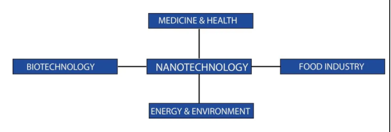
Figure 1 Utility of nanotechnology in various areas………2
Figure 2 Bottom-up and top-down approaches………3
Figure 3 Nanotechnology approaches for cellular responses………...4
Figure 4 Self-assembling monomers and self-assembled structures………...6
Figure 5 Schematics of self-Assembly of peptide amphiphiles………...8
Figure 6 Supramolecular GAG-like self-assembled glycopeptide nanofibers induce chondrogenesis and cartilage regeneration……….10
Figure 7 Glycosaminoglycan-Mimetic signals direct the Osteo/Chondrogenic differentiation of mesenchymal stem sells in a three-dimensional peptide nanofiber extracellular matrix mimetic environment ……….11
Figure 8 Local delivery of anti-cancer drug doxorubicin in a mice breast cancer model ………..12
Figure 9 Generation of cellular immune response against tumor antigens…………16
Figure 10 Interaction of dendritic cells with CD4 and CD8 T cells………..17
Figure 11 DC-SIGN receptor on immature DCs………...19
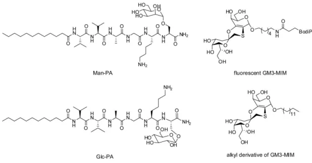
Figure 12 Chemical structures of molecules used in the study……….27
Figure 13 Liquid chromatograms and mass spectra (LC-MS) of amphiphilic glycopeptides. Man-PA, [M+H]+ (calculated) = 903.5688, [M+H]+ (observed) =
xi
903.5729; Glc-PA, [M+H]+ (calculated) = 903.5688, [M+H]+ (observed) = 903.5758………..27
Figure 14 STEM images of (A) Man-PA (B) Glc-PA………...28
Figure 15 Solvent exchange (DCM:H2O) process for the integration of the GM3-MIM molecule into peptide structures. (B) Fluorescence and (C) UV spectra of GM3-MIM and GM3-MIM integrated glycopeptide solutions dissolved in DCM:MeOH (7:1) and water, respectively………30
Figure 16 CD spectra of amphiphilic glycopeptide solutions, MIM and GM3-MIM integrated glycopeptide systems………31
Figure 17 STEM images of (A) Man-PA/GM3-MIM (B) Glc-PA/GM3-MIM……32
Figure 18 TEM mapping of fluorescent GM3-MIM integrated Man-PA nanofibers, S-pre edge (left), sulfur mapping (middle), oxygen mapping (right)……….33
Figure 19 Zeta potentials of Man-PA and Glc-PA………33
Figure 20 Cellular viability of THP-1 cells treated with different concentrations of Man-PA and Glc-PA, and TCP for 24 h. Values represent mean ± SEM (****p<0.0001)………...35
Figure 21 Schematic representation of the transition from human monocytes to mature dendritic cells (mDC)………..36
Figure 22 Differentiation of THP-1 monocytes into iDCs in terms of (A) de novo surface CD86 expression and (B) morphological changes upon differentiation into iDC………..36
xii
Figure 23 Uptake and morphological changes of iDCs when treated with Man-PA/GM3-MIM, Glc-PA/GM3-MIM and only GM3-MIM for 24 h. Blue indicating To-Pro nuclei staining, red indicating Phalloidin microtubule staining and green indicating bodipy conjugated GM3-MIM………...38
Figure 24 GM3-MIM MFI analyses for Man-PA/GM3-MIM, Glc-PA/GM3-MIM and only GM3-MIM for 24 h. Values represent mean ± SEM (****p<0.0001)……39
Figure 25 Surface expression MFI values of (A) CD86, (B) CD83 and (C) MHC-II under Man-PA/GM3-MIM, Glc-PA/GM3-MIM and GM3-MIM treatment for 24 h. Values represent mean ± SEM (****p<0.0001)……….41
Figure 26 Morphological analyses of the differentiation in iDCs when treated with Man-PA/GM3-MIM, Glc-PA/GM3-MIM, only GM3-MIM, only Man-PA and only Glc-PA for 24 h. Scale bars are 30 µm………...42
Figure 27 Schematic illustration of cellular interaction with ECM………...48
Figure 28 Chemical structures of peptide amphiphile molecules (LN-PA, KK-PA and EE-PA)……….59
Figure 29 Liquid chromatography and mass spectroscopy of LN-PA (A), KK-PA (B) and EE-PA (C). Mass spectrometry of LN-PA; [M+H]+ (calculated): 1292.93, [M+H]+ (observed): 1293.93, [M+2H]+2/2 (calculated): 646.96, [M+2H]+2/2 (observed): 646.97, [M+3H]+3/3 (calculated): 431.64, [M+2H]+2/2 (observed): 431.65. Mass spectrometry of KK-PA; [M+H]+ (calculated): 782.58, [M+H]+ (observed): 782.59, [M+2H]+2/2 (calculated): 391.79, [M+2H]+2/2 (observed):
xiii
391.79. Mass spectrometry of EE-PA; [M-H]- (calculated): 782.47, [M-H] -(observed): 782.48……….….60
Figure 30 CD spectra of LN-PA/EE-PA and KK-PA/EE-PA nanofiber systems….61
Figure 31 Equilibrium storage and loss moduli of LN-PA/EE-PA and KK-PA/EE-PA hydrogels prepared in water at pH 7……….62
Figure 32 Microscopic characterization of peptide nanofibers. SEM images of a) LN-PA/EE-PA and b) KK-PA/EE-PA………63
Figure 33 Zeta potential measurements of the peptide nanofibers………63
Figure 34 PC-12 cells cultured on a) LN-PA/EE-PA and b) KK-PA/EE-PA nanofibers (2D) for 7 days, scale bars: 100 µm. c) Quantification of neurite length and percentage of neurite bearing cells for 7 days. Values represent mean ± SEM (****p<0.0001)………...64
Figure 35 PC-12 cells cultured on LN-PA/EE-PA (a) and KK-PA/EE-PA (b) nanofibers (2D) for 7 days without NGF addition, scale bars: 100 µm……….65
Figure 36 Gene expression analyses of β-III Tubulin and SYN1 on day 7 in 2D nanofibers and 3D hydrogels with and without the addition of NGF, expression level of each gene was normalized to GAPDH. a) Gene expression levels of PC-12 cells cultured on LN-PA/EE-PA nanofibers and hydrogels. b) Gene expression levels of PC-12 cells on KK-PA/EE-PA nanofibers and hydrogels. Values represent mean ± SEM (***p<0.001, **p<0.01, *p<0.05)……….67
xiv
Figure 37 Gene expression analyses of β-III tubulin and SYN1 on day 7 in 3D hydrogels with and without the NGF addition. Expression level of each gene was normalized to GAPDH. Values represent mean ± SEM (****p<0.0001, ***p<0.001, **p<0.01)………68
Figure 38 β-III tubulin expression of cells cultured in 3D hydrogels with and without NGF addition on day 7. The density of the bands were analyzed by Image J and normalized to GAPDH. Values represent mean ± SEM (****p<0.0001, ***p<0.001)………69
Figure 39 Neurite extension and morphological characteristics of PC-12 cells cultured on 2D and 3D LN-PA/EE-PA with and without the addition of NGF, scale bars: 40 µm……….70
Figure 40 Neurite extension and morphological characteristics of PC-12 cells cultured on 2D and 3D KK-PA/EE-PA with and without the addition of NGF, scale bars: 40 µm……….71
xv
Abbreviations
ANOVA Analysis of variance
CD Circular dichroism
DCM Dichloromethane
DIEA N,N-diisopropylethylamine
DMEM Dulbecco's modified Eagle's medium
DMF N,N-Dimethylformamide
ECM Extracellular matrix
FBS Fetal bovine serum
Fmoc 9-Fluorenylmethoxycarbonyl
GAPDH Glyceraldehyde 3-phosphate dehydrogenase HBTU N,N,N′,N′-Tetramethyl-O-(1H-benzotriazole-1-yl)
uronium hexafluorophosphate
HPLC High pressure liquid chromatography LC-MS Liquid chromatography-mass spectroscopy
PA Peptide amphiphile
PBS Phosphate buffered saline
qRT-PCR Quantitative real-time polymerase chain reaction
SEM Scanning electron microscopy
TCP Tissue culture plate
TEM Transmission electron microscopy
TFA Trifluoroacetic acid
TIS Triisopropyl silane
1
Chapter 1
1. Introduction
1.1. Nanomaterials for Biomedical Applications
Nanotechnology is an area which investigates nanomaterials and develops new functional materials for various applications. Improvements in nanotechnology have enabled the control of physical, chemical and biological properties of nanomaterials, eventually leading to the production of improved functionality for nanomaterials. Design and usage of nanomaterials provide important opportunities for biomedical applications, especially for the diagnosis and treatment of several disease conditions. Understanding the properties of natural materials at the nanoscale can facilitate the design of such materials and their usage in various industries such as medicine biotechnology, information technologies, textile etc. [1, 2]. These applications can vary from; construction of scaffolds for cellular studies and drug delivery, to growth factor vehicles, imaging or biosensors etc (Figure 1). Synthesized nanomaterials should carry out their biological functions but should not change the biological activity of unrelated molecules; they have to preserve their physical features upon chemical modification and they have to be nontoxic [3].
2
Figure 1 Utility of nanotechnology in various areas.
Nanoscale indicates a scale between 0.1 and 100 nm and there are 2 main techniques to synthesize nanoscale materials; top-down and bottom-up approaches (Figure 2) [4]. In top-down method, bulk materials are used for the production of nanoscale materials. This technique enables the production of nanomaterials with specific size and shape. Lithography techniques enable the tailoring of bulk materials into nanostructures [5]. On the other hand, bottom-up approach is based on obtaining nanoscale materials by using small molecules as building blocks through supramolecular interactions [2]. Even though there are certain drawbacks and benefits for both synthesis techniques, the advancements in technology can enable to overcome the current weaknesses.
3
Figure 2 Bottom-up and top-down approaches. (Adapted from Ref. 4 with permission from Nature Publishing Group)
Recently there has been much advancement for the usage of nanotechnology for the cellular studies. Signals obtained from extracellular matrix, soluble signals or molecules participating in cell-cell interactions can be utilized through nanotechnology and enable to direct cellular differentiation, self renewal, migration etc. [6] (Figure 3). Stem cells hold high potential for the treatment of a variety of diseases such as degenerative diseases, aging etc. Therefore, nanomaterials conjugated with bioactive epitopes provide directed cellular behaviors, which then can be translated into the medicine.
4
Figure 3 Nanotechnology approaches for cellular responses. (Adapted from Ref. 6 with the permission of Wiley)
1.2. Self-Assembling Nanomaterials
Molecular self-assembly is a process by which molecules organize together into more complex structures through non-covalent interactions. Peptides, proteins and
5
lipids are well known self-assembly monomers [7] and actin filaments, viral capsid proteins, and cell membrane’s lipid bilayer are examples of self-assembly in nature [8]. Self-assembled nanoscale materials can be designed functionally through bottom-up fabrication techniques [9, 10] that can then be used for different purposes such as biomedical applications, drug delivery and regenerative medicine [11].
Proteins are organic molecules in living organisms and participate in many processes inside the cell; these proteins constantly interact with other proteins or molecules and carry out their functions according to these interactions. Amino acids are the building blocks of proteins and each amino acid has different characteristics that distinguish them from each other with regards to their functional groups, which is essential for protein functionality and structure [12]. Self-assembled nanostructures can be composed of natural or synthetic amino acids which are able to self-assemble depending on their intrinsic chemical properties such as hydrophobicity, charge and size (Figure 4). These intrinsic properties can be utilized for self-assembling peptides. However, there are also external factors affecting the self assembly such as pH, temperature, ionic strength, light etc. [13].
6
Figure 4 Self-assembling monomers and self-assembled structures. (Adapted from Ref. 6 with permission from Elsevier)
1.3. Self-assembling Peptide Amphiphiles
Peptide amphiphiles (PAs) are produced through solid phase peptide synthesis method, enabling accurate control on the peptide sequence. It provides the synthesis of peptides at high purities and without harsh chemical conditions [14]. Synthesis of the peptide starts with the conjugation of the first amino acid from its C-terminal to the solid support and subsequent attachment of amino acids to the N-terminal end of the alpha amino group [15]. Side reactions are prevented through the protection of amino acid’s functional side chains. The end product, peptide amphiphile, can then organize into ordered structures through non-covalent interactions like electrostatic
7
interactions, hydrogen bonding and hydrophobic intractions and form supramolecular structures at different pH values [16]. Peptide amphiphile molecules contain hydrophobic fatty acids like lauric acid, they facilitate hydrophobic collapse and allow hydrophilic region to interact with water and eventually from micelle structures [17, 18]. Since there is hydrogen bonding interaction between these structures they are usually ordered into high ratio nanosystems such as nanofibers. Peptide amphiphiles have been extensively used for their biocompatibility and biodegradability [19]. In addition, these molecules can be designed to form nanofibers and micelles (Figure 5) and can be tailored with a variety of functional groups and residues for cellular targeting [20, 21]. Oppositely charged peptide amphiphile molecules can also be used for the formation of assembly through the coassembly of these peptide amphiphiles due to electrostatic interactions [22]. Additionally coassembly provide the dilution of bioactive epitope density through the differential mixing of oppositely charged PAs [23].
Self-assembled peptide nanofibers can be developed for 2D and 3D cell culture studies because they can be designed to mimic the microenvironment of cells due to their ability to support cell migration, differentiation and survival [19]. Since these structures are made up from natural amino acids, they are biocompatible and biodegradable, which makes them essential candidates for regenerative medicine purposes.
8
Figure 5 Schematics of Self-Assembly of Peptide Amphiphiles. (Adapted from Ref. 17 with permission from Elsevier.)
1.4. Regenerative Medicine
Regenerative medicine is one of the main areas in healthcare and advances in nanobiotechnology improve this field from different aspects. Bioinspired molecules are synthesized for many applications such as extracellular matrix mimicking, drug delivery, wound healing etc. Self-renewal and being able to differentiate into other cell types make stem cells suitable candidates for regenerative medicine applications. Therefore, scaffolds should be designed to support cellular viability and adhesion,
9
mimic the elastic properties of the target tissue and be nonimmunogenic to the host [24]. Peptide amphiphiles decorated with bioactive signals was shown to be effective in terms of supporting viability, adhesion and directing stem cell differentiation [25, 26].
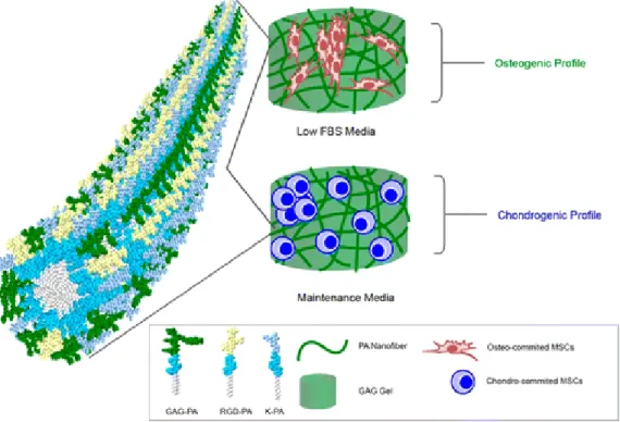
Peptide amphiphiles can form nanofibers which can be used as scaffolds to mimic the ECM. They have high water content and form a porous network which is similar to the ECM. Mimicking ECM structure in terms of functionality and complexity holds high potential for regenerative medicine [27]. Peptide amphiphile nanofibers have been widely used in 2D and 3D cellular culture studies as scaffolds [28] and which make them suitable candidates for regenerative medicine applications due to their biocompatibility, biodegradability and incorporation of bioactive signals [29, 30] which enables the synthesis of scaffolds for different applications for guided differentiation; neural differentiation [31], adipogenesis [32], osteogenesis [33], chondrogenesis [26], wound healing [34]. Glycopeptides have many functional roles in tissue engineering and the bioactive region of the peptide amphiphile molecules can also be functionalized by using sugar moieties such as glucose. It has been shown by our group that these sugar conjugated peptides can modulate the differentiation of cells [35] (Figure 6) as well as can be used for targeted delivery [20].
10
Figure 6 Supramolecular GAG-like self-assembled glycopeptide nanofibers induce chondrogenesis and cartilage regeneration. (Adapted from Ref. 37 with permission from American Chemical Society)
Petide amphiphiles have also been shown to be affective in terms of neural regeneration and differentiation. Non-covalent functionalization of adamantine-conjugated IKVAV epitope with electrospun cyclodextrin nanofibers was shown to be inducing neural differentiation as well as guiding neurite extension. These nanofibers were biocompatible for the cells and also having adhesive properties which provided an ECM like environment for the cells. Presenting a bioactive epitope on the surface of electrospun nanofibers enhanced the directional neurite extension and neural differentiation of PC-12 cells [31].
Peptide amphiphiles having different bioactive moieties can also be utilized for cellular differentiation purposes. 3D construction of peptide amphiphiles into gel like structures provide a more in vivo like environment for the cells and enables directed differentiation through specific ECM mimetic signals [28] (Figure 7).
11
Figure 7 Glycosaminoglycan-Mimetic signals direct the Osteo/Chondrogenic differentiation of mesenchymal stem sells in a three-dimensional peptide nanofiber extracellular matrix mimetic environment. (Adapted from Ref. 30 with permission from American Chemical Society)
1.5. Drug Delivery
Peptide amphiphiles are potential candidates to be used as delivery agents, through the modification of the bioactive epitope region, peptide amphiphiles can be synthesized to target cells and deliver agents into the cells [20, 36]. Their internalization ability into cells also makes them essential candidates for drug delivery applications. Covalent conjugation of drugs to peptide amphiphile molecules also enables the preservation of the stability of the drug in blood stream
12
and increase the effectivity of the drug molecule [37]. Additionally these molecules can form supramolecular nanofiber gels and can be used for local delivery of drugs [38] (Figure 8).
Figure 8 Local delivery of anti-cancer drug doxorubicin in a mice breast cancer model. (Adapted from Ref. 29 with permission from Royal Society of Chemistry)
Overall nanomaterials offer various improvements in many different areas. Although there are many types of nanomaterials developed for various biological applications, their clinical translations are limited. In the production of nanomaterials there are certain aspects that need to be covered in more detail such as the stability of the nanomaterial should be high in different acidic and basic conditions, side effects and
13
side products should be eliminated, accumulation of the nanoparticle should be limited and biodegradable molecules should be designed for therapeutic purposes in order to prevent metabolic problems in organs. In this thesis work I have utilized biomaterials for two different purposes: for the activation of dendritic cells and as a 3D scaffold for neural tissue engineering.
14
Chapter 2
2. Objectives
This work is partially described in the following publication[39]:
G. Gunay, M. Sardan Ekiz, X. Ferhati, B. Richichi, C. Nativi, A. B. Tekinay, et al., "Antigenic GM3 Lactone Mimetic Molecule Integrated Mannosylated Glycopeptide Nanofibers for the Activation and Maturation of Dendritic Cells," ACS Applied Materials & Interfaces, 2017.
The ability of dendritic cells to coordinate innate and adaptive immune responses makes them essential targets for vaccination strategies. Presentation of specific antigens by dendritic cells is required for the activation of the immune system against many pathogens, and nanoscale materials can be designed for active targeting of dendritic cells. In this work, we integrated an immunogenic, carbohydrate melanoma-associated antigen-mimetic GM3-lactone molecule into mannosylated peptide amphiphile nanofibers to target dendritic cells through DC-SIGN receptor, as it is well known that the DC-SIGN receptor has affinity to highly mannosylated structures and facilitates their uptake. Human monocytic THP-1 cell line was
15
cultured in IL-4 and GM-CSF and differentiated into immature dendritic, previous studies indicated the expression of DC-SIGN in these cells. Overall aim was to activate and mature immature dendritic cells into mature dendritic cells through the delivery of GM3-MIM into cells by using mannosylated PA molecules.
2.1. Introduction
2.1.1. Dendritic Cells and Adaptive Immunity
Dendritic cells (DCs) were discovered in 1973 [40], and are part of the mammalian immune system. Dendritic cells are professional antigen-presenting cells; they habe ability to sense and capture microorganisms, antigens and present these processed antigens to T & B cells through MHC molecules [41]. This presentation initiates the adaptive immune response through the activation of T cells to specific antigens. The activation of dendritic cells and their capability to induce other cells depend on the state of their maturation, which is induced through the up-regulation of certain co-stimulatory molecules [42].
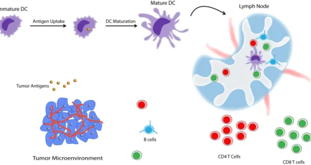
16
Figure 9 Generation of cellular immune response against tumor antigens.
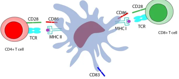
In their immature state, DCs sample antigens in their native environment. Upon antigen sensing and uptake, they start to mature, migrate to lymph nodes, and present the antigenic peptides for the induction of an adaptive immune response [43] (Figure 9). Maturation of dendritic cells is vital because they cannot initiate an immune response in their immature state [44]. In order for a dendritic cell to activate T cells properly, T cells antigen receptors (TCRs) must recognize the antigens which is presented on the surface of antigen-presenting cells like dendritic cells. These antigens (which may be peptides, nucleic acids or glycans) are typically presented in association with major histocompatibility complex (MHC) molecules [45]. MHC class I molecules present antigens from intracellular sources, while MHC class II molecules present peptides from exogenous sources; their targets are CD8+ and CD4+ T cells, respectively [46]. However, the signal between MHC complex and TCRs is not by itself enough for a proper T cell activation and subsequent
17
proliferation, as CD80 or CD86 molecules on the surface of dendritic cells should also interact with the CD28 molecule on T cells to initiate the activation process [47, 48]. Another marker that is very important for dendritic cell maturation is CD83, which is expressed on mature human dendritic cells and upregulated during the transition from the immature state to mature state [49-51] (Figure 10).
Figure 10 Interaction of dendritic cells with CD4 and CD8 T cells.
In terms of activating dendritic cells, all of these co-stimulatory molecule expressions are significant for complete activation and the ability of these cells to initiate immune response makes them vital for antigen-specific immunotherapy applications [52]. DCs have immunostimulatory functions, which have been confirmed both in in vivo and in vitro conditions [53], DCs pulsed with proteins [54] or with synthetic peptides [55] have been shown to induce antigen-specific effector T cells.
18 2.1.2. C-Type Lectins
Host recognition by dendritic cells occurs through several receptor families such as C-type lectin receptors (CLRs), which are mainly responsible for carbohydrate recognition [56]. These receptors are generally expressed on the immature state and down-regulated upon activation. The specificity of carbohydrate recognition has made CLRs very good vaccine targets for improved antigen presentation [57]. Among CLRs, dendritic cell intracellular adhesion molecule 3 (ICAM-3) grabbing non-integrin (DC-SIGN) exhibits considerable affinity to mannose residues and is mainly expressed on immature dendritic cells [58, 59] (Figure 11). It modulates the uptake of specific pathogens through its interactions with mannosylated structures [58, 60, 61]. Ligands that are internalized through DC-SIGN receptor are processed and presented to CD4+ T cells following their binding to MHC-II molecules [62].
19 Figure 11 DC-SIGN receptor on immature DCs.
2.1.3. Carbohydrate Associated Antigens
Discovery of human cancer antigens enabled scientists to develop new immunotherapy strategies [63, 64]. Melanoma, as a major type of cancer, is widely appreciated to be a promising target for immunotherapy applications [65]. GM3 ganglioside is a major glycosphingolipid that is overexpressed in metastatic melanoma cells, and there is an abundance of GM3 ganglioside studies that focus on its potential to be used as a carbohydrate-based, melanoma-associated antigen for cancer diagnosis [66]. A metabolite of GM3 ganglioside, called GM3 lactone, is also present in melanoma cells; however, due to its low amounts it is below the recognition threshold and does not induce an immune response. However, GM3 lactone is more immunogenic than GM3 ganglioside and can induce stronger immune responses at equal concentrations. A previous study had shown that permannosylated dendrons containing an antigenic GM3 lactone mimetic is able to induce dendritic cell activation through DC-SIGN internalization [67].
20
Recently, antigens delivered through nanoscale materials have been shown to provide considerable advantages over soluble antigen administration, due to the stimulation of antigen uptake and cellular activation by surface-functionalized delivery vectors. Gold and PLGA nanoparticles, dendrimers and liposomes have previously been functionalized for in vitro dendritic cell targeting purposes [67-69].
In this project we targeted dendritic cells by using glycopeptides and delivered a mimetic of GM3-lactone (GM3-MIM) and analyzed the activation and maturation levels of immature dendritic cells.
21 2.2. Experimental Section
2.2.1. Materials
9-fluorenylmethoxycarbonyl (Fmoc) protected amino acids, [4-[α-(2’,4’-dimethoxyphenyl) Fmoc aminomethyl] phenoxy] acetamidonorleucyl-MBHA resin (Rink amide MBHA resin) and 2-(1H-Benzotriazol-1-yl)-1,1,3,3-tetramethyluronium hexafluorophosphate (HBTU) were purchased from NovaBiochem. Fmoc-Ser[β-Glc(OAc)4]-OH was purchased from AAPPTec. Lauric acid and N,N- Diisopropylethylamine (DIEA) were purchased from Merck. Other chemicals were purchased from Alfa Aesar or Sigma-Aldrich and used without any purification. Deionized water (resistance of 18 MΩ.cm) was used during the experiments.
2.2.2. Synthesis of Amphiphilic Glycopeptides
Protected glycopeptides (Man-PA and Glc-PA) were constructed on MHBA Rink Amide resin. All amino acid couplings were performed with 2 equivalents of Fmoc protected amino acid, 1.95 equivalents of HBTU and 3 equivalents of N,N-diisopropylethylamine (DIEA) in DMF for 3 h. Fmoc deprotections were performed with 20% piperidine/ dimethyl formamide (DMF) solution for 20 min. Cleavage of the peptides from the resin and deprotection of acid labile protecting groups were carried out with a mixture of trifluoroacetic acid (TFA) : triisoproplysilane (TIS) : water in the ratio of 95 : 2.5 : 2.5 for 2 h. Excess TFA was removed by rotary evaporation. The remaining residue was triturated with ice-cold diethyl ether and the resulting white pellet was freeze-dried. Deacetylation reaction was carried out in solution. For the cleavage of acetyl groups, 210 mg of protected glycopeptide (1 eq.)
22
was dissolved in 105 ml of anhydrous methanol. 2M of NaOMe (4.4 eq.) was dissolved in methanol and poured into the solution. After adjusting pH to 8-8.5, the reaction was carried out at room temperature for 2-3 h. The solution was neutralized with a few drops of acetic acid in order to terminate the reaction. The solvent was removed by vacuo. After water addition, it was frozen at -80°C and freeze-dried. Before and after the deacetylation reaction, glycopeptides were identified and analyzed by reverse phase HPLC on an Agilent 6530 accurate-Mass Q-TOF LC/MS equipped with an Agilent 1200 HPLC. An phenomenex Luna 3μ C8 100A (50 x 3.00 mm) column as stationary phase and water/acetonitrile gradient with 0.1% volume of formic acid as mobile phase were used to identify peptide amphiphile. They were purified on Agilent 1200 by using a Zorbax prepHT 300CB-C8 column with a water–acetonitrile (0.1% TFA) gradient.
2.2.3. Synthesis of Fluorescent GM3-MIM Derivatives (GM3-MIM-Bodipy) The alkyl derivative of GM3-MIM was prepared and provided by Prof. Cristina Nativi according to the previously reported procedure [70].
2.2.4. Preparation of GM3-MIM Integrated Glycopeptide Nanofibers
1 mM of Man-PA and Glc-PA (2 ml) were dissolved in water and heated upto 50°C during sonication. 1.33 mM of GM3-MIM was dissolved in DCM:MeOH mixture (7:1 v/v %, 610 μl) and added into each glycopeptide solution. They were sonicated and vortexed for an hour at 50°C. While the mixture was initially turbid, it became transparent as the solvents evaporated. Final GM3-MIM concentration to PA concentration ratio was adjusted to 1:2.5.
23
2.2.5. Characterization of Fluorescent GM3-MIM Integrated Glycopeptide Nanofibers
Fluorescence spectra of GM3-MIM and GM3-MIM integrated glycopeptide nanofibers were recorded by a Varian Cary Eclipse spectrophotometer with an excitation wavelength of 503 nm. Varian Cary 100 UV-VIS spectrophotometer was used to record absorption spectra of GM3-MIM-BodiPy and GM3-MIM integrated glycopeptide nanofibers. For UV and fluorescence measurements, DCM:MeOH mixture (7:1, v/v %) was used to dissolve GM3-MIM-BodiPy, and water was used as the solvent for GM3-MIM integrated glycopeptide nanofibers. A Jasco J-815 CD spectrophotometer was used to investigate the secondary structures of PA molecules and GM3-MIM integrated PA nanofibers. All samples were measured at physiological pH at 0.25 mM concentration. 300 µL of the sample was transferred into a 1 mm quartz cuvette and spectra were obtained at room temperature from 300 nm to 190 nm with a data interval of 1 nm and a scanning speed of 100 nm/min. TEM images were obtained with a FEI Tecnai G2 F30 TEM at 200 kV. A high-angle annular dark field (HAADF) detector was used for images taken in STEM mode. 1 mM of Glc-PA, Man-PA and GM3-MIM integrated glycopeptide nanofiber systems were first diluted to 150 μM and then dropped on 300-mesh carbon TEM grids. Samples were allowed to stay at room temperature for 3 min, stained by 2 wt % uranyl-acetate staining for another 1-2 min and air dried prior to STEM imaging. Zeta potential of the nanofibers was measured by ZetaSizer. A Malvern Nanosizer/ZetaSizer Nano-ZS ZEN 3600 (Malvern Instruments, USA) instrument was used for the analysis. Measurements were performed in quartz cuvettes and
24
repeated at least three times. Samples were prepared by dissolving each component in water at a concentration of 0.25 mM.
2.2.6. Cellular Viability
THP-1 human monocytes were kindly provided by Prof. E. Erbay of Bilkent University. Cells were seeded onto 96 well plates at a density of 5000 cells/well within media containing Man-PA and Glc-PA. Viability was assessed at 150 µM and 250 µM for both peptide molecules. At the end of 24 h, cells were washed with 1X PBS and were stained with Calcein-AM and ethidium homodimer for 30 min at room temperature in dark. Then, images of the cells were taken and quantified by using
Image J program.
2.2.7. Differentiation of THP-1 Human Monocytes into Immature Dendritic Cells
THP-1 human monocyte cell line was differentiated into immature dendritic cells in RPMI medium containing 10% FBS, rhIL-4 (100 ng/mL) and rhGM-CSF (100 ng/mL) for 5 days. Medium was exchanged with fresh cytokines after 2 days.
2.2.8. Internalization of Fluorescent GM3-MIM Integrated Glycopeptide Nanofibers
13 mm glass coverslips were placed in 24-well plates and 5 x 10⁴ THP-1 cells were seeded and differentiated into immature dendritic cells as previously stated. After 5 days, the PA molecules were administered and cells were incubated for 24 h. After incubation, cells were washed with PBS two times, fixed with 4% paraformaldehyde, and permeabilized with 3% Triton-X and stained with phalloidin and TO-PRO-3.
25
Cells were visualized by using Laser Scanning Confocal Microscope (LSM 510, Zeiss).
2.2.9. Surface Marker Analyses of Dendritic Cell Activation
iDCs were obtained from THP-1 monocytes as mentioned above. At the end of 5 days, cells were treated with; Man-PA, Glc-PA, Man-PA/GM3-MIM, Glc-PA/GM3-MIM, and only GM3-MIM for 24 h. Total peptide concentration for each group was 150 µM and GM3-MIM amount was 40 µg. At the end of 24 h, media were discarded, cells were centrifuged at 2500 rpm for 5 min and washed with 1X PBS and then centrifuged again. Then, pellet was dissolved in 1X PBS and cells were stained with CD86 (PerCP), CD83 (Phycoerythrin) and HLA-DR (MHC-II) (PE / Cy5) antibodies for 15 min at room temperature. 10.000 cells were recorded for each condition during flow cytometry analyses.
2.2.10. Scanning Electron Microscopy (SEM) Imaging
SEM imaging was carried out in order to investigate the morphological changes during dendritic cell maturation. 5 x 10⁴ THP-1 cells were seeded onto coverslips in 24 well plates and differentiated into immature DCs as previously stated. Treatment was carried out for 24 h and at the end of 24 h cells were washed once with 1X PBS, fixed with 2% gluteraldehyde/PBS and 1 wt % OsO4 for 1 h each. After fixation, cells were washed with ddH2O and dehydrated by using increasing ethanol concentrations; 20%, 40%, 60%, 80% and 100% v/v. Critical point dryer (Autosamdri 815B equipment from Tousimis) was used afterwards and coverslips were coated with 4 nm Au/Pd. SEM (FEI Quanta 200 FEG) images were taken through using Everhart–Thornley Detector (ETD) at high vacuum mode at 5 keV beam energy.
26 2.3. Results and Discussion
2.3.1. Design, Synthesis and Characterization of Glycopeptide Conjugates
In this study, mannose-PA (Man-PA) and glucose-PA (Glc-PA) were used for the conjugation of a GM3-lactone mimetic molecule (GM3-MIM) and targeting of immature dendritic cells (Figure 12). The purity of the synthesized peptides was assessed through liquid chromatography-mass spectrometry (LC-MS) (Figure 13). Purity of synthesized peptides was higher than 90%, and GM3-MIM or GM3-MIM Bodipy was integrated into glycopeptides following the confirmation of peptide purity.
27 5 10 15 20 Man-PA Time (min) In te n s ity (a .u ) 5 10 15 20 Glc-PA Time (min) In te n s ity ( a .u ) [M+H] [M+H] + +
Figure 12 Chemical structures of molecules used in the study.
Figure 13 Liquid chromatograms and mass spectra (LC-MS) of amphiphilic glycopeptides. Man-PA, [M+H]+ (calculated) = 903.5688, [M+H]+ (observed) = 903.5729; Glc-PA, [M+H]+ (calculated) = 903.5688, [M+H]+ (observed) = 903.5758.
28
Before the integration of fluorescent GM3-MIM into the glycopeptides, the secondary structure and morphological properties of the amphiphilic glycopeptides were investigated at physiological pH. Scanning transmission electron microscopy (STEM) was performed in order to analyze the morphological characteristics of glycopeptides and both glycopeptide samples, regardless of the type of sugar they contain, were found to form high-aspect-ratio nanofibers with diameters in the order of 9-11 nm and lengths reaching several micrometers (Figures 14A and 14B),
Figure 14 STEM images of (A) Man-PA (B) Glc-PA.
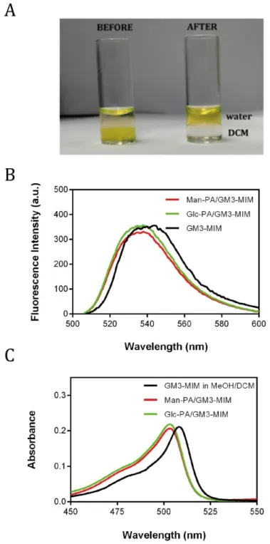
GM3-MIM has poor solubility in water; consequently, molecule was integrated into glycopeptide nanofibers through solvent exchange process to improve its bioavailability (Figure 8). GM3-MIM was solubilized in DCM:MeOH (7:1, v/v %) and glycopeptide solutions were dissolved in slightly acidic water. Glycopeptide solutions were sonicated before the addition of GM3-MIM at 50 °C at pH 6. The rationale behind this step was to disrupt the self-assembled fibers and promote the
29
generation of glycopeptide molecules as single units, allowing the integration of GM3-MIM into fiber structures during re-assembly. Due to the lysine residues they bear, glycopeptides carry an overall positive charge that increases their solubility in water and promotes their integration during self-assembly. GM3-MIM containing solution was added to glycopeptides separately, and the mixture was heated and sonicated to allow the interaction of the hydrophobic tail region of GM3-MIM with the hydrophobic amino acids and hydrocarbon chain region of glycopeptides. This procedure promoted the transfer of GM3-MIM into water. GM3-MIM displays an amphiphilic structure and is expected to have its hydrophobic tail region face the fiber core following integration into peptide nanofibers, while the hydrophilic ring structure would face the outer region.
GM3-MIM integration was visualized through the use of Bodipy dye-conjugated GM3-MIM molecules (Figure 15A). Quantitative analysis of the GM3-MIM transfer was carried out with fluorescence and UV spectra measurements. GM3-MIM integrated glycopeptides were blue-shifted compared to the non-conjugated GM3-MIM group, as observed both visually and through spectral measurements (Figure 15B and 15C). UV spectra results also provided similar absorbance values for integration into both glycopeptide groups, and suggested that the efficiency of the integration was very close to 100%.
30
Figure 15 Solvent exchange (DCM:H2O) process for the integration of the GM3-MIM molecule into peptide structures. (B) Fluorescence and (C) UV spectra of GM3-MIM and GM3-MIM integrated glycopeptide solutions dissolved in DCM:MeOH (7:1) and water, respectively.
31
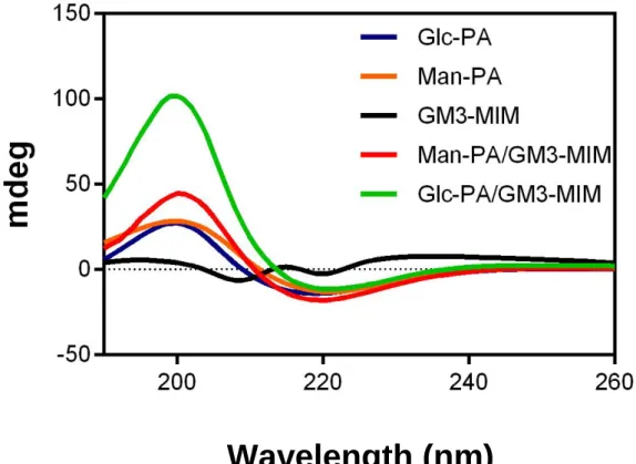
After achieving the successful transfer of GM3-MIM, glycopeptides containing the antigen were analyzed for their morphological characteristics through CD and STEM. Diluted solutions of Man-PA and Glc-PA were studied by circular dichroism (CD) spectroscopy and the results revealed that both pure systems were oriented in a β-sheet conformation, exhibiting a positive peak at 202 nm and a negative peak at 218 nm (Figure 16).
Wavelength (nm)
Figure 16 CD spectra of amphiphilic glycopeptide solutions, MIM and GM3-MIM integrated glycopeptide systems.
32
Even though bundling to a higher degree was observed after the integration of antigen in STEM images, self-assembly still resulted in the formation of nanofibers with similar diameters, which suggests that there are no significant changes during PA assembly in the presence and absence of the antigen (Figure 17A and 17B). In addition, the antigen contained sulfur and its integration into glycopeptides could also be analyzed through sulfur and oxygen mapping, which was performed by using TEM. This analysis confirmed that GM3-MIM antigen was present in glycopeptides (Figure 18).
33
Figure 18 TEM mapping of fluorescent GM3-MIM integrated Man-PA nanofibers, S-pre edge (left), sulfur mapping (middle), oxygen mapping (right).
Charge is a very important factor that may interfere with the internalization efficiency; therefore we analyzed glycopeptides for their charge properties by using Zeta potential measurements. The results indicated a similar positive net charge for both glycopeptides around +27 mV (Figure 19).
2 μm
34
2.3.2. Cell Viability and iDC Differentiation from THP-1 Monocytes
THP-1 human monocyte cells were used for in vitro analyses. Cells were treated with different concentrations of Man-PA and Glc-PA. No significant difference in terms of viability was found between TCP and glycopeptides at 150 µM (Figure 20), indicating the biocompatibility of both peptide amphiphiles at this concentration. However, toxicity was observed at higher concentrations, possibly due to the stress formed by the accumulation of peptides on the cell membrane.
35
Figure 20 Cellular viability of THP-1 cells treated with different concentrations of Man-PA and Glc-PA, and TCP for 24 h. Values represent mean ± SEM (****p<0.0001).
After ensuring the biocompatibility of the peptide amphiphiles, human THP-1 monocytes were differentiated into immature dendritic cells (iDC) as previously mentioned (Figure 20 and Figure 21).
36
Figure 21 Schematic representation of the transition from human monocytes to mature dendritic cells (mDC).
Figure 22 Differentiation of THP-1 monocytes into iDCs in terms of (A) de novo surface CD86 expression and (B) morphological changes upon differentiation into iDC.
After differentiation of THP-1 monocytes into iDCs, Man-PA/GM3-MIM, Glc-PA/GM3-MIM, GM3-MIM, Man-PA and Glc-PA were applied to cells for 24 h. At the end of 24 h, light microscope images of the cells were taken in order to examine the morphological changes. Cellular morphology was more indicative of maturation
37
in Man-PA/GM3-MIM compared to the other groups, indicating the higher delivery of GM3-MIM into iDCs through Man-PA.
2.3.3. Internalization of Fluorescent GM3-MIM Integrated Glycopeptide Nanofibers
After examining the morphological differences, the uptake of PA molecules were assessed through confocal microscopy visualization and flow cytometry quantification. Concentrations were optimized according to the live-dead experiment results. Since GM3-MIM molecule was conjugated with Bodipy fluorescent dye, internalization as well as the morphological changes due to the internalization could be directly visualized.
Confocal microscopy images indicated higher uptake and a more mDC-like morphology in the Man-PA/GM3-MIM group compared to other groups (Figure 22). Results indicate the importance of using Man-PA for the delivery of GM3-MIM into the cells.
38
Figure 23 Uptake and morphological changes of iDCs when treated with Man-PA/GM3-MIM, Glc-PA/GM3-MIM and only GM3-MIM for 24 h. Blue indicating To-Pro nuclei staining, red indicating Phalloidin microtubule staining and green indicating bodipy conjugated GM3-MIM.
39
Quantitative analysis of GM3-MIM internalization was carried out by using flow cytometry. Cells were treated with Man-PA/GM3-MIM, Glc-PA/GM3-MIM, or GM3-MIM for 24 h. Mean fluorescent intensity (MFI) of GM3-MIM for each group was compared between each other and results indicated a significantly greater internalization in the Man-PA/GM3-MIM group compared to other groups (Figure 23). Additionally, there was a significant difference between the Glc-PA/GM3-MIM group and the GM-3/MIM group, which suggests that even though there is no specificity of glucose to DC-SIGN, the nanofiber system itself promotes better internalization (Figure 23).
Figure 24 GM3-MIM MFI analyses for Man-PA/GM3-MIM, Glc-PA/GM3-MIM and only GM3-MIM for 24 h. Values represent mean ± SEM (****p<0.0001).
2.3.4. Surface Marker Analyses of Dendritic Cell Activation
Activation and maturation state of DCs were measured by analyzing the expression of CD86, CD83 and MHC-II receptors. Increased expression of these molecules was
40
previously shown to be associated with the maturation and activation status of dendritic cells [67, 71]. CD86 is a co-stimulatory molecule that interacts with CD28 on T cells and is required for proper activation [47]. Man-PA/GM3-MIM group significantly increased the expression of CD86 compared to other groups, as measured by flow cytometry (Figure 24A). CD83 is another activation marker for DCs and its expression is up-regulated during the transition from immature state to mature state [49] . Man-PA/GM3-MIM also significantly increased the expression of CD83 compared to the other compounds (Figure 24B), and the same pattern was observed for the expression of MHC-II (Figure 24C), which is a molecule essential for the proper presentation of antigens [62]. Expression profiles obtained by flow cytometry analysis were in agreement with fluorescent imaging (Figure 22) and flow cytometry analyses (Figure 23) of GM3-MIM internalization. In summary, considering the necessity of the up-regulation of these molecules for DC activation and maturation, Man-PA/GM3-MIM was shown to have a significant effect on inducing the activation and maturation of DCs in terms of surface expression of CD86, CD83 and MHC-II receptors.
41
Figure 25 Surface expression MFI values of (A) CD86, (B) CD83 and (C) MHC-II under Man-PA/GM3-MIM, Glc-PA/GM3-MIM and GM3-MIM treatment for 24 h. Values represent mean ± SEM (****p<0.0001).
42
2.3.5. Morphological Analyses of Dendritic Cell Activation
Morphological changes occuring during the activation of iDCs into mDCs were also analyzed with SEM imaging. Cells treated with Man-PA/GM3-MIM matured to a greater extent than other groups, although mDC-like morphologies were also observed in Glc-PA/GM3-MIM and only GM3-MIM groups. Additionally, the effects of Man-PA and Glc-PA in the absence of GM3-MIM loading were also analyzed in order to ensure that the maturation of the cells was due to antigen delivery rather than the PA molecules (Figure 26).
Figure 26 Morphological analyses of the differentiation in iDCs when treated with Man-PA/GM3-MIM, Glc-PA/GM3-MIM, only GM3-MIM, only Man-PA and only Glc-PA for 24 h. Scale bars are 30 µm.
43 2.4. Conclusions and Future Perspectives
In this study, peptide amphiphile molecules bearing mannose moieties were used for the targeting of the DC-SIGN receptor and the delivery of an immunogenic mimetic of GM3-lactone antigen to induce DC maturation and activation [39]. GM3-MIM presented on Man-PA nanofibers were more efficient than Glc-PA/GM3-MIM and GM3-MIM in terms of antigen internalization, stimulation of CD86, CD83 and MHC-II expression and the induction of a mature DC-like morphology, suggesting that mannose/DC-SIGN interactions are primarily responsible for uptake. Considering the instability of GM3 lactone molecule and its low concentration under physiological conditions, Man-PA is a promising delivery vehicle for the internalization of this immunogenic antigen through DC-SIGN receptors. We showed that the targeted delivery of antigens by using Man-PA to activate iDCs can play important roles in inducing cellular immunity against cancer conditions, and that GM3-MIM can be delivered to the immune system through carrier nanofibers.
Peptide amphiphiles are biocompatible and biodegradable materials with a broad range of potential applications for tissue-specific targeting and delivery. Unlike conventional delivery vesicles, such as liposomes, PA systems do not need to be functionalized with targeting molecules and can naturally localize to tissues of interest due to the bioactive epitopes expressed on their surfaces. These epitope sequences can also be designed to interact with specific cell surface receptors, promoting entry into target cell types. In the present study, the integration of GM3-MIM into the peptide nanofiber structure enabled efficient internalization of the molecule. Considering that highly mannosylated structures are suitable candidates
44
for DC-SIGN receptor targeting, using sugar moieties for delivery purposes holds high potential for future studies.
45
Chapter 3
3. Three-dimensional laminin mimetic peptide nanofiber
gels for in vitro neural differentiation
This work is partially described in the following publication:
Effect of 2D and 3D Culturing on in vitro Neural Differentiation Under Bioactive Signaling
Melike Sever, Gokhan Gunay, Mustafa O. Guler*, Ayse B. Tekinay* (Under revision in Biotechnology Journal)
46
The extracellular matrix (ECM) provides biochemical signals and structural support for cells and its functional imitation is a fundamental aspect of biomaterial design for regenerative medicine applications. In this study, we investigated the stimulation of neural differentiation by a laminin-derived epitope in two-dimensional (2D) and three-dimensional (3D) environments. The 3D gel system was found to be superior to its 2D counterpart for the induction of neural differentiation, even in the absence of a crucial biological inducer, the nerve growth factor (NGF). In addition, cells cultured in 3D gels exhibited a spherical morphology that is consistent with their natural form under in vivo conditions. Overall, the present study underlines the impact of bioactivity, dimension and NGF addition, as well as the cooperative effects thereof, on the neural differentiation of PC-12 cells. These results underline the significance of 3D culture systems in the development of scaffolds that closely replicate in vivo environments for the formation of cellular organoid models in vitro.
3.2. Introduction
The extracellular matrix (ECM) provides biochemical cues and structural support for cells and mimicking the ECM for the investigation of cellular responses has been a great challenge for scientists. Design of biomaterials aims to mimic the native environment of cells (Figure 26) and the interactions of these biomaterials with cells could vary accordingly with the culturing technique. Recently, 3D culturing studies indicated that the cellular responses might significantly change compared to 2D culturing in terms of cellular morphology, viability [72, 73], proliferation [74], signaling [75, 76], differentiation [77, 78] and release molecules [79, 80].
47
3D hydrogels such as Matrigel®, collagen and alginate are extensively used in cell culture studies, and their similarity to the natural extracellular matrix contributes strongly to their bioactivity in regenerative medicine applications as delivery systems of bioactive agents and their usage in wound healing and cellular transplantation purposes [81]. 3D Collagen gels on the other hand are very suitable systems for mimicking connective tissue [82]. Matrigel is another 3D gel used for regenerative medicine applications; it contains laminin, collagen IV and entactin in high amounts [83], providing a wide range of applications for cellular studies. 3D collagen gels, for example, are highly suitable for mimicking the connective tissue, while Matrigel® is applicable for the culture of a broad range of cells and tissues due to its incorporation of basement membrane elements such as laminin, collagen IV and entactin. However, these materials present significant batch-to-batch variation and do not necessarily provide the full range of ECM-mimetic signals found in natural tissue environments [84, 85]. Consequently, there is considerable potential in developing easy-to-use and tailorable 3D culture systems through the use of synthetic biomaterials.
48
Figure 27 Schematic illustration of cellular interaction with ECM.
Peptide amphiphile systems have been extensively used in neural regeneration applications, and bioactive epitopes derived from ECM proteins, such as the laminin-derived IKVAV and YIGSR, fibronectin-laminin-derived RGD, and tenascin-C-laminin-derived VFDNFVLK sequences, have been shown to induce neural differentiation, attachment and migration [86-89]. The imitation of ECM proteins is especially important in this context, as integrin receptors and similar mechanotransduction elements are essential for cellular adhesion, homing and differentiation [90]. Many types of integrins, in turn, are directly bound to ECM components [91] and initiate signaling cascades through focal adhesion kinase (FAK) and phosphoinositide 3-kinase pathways. β1 integrins, for example, were shown to be effective for stem cell differentiation and self-renewal [92]. Previous studies also indicated that β1 integrins regulate the self-renewal and differentiation of neural stem cells (NSCs) through
49
Notch and EGF receptors [93]. Laminin is another major ECM component of both central and peripheral nervous systems (CNS and PNS) and serves as an important mediator of axonal outgrowth [94]. IKVAV is one of the most extensively studied bioactive epitopes and is located on the α-laminin chain [95], which is essential for the ability of laminin to regulate the overall cellular fate [96]. IKVAV sequence, derived from laminin induces neural differentiation [97], axonal growth after spinal cord injury in vivo [98] and neural differentiation of PC-12 cells in vitro [31, 87] as a part of peptide nanofiber system. However, no 3D study has so far demonstrated the effect of a laminin-mimetic environment on neuronal differentiation, which is a critical step for the translation of biomimetic scaffolds to clinical applications. Cell behavior is influenced strongly by the interactions that occur at the cell-biomaterial interfaces, and the development of novel cell-biomaterials that can exercise precise control over cell function are of fundamental importance for tissue regeneration. Consequently, physical properties should also be considered in addition to biological and chemical properties when biomaterials designing for tissue-engineering applications. Parameters such as stiffness and dimensionality are important for scaffold functionalization to induce neural differentiation. When designing a scaffold for neural differentiation, the mechanical properties of the scaffold should be similar to that of brain tissue, the elastic modulus of which is below 1 kPa [99]. Stiffness is also important for differentiation into different cell types. For instance, NSCs prefer to differentiate into neurons under an intermediate substrate stiffness (500 Pa), while increased stiffness favors differentiation into the astrocyte lineage [100]. Dimensionality is another important parameter, and although two-dimensional (2D) cell cultures are commonly used in differentiation studies,
50
three-dimensional cell cultures are important in vitro models to fill the gap between 2D cell culture experiments and in vivo studies. 3D models are especially important for studying the regeneration of neural tissue, which has a very low regeneration capacity that may be improved by closely mimicking the native extracellular matrix of neural cells to provide support and enhance the diffusion of oxygen and nutrients [101].
In this chapter, the neural differentiation potential of PC-12 cells was investigated on 2D and 3D laminin-mimetic environments produced through the self-assembly of peptide nanofibers. Effects of bioactivity, dimensionality and NGF addition on neural differentiation were analyzed by monitoring the expression of neurogenic markers and proteins, and scanning electron microscopy imaging was used to observe the overall morphology of cells in 2D and 3D scaffolds. Overall, the present study compares the neural differentiation potential of PC-12 cells under different culturing conditions and highlights the importance of presenting bioactive IKVAV sequence on 3D hydrogels.