DEVELOPMENT OF A SPECIALIZED ZEBRAFISH
XENOTRANSPLANTATION DATABASE AND
ESTABLISHMENT OF ALU-BASED TUMOR DNA
QUANTIFICATION METHOD IN ZEBRAFISH:
FOCUS ON MODELS OF OVEREXPRESSION AND
MICROENVIRONMENT
A DISSERTATION SUBMITTED TO
THE GRADUATE SCHOOL OF ENGINEERING AND SCIENCE OF
BILKENT UNIVERSITY
IN PARTIAL FULFILLMENT OF THE REQUIREMENTS FOR
THE DEGREE OF DOCTOR OF PHILOSOPHY
IN
MOLECULAR BIOLOGY AND GENETICS
BY SENİYE TARGEN
SEPTEMBER 2020
DEVELOPMENT OF A SPECIALIZED ZEBRAFISH
XENOTRANSPLANTATION DATABASE AND ESTABLISHMENT OF ALU-BASED TUMOR DNA QUANTIFICATION METHOD IN ZEBRAFISH: FOCUS
ON MODELS OF OVEREXPRESSION AND MICROENVIRONMENT By Seniye Targen
September 2020
We certify that we have read this dissertation and that in our opinion it is fully adequate, in scope and in quality, as a thesis for the degree of Doctor of Philosophy.
_____________________
Özlen Konu Karakayalı (Advisor)
_____________________ Michelle Marie Adams
______________________ Murat Alper Cevher
______________________ Sreeparna Banerjee
______________________ Ceren Sucularlı
Approved for the Graduate School of Engineering and Science:
__________________ Ezhan Karaşan
ABSTRACT
DEVELOPMENT OF A SPECIALIZED ZEBRAFISH
XENOTRANSPLANTATION DATABASE AND ESTABLISHMENT OF ALU-BASED TUMOR DNA QUANTIFICATION METHOD IN ZEBRAFISH:
FOCUS ON MODELS OF OVEREXPRESSION AND MICROENVIRONMENT
Seniye Targen
Ph.D. in Molecular Biology and Genetics Supervisor: Özlen Konu Karakayalı
September 2020
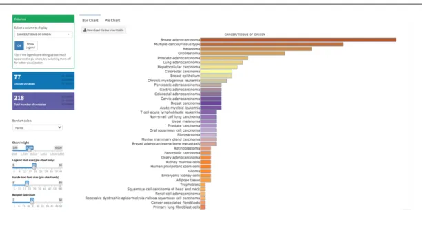
Successful xenotransplantation of human cancer cells into zebrafish host marked a new era in cancer research enabling high throughput in vivo screens. Zebrafish xenotransplantation literature continues to rapidly accumulate, and this necessitates the development of an interactive database for accommodating the collective data for fined-tune search, visualization and statistical representation purposes. Herein, I have introduced an interactive database, ZenoFishDb v1.1 (https://konulab.shinyapps.io/zenofishdb), housing manually curated details on molecularly-modified cell transplantations, PDXs, stem cell and cancer stem cell transplantation studies as well as transplantation studies bearing modified host details. The database projects collected data in a table format via various attributes and provides graphical representation of the curated details as well as statistical analyses yielding information on incorporated numbers and frequencies of selected attributes; hence can be used for reviews and designing new experiments. Zebrafish PDX studies are separately conceptualized and displayed through ZenoFishDb v1.1. Development of the ZenoFishDb v1.1 leads to a better understanding of tumor analysis methods such as assessment of proliferation and/or tumor growth in xenotransplantation studies and further marks the need for development of novel methods for precise quantification of tumor size. In the light of these findings, I have helped establish a novel qRT-PCR-based proliferation assessment method for xenografts in zebrafish, adapted from previous mouse xenotransplantation studies. Herein, the use and precision of ALU
repeat-based quantification of transplanted liver cancer cells in genotyped zebrafish ache mutants and wildtype siblings was shown exemplifying microenvironment as an important factor for tumor growth. I further demonstrated the power of ALU repeat-based quantification in Mineralocorticoid Receptor (MR) overexpressing breast cancer cells (MCF7) injected to the transparent casper zebrafish as a case study. First, I demonstrated that MR expression and signaling was important in breast cancer biology and prognosis based on in silico TCGA and custom RNA sequencing as well as other in vitro and ex vivo assays. I further showed that results obtained from ALU repeat-based quantification of tumor growth in MR-overexpressing MCF7 cells paralleled fluorescent image-based intensity measurements while the former being relatively less time-consuming and more high-throughput. In this study, accurate quantification of MR overexpression in xenografts was also successfully performed by a cDNA-specific primer pair; and the rate of tumor growth based on image analysis, did not correlate with the amount of MR DNA in casper fish xenografts. However, MCF7 cells overexpressing MR exhibited lower cell viability in vitro although some of these effects were due to empty vector (EV) integration. Accordingly, tumor size in xenografts of naïve, EV- and MR-transfected MCF7 cells injected into pigmented AB larvae were quantified for ALU-repeats yet no significant difference was observed due to high within-group variability in vivo. Future studies are needed to assess the role of varying the volume and placement of injected cells along with the amount of MR gene transfected on tumor growth in vivo.
Key words: Zebrafish, Xenograft, Database, ALU repeat, Tumor quantification, Microenvironment, Molecular Modification, Mineralocorticoid Receptor.
ÖZET
ZEBRA BALIĞI KSENOTRANSPLANTASYON ÇALIŞMALARINI BARINDIRAN VERİ TABANI GELİŞTİRİLMESİ VE ALU-DAYANAKLI
TÜMÖR DNA ÖLÇÜMÜNÜN OVEREKSPRESYON VE MİKROÇEVRE ÖRNEKLENDİRİLMELERİ İLE ZEBRA BALIĞI KSENOGRAFT
MODELLERİNDE UYGULANMASI
Seniye Targen
Moleküler Biyoloji ve Genetik Doktora Programı Tez Danışmanı: Özlen Konu Karakayalı
Eylül 2020
Literatürde zebra balığına uyarlanan hücre ve doku transplantasyon çalışmaları gün geçtikçe artmakta ve buna bağlı olarak bu çalışmaların kolektif olarak, görüntülenebilir ve güncellenebilir bir şekilde aktarılması gereklilik arz etmektedir. Buna istinaden, zebra balığı üzerinde uygulanan moleküler modifikasyona uğramış hücre hatları transplantasyonlarının, hasta dokusu ve primer kültür transplantasyonlarının, kök hücre ve kanser kök hücresi transplantasyonu çalışmalarının, farklı zebra balığı modellerine uygulanmış transplantasyonları barındıran çalışmalarının ve detaylarının veri tablosu halinde, grafikler halinde ve istatistiksel bulgularının da tablo halinde yansıtıldığı, ZenoFishDb v1.1 (https://konulab.shinyapps.io/zenofishdb) isimli bir veri tabanı geliştirilmiştir. Bu veri tabanında primer hüre kültürü ve ksenograft çalışmalarına ayrıcalıklı olarak yer verilmiş ve de farklı veri tablo ve grafik öğeleriyle gösterilmeleri sağlanmıştır. Bu veri tabanının geliştirilmesi ile alandaki eksikler fark edilip, örneğin tümör büyüme çalışmalarındaki uygulamalar incelenip, tümör büyümesi ölçümü için daha hassas ve etkili yeni bir yöntem geliştirilmiştir. Bu metot kantitatif PCR (qRT-PCR) kullanılarak tümör hücresi enjekte edilmiş zebra balığı DNA çıkarımı sonrası insanda bulunan ALU dizilerinin niteliksel olarak değerlendirmesine dayalıdır ve daha önce fare ksenograft çalışmalarında denenmiş bir uygulamadır. Burada, ALU dizisi dayanaklı kantitatif PCR metodunun hassasiyetini ve etkinliğini, karaciğer kanseri hücresi nakledilmiş ache mutantı ve normal zebra balıklarından elde edilen DNA üzerinde test
ederek ölçtük. Başka bir örnek uygulamayı ise gen modifikasyonuna maruz kalmış meme kanser hücresi (MCF7) enjekte edilmiş zebra balıklarında test ettik. Öncelikle, modifikasyona uğrayan genin, mineralokortikoid reseptörünün (MR), meme kanserindeki önemi in silico, in vitro ve ex vivo bulgularımızla saptanmıştır. Sonraki çalışmamız ise, ALU dizisi bazlı kantitatif PCR çalışmaları sonucu ile floresan imaj bazlı ölçümleri kıyaslamak olmuştur ve ALU dizisi bazlı tümör DNA nitelendirme tekniğinin çok daha az zaman alan ve çok daha hassas bir yöntem olduğu MR ifadesi artırılmış MCF7 hücresi enjekte edilmiş casper balıkları üzerinde test edilip, saptanmıştır. MR transfekte edilmiş hücre barındıran balıklarda MR DNA ifadesinin ölçümü MR cDNA spesifik primer çifti ile başarı ile test edilmiştir. Floresan bazlı tümör oranı ile MR DNA miktarının korelasyonu test edilmiş fakat aralarındaki fark istatistiksel olarak anlamlı bulunmamıştır. Bu deneyler paralelinde, in vitro çalışmalarda, MR ifadesi artırılmış MCF7 hücrelerinin hücre kültüründe kontrol vektörü enjekte edilmiş (EV) hücrelere göre daha az çoğaldığı saptanmıştır fakat bu etkinin bir kısmı da EV vektörü entegrasyonundan kaynaklanmaktadır. Son olarak, ALU dizisi bazlı qRT-PCR yöntemi ile tümör çoğalması ölçülen naif, EV barındıran ve MR barındıran hücre enjekte edilmiş pigmentli AB tipi zebra balık grupları arasında var olan yüksek varyasyon sebebinden ötürü, istatistiksel olarak anlamlı bir değişim kaydedilmemiştir. Bu çalışmaların devamında, farklı hacim/sayı ve lokasyonlara yapılacak enjeksiyonların gruplarının ve de farklı dozlarda MR transfekte edilmiş hücrelerin enjekte edildiği grupların tümör büyümesi üzerindeki etkilerinin test esilmesi elzemdir.
Anahtar sözcükler: Zebra balığı, Ksenograft, Veri tabanı, ALU dizisi, Tümör ölçümü, Mikroçevre, Moleküler modifikasyon, Mineralokortikoid reseptörü.
ACKNOWLEDGEMENTS
First of all, I would like to express my utmost gratitude towards my PhD supervisor, Ozlen Konu. Although, not possible to fit all six years in two short lines of sentences, she has opened the doors of her laboratories, assigned me with my project and put her trust in me. On top of this, she has been very understanding, encouraging and supportive mentor during this journey.
I also would like to thank Dr. Sreeparna Banerjee and Dr. Murat Cevher for being part of my TIK comittee for all these years and for sharing their most valuable experiences and constructive comments towards my project. I would also like to thank Dr. Michelle Adams and Dr. Ceren Sucularlı for kindly accepting my invitation and sparing their time for evaluating my thesis and being part of my thesis defense committee.
I would like to thank Tuğberk Kaya for R programming in creating the database and Damla Güneş, Ender Avcı and Ayşe Gökçe Keşküş, for their contribution to the manual curation for the database. I would like to thank Bircan Çoban for providing protein expression of MR in breast cancer cell lines and Olivier Staub for helpful discussions and Nourdine Faresse and Onno C. Meijer for hosting me in their laboratories and introducing me to mineralocorticoid receptor and aldosterone signaling in the context of COST BM1301 ADMIRE. I would like to thank Ayşe Gökçe Keşküş for her collaboration in establishing the ALU-based DNA quantification method in zebrafish. I would like to thank EMBL Gene Core Facility at Heidelberg Germany and Vladimir Benes and his team for conducting the RNAseq experiments, and Fatma Betül Dinçaslan, Ayse Gökce Keşküş and Ronaldo Leka analyzing the raw RNAseq data and their help with the PCA analysis. I would like to thank Bilkent University Zebrafish Facility technician Tülay Arayıcı for technical help and Dr. Gulcin Cakan (IBG, Izmir) for contributions to the development of our zebrafish stocks in the facility.
I would like to thank to former and present Konu lab members namely, Murat Yaman, Basak Özgürsoy, Bircan Coban, Ayşe Gökçe Keşküş, Damla Güneş, Şahika Çıngır Köker, Melike Tombaz, Said Rafet Tiryaki, Ronaldo Leka, Burçin İrem Arıcı as well
as each and every one of the Bilkent MBG members who were always there for me and for they have become my family in Ankara.
I would like to thank my family, my father Tözüm Targen, my mother Bilge Targen, my sister Neşe Targen for their unconditional love and support by all means. I thank to my very dear friends; Şebnem Kuter, Pelin Makas, Hürkan Karas and Hazal Özaktan for always trying to keep me positive and literally forcing me to cheer up during the challenging times of my studies. I thank to Onat Şahdalaman for caring for me in difficult moments and sharing beautiful moments with me during my journey in Ankara.
I would like to thank to Bilkent MBG department for providing us a nice and well-equipped laboratory to pursue our experiments. In vitro and ex-vivo Mineralocorticoid Receptor (MR) signaling experiments were supported by a research grant (to Ozlen Konu) from TUBITAK 114S226 and cost action BM1301. I have received PhD scholarship from TUBITAK 114S226 and was funded by COST for attending conferences and workshops.
Table of Contents
List%of%Figures%...%xii! List%of%Tables%...%xiv! CHAPTER%1...1! INTRODUCTION%...1! 1.1.%Xenotransplantation%applications%in%zebrafish%model%organism%...1! 1.1.1.%The%nature%of%existing%zebrafish%xenograft%models%...3! 1.1.1.1.#Immortalized#cell#line#injections#...#3! 1.1.1.2.#PDXs#of#zebrafish#...#4! 1.1.1.3.#Stem#cell#and#cancer#stem#cell#injections#...#5! 1.1.2.#MolecularlyAmodified#cell#injections#...#7! 1.1.2.1.!Tag!expression!vectors!...!7! 1.1.2.2.!Overexpression!models!...!8! 1.1.2.3.!RNA!interference!applications!...!9! 1.1.2.4.!Mutations!...!10! 1.1.2.5.!Gene!editing!tools!...!10! 1.1.3.#Zebrafish#xenograft#host#modifications#...#12! 1.1.4.#Biological#assessment#and#validation#methods#in#zebrafish#xenografts#...#13! 1.1.4.1.!Tumor!growth,!proliferation!and!apoptosis!...!13! 1.1.4.2.!Invasion,!extravasation,!dissemination!and!metastasis!...!18! 1.1.4.3.!Angiogenesis...!19! 1.1.5.#Technical#perspectives#of#engraftment#procedure#in#zebrafish#model#organism#20! 1.1.5.1.!Site!of!injection!...!20! 1.1.5.2.!Injected!cell!numbers!...!21! 1.1.5.3.!Cell!tracking!sources!...!22! 1.1.6.#Review#of#xenograft#databases#of#fish#and#murine#models#present#in#the# literature#...#22! 1.1.6.1.!Mouse!Models!of!Human!Cancer!Database!...!23! 1.1.6.2.!PDX!Finder!...!24! 1.1.6.3.!PatientTDerived!Models!Repository!Database...!27! 1.1.6.4.!PDXliver!...!30! 1.2.#Mineralocorticoid#receptor#(MR)#signaling#and#cancer#...#32! 1.2.1.!MR!signaling!at!a!glance!...!32! 1.2.2.!Aldosterone!and!MR!signaling!in!epithelial!tissues!...!34! 1.2.3.!Mineralocorticoid!receptor!and!aldosterone!signaling!in!epithelial!cancers!...!36! 1.2.4.!Mineralocorticoid!and!glucocorticoid!receptors!and!breast!cancer!...!39! 1.2.5.!MR!overexpression!studies!...!42! 1.3.#Aims#and#rationale#...#44! CHAPTER%2...46! MATERIALS%AND%METHODS%...46!2.1.%Development%of%ZenoFishDb%v1.1%zebrafish%xenotransplantation%database%...46! 2.1.1.!Manual!data!curation!guidelines!...!46! 2.1.2.!ZenoFishDb!v1.1!was!developed!using!R!Shiny!...!51! 2.2%In%vitro%and%in%vivo%studies...52! 2.2.1.#Materials#...#52! 2.2.1.2.!List!of!laboratory!solutions,!reagents!and!kits!...!52! 2.2.1.2.!Primer!lists...!53! 2.2.1.3.!DNA!constructs!...!54! 2.2.2.#Methods#...#54! 2.2.2.1.!Cell!culture!studies!...!54! 2.2.2.2.!Transfection!protocol!for!overexpression!studies!on!sixTwell!plates!...!54! 2.2.2.3.!Transfection!protocol!for!overexpression!studies!on!ninetyTsix!well!plates!...!55! 2.2.2.4.!Hormone!treatment!conditions!...!55! 2.2.2.5.!RNA!extraction...!56! 2.2.2.6.!DNAseI!treatment!...!57! 2.2.2.7.!cDNA!synthesis!...!57! 2.2.2.8.!Ex#vivo!cDNA!array!dilution!...!58! 2.2.2.9.!RNA!quality!assessment!...!58! 2.2.2.10.!RNA!sequencing!protocol!and!normalization!...!58! 2.2.2.11.!Quantitative!real!time!polymerase!chain!reaction!(qRTTPCR)!...!59! 2.2.2.12.#In%silico#gene#expression#analysis#...#60! 2.2.2.13.#Survival#analysis#by#GEPIA#2#and#KM#Plotter#...#60! 2.2.2.14.#MTT#cell#viability#assay#...#61! 2.2.2.15.#Zebrafish#xenotransplantation#studies#...#61! 2.2.2.15.1.!Xenografting!MRToverexpressing!breast!cancer!cells!and!liver!cancer!cells...!61! 2.2.2.16.#Zebrafish#DNA#extraction#...#62! 2.2.2.17.#ALU#repeatAbased#human#DNA#quantification#in#zebrafish#xenografts#...#63! 2.2.2.18.#Use#of#Image#J#as#an#alternative#tumor#growth#quantification#method#in# casper%zebrafish#xenografts#...#63! 2.2.2.19.#Statistical#analysis#...#64! CHAPTER%3...65! RESULTS%...65! 3.1.#Overview#of#ZenoFishDb#v1.1#database#...#65! 3.2.#Structure#of#the#ZenoFishDb#v1.1.#...#66! 3.3.#The#cancer#and#tissue#types,#cell#lines#and#cell#species#incorporated#onto# ZenoFishDb#v1.1#...#70! 3.4.#ZenoFishDb#v1.1#houses#molecularlyAmodified#cells,#stem#cells#and#PDXs#as# injection#materials#...#71! 3.5.#Biological#pathway#analysis#of#molecularly#modified#genes#accessible#through# ZenoFishDb#v1.1#...#73! 3.6.#ZenoFishDb#v1.1#reveals#biological#assessments#performed#on#zebrafish#xenograft# models#...#76! 3.7.#ZenoFishDb#v1.1#identifies#most#distinguished#modified#hosts#for# xenotransplantation#studies...#77!
3.8.#ZenoFishDb#v1.1#delivers#technical#details#of#xenotransplantation#procedures#....#79! 3.9.#ALU#quantification#of#liver#cancer#xenografts#in#zebrafish#ache#mutants#is# advantageous#over#imaging...#81! 3.10.#CASE#STUDY:#Analysis#of#MR#signaling#in#breast#cancer#using#in%vitro,#ex%vivo#and# in%vivo#models#...#84! 3.1.1.!MR!and!GR!expression!in!breast!cancer!cell!lines...!84! 3.1.2.!MR!and!GR!expression!in!breast!cancer!tissue!on!ex#vivo!cDNA!panel!...!85! 3.1.3.#In#silico!analysis!of!MR!and!GR!expression!...!86! 3.1.4.!Survival!analysis!based!on!MR!and!GR!expressions!on!two!different!platforms:!GEPIA!and! KMTPlotter!...!87! 3.1.5.!Establishment!and!whole!transcriptome!analysis!of!MRToverexpressing!ZRT75T1!and! MCF7!cell!lines!in!the!presence!or!absence!of!Aldosterone!and/or!Spironolactone...!95! 3.1.6.!RNAseq!expression!profiling!and!Principal!Component!Analysis!show!MR!overexpression! with!or!without!ALDO!alter!cell!transcriptome!...!96! 3.1.7.!Pathways!modulated!by!overexpression!of!MR!in!MCF7!cells!without!ALDO!and/or!SPIRO !...!99! 3.1.8.!Cell!viability!assessment!of!naïve!and!MRToverexpressing!MCF7!cell!lines!in!the!presence! or!absence!of!ALDO!...102! 3.1.9.!Establishing!zebrafish!xenograft!model!of!MRToverexpressing!MCF7!cell!line!...106! 3.1.9.1.!Comparison!of!fluorescence!imageTbased!and!ALUTbased!tumor!proliferation! assessment!methods!in!MRToverexpressing!MCF7!cell!line!transplanted!casper!fish!...106! 3.1.9.2.!ALUTbased!tumor!quantification!assessment!of!zebrafish!xenograft!model!of!MRT overexpressing!MCF7!cell!lines!...112! CHAPTER%4...%115! CONCLUSIONS,%DISCUSSION%AND%FUTURE%PERSPECTIVES%...%115! 4.1.#ZenoFishDb#v1.1#...#115! 4.2.#Development#of#a#high#throughput#tumor#size#estimation#methodology#using#ALU# quantification#in#zebrafish#...#117! 4.3.#MR#and#GR#expression#and#breast#cancer#...#119! 4.4.#Aldosterone#and#MR#signaling#in#breast#cancer#...#121! 4.5.#Assessing#role#of#MR#overexpression#on#growth#of#MCF7#cells#in%vivo#using# zebrafish#xenografts#...#123! 4.6.#Future#perspectives#...#125! REFERENCES%...%128! COPY%RIGHT%PERMISSIONS%...%146!
List of Figures
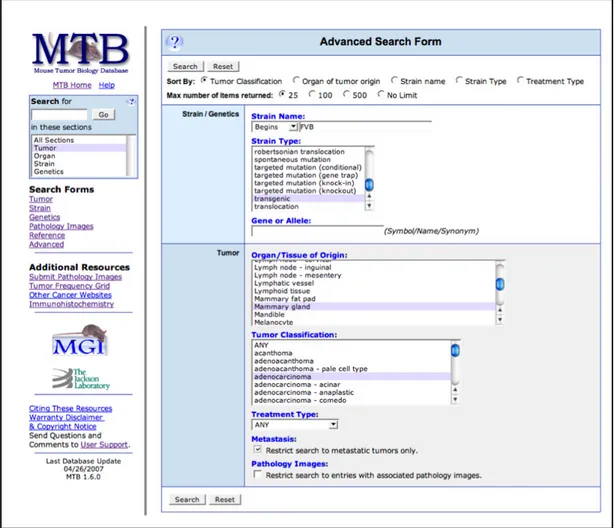
Figure 1. 1 Schematic representation of a standard zebrafish xenotransplantation procedure applied on zebrafish embryos and adult fish. ...1! Figure 1. 2 Comparison of ALU-based detection system and laser scanning cytometry system reflecting the number of cells present in colon cancer cell-spiked immune deficient mice blood. ... 16! Figure 1. 3 Linear regression analysis of ALU-based detection method (AluYb8) performed across different species.. ... 17! Figure 1. 4 Schematic representation of different injection sites for xenotransplantation studies in 48hpf fish. ... 21! Figure 1. 5 Screenshot of Mouse Tumor Biology database advanced search page. ... 24! Figure 1. 6 Screenshot of PDX Finder search page. ... 27! Figure 1. 7 Screenshot of PDXliver browse and search page. ... 31! Figure 1. 8 Aldosterone-mediated transactivation of mineralocorticoid receptor. ... 33! Figure 1. 9 Aldosterone and mineralocorticoid signaling in a polarized renal cell. ... 35! !
Figure 3. 1 ZenoFishDbv1.1 Homepage. ... 66! Figure 3. 2 ZenoFishDb v1.1 database displaying the Data Table module. ... 67! Figure 3. 3 Screenshot of the ZenoFishDb v1.1 Visualisation page. ... 68! Figure 3. 4 PDX Details module displays patient data, tumor data and zebrafish injection details of patient-derived xenografts listed on ZenoFishDb v1.1. ... 69! Figure 3. 5 Screenshot of the PDX Charts module on ZenoFishDb v1.1. ... 70! Figure 3. 6 Graphical representation of registered cancer and tissue types on ZenoFishDb v.1.1. ... 71! Figure 3. 7 Molecular modifications represented on ZenoFishDb v1.1. ... 72! Figure 3. 8 Stem cell properties of stem cell (SC) and cancer stem cell (CSC) xenograft models displayed on ZenoFishDb v1.1.. ... 73! Figure 3. 9 GO PANTHER Pathway analysis of molecularly modified genes.... 74! Figure 3. 10 STRING analyses of molecularly-modified genes. ... 75! Figure 3. 11 List of biological assessments performed on zebrafish xenograft studies incorporated onto ZenoFishDb v1.1. ... 77! Figure 3. 12 Host modifications and host modification details search through the Visualization and Data Table modules. ... 78! Figure 3. 13 ZenoFishDb v1.1 reveals the injection sites of all zebrafish xenotransplantation studies. ... 80! Figure 3. 14 ZenoFishDb v1.1 reveals the categorized injection cell numbers of all zebrafish xenotransplantation studies... 81! Figure 3. 15 Correlation of Alu Ct and Human DNA (pg) injected in zebrafish model organism. ... 82! Figure 3. 16 ALU-based tumor proliferation comparison among ache mutant and wildtype fish injected with liver cancer cell lines. ... 83!
Figure 3. 17 Mineralocorticoid and glucocorticoid expression across breast cancer cell lines. ... 85! Figure 3. 18 MR and GR mRNA expression in ex vivo breast cancer cDNA arrays. ... 86! Figure 3. 19 MR and GR are co-expressed in breast cancer tissue.. ... 87! Figure 3. 20 Kaplan-Meier plots illustrating survival graphs of MR and GR in breast cancer.. ... 88! Figure 3. 21 KM Plotter microarray-based overall survival (OS) and relapse free survival (RFS) analysis for MR in breast cancer patients. ... 89! Figure 3. 22 Association of stratified MR expression and RFS across intrinsic subtypes of breast cancer... 90! Figure 3. 23 Association of stratified MR expression and OS across intrinsic subtypes of breast cancer.. ... 91! Figure 3. 24 KM-Plotter microarray-based overall survival (OS) and relapse free survival (RFS) analysis for GR in breast cancer patients.. ... 92! Figure 3. 25 Association of stratified GR expression and OS across intrinsic subtypes of breast cancer... 93! Figure 3. 26 Association of stratified GR expression and RFS across intrinsic subtypes of breast cancer.. ... 94! Figure 3. 27 MR overexpression model in breast cancer cell lines. ... 95! Figure 3. 28 Principal component analysis of MR-overexpressing MFC7 cell lines in the presence of aldosterone and/or spironolactone. ... 96! Figure 3. 29 Principal component analysis of MR-overexpressing MFC7 and ZR-75-1 cell lines in the presence and absence of aldosterone.. ... 97! Figure 3. 30 Confirmation of MR downstream target genes in MR-Overexpressing MCF7 cells in the presence of 24-hour aldosterone and/or spironolactone treatment. ... 98! Figure 3. 31 Confirmation of MR downstream target genes in MR-overexpressing ZR-75-1 cells in the presence of 24-hour aldosterone and/or spironolactone treatment. ... 99! Figure 3. 32 GO Slim and PANTHER analysis of MR-induced upregulated genes in MCF7 cell line.. ... 100! Figure 3. 33 GO Panther pathway analysis of upregulated genes in the presence of MR overexpression in MCF7 cells. ... 101! Figure 3. 34 Effect of five-day aldosterone exposure on naïve MCF7 cell viability. ... 102! Figure 3. 35 Viability effect of transiently MR-overexpressing cells on MCF7 supplemented with 5% charcoal-striped FBS-supplemented media. ... 103! Figure 3. 36 Viability effect of transiently MR-overexpressing cells on MCF7 cells supplemented with standard-FBS supplemented media. ... 104! Figure 3. 37 Effect of aldosterone exposure on transiently MR-overexpressing cells on MCF7 cell viability. ... 105! Figure 3. 38 Fluorescent microscope images of zebrafish xenograft model of MR-overexpressing MCF7 cells (FISH1-FISH7). ... 108! Figure 3. 39 Fluorescent microscope images of zebrafish xenograft model of MR-overexpressing MCF7 cells (FISH8-FISH14). ... 109! Figure 3. 40 Assessment of tumor growth at 24 hpi and 72 hpi by measuring CTCF.. ... 110!
Figure 3. 41 Comparison of ALU-based tumor quantification and corrected total cell fluorescence-based tumor quantification in 3 dpi MR-Overexpressing casper zebrafish strain. ... 110! Figure 3. 42 Correlation analysis of raw ache Ct and DNA concentration (log2) in 3 dpi MR-overexpressing casper zebrafish strain.. ... 111! !
!
Figure 3. 43 Correlation analysis of raw MR Ct and ache normalized ALU DNA in 3 dpi MR-overexpressing casper zebrafish strain... ... 111! Figure 3. 44 Correlation analysis of raw MR Ct and CTCF difference among 72hpi and 24hpi. ... 112! Figure 3. 45 ALU-based tumor quantification in MR-overexpressing MCF7 cell line experimental setup. ... 113! Figure 3. 46 Correlation analysis of ache Ct values and total DNA extracted from zebrafish.. ... 114!
List of Tables
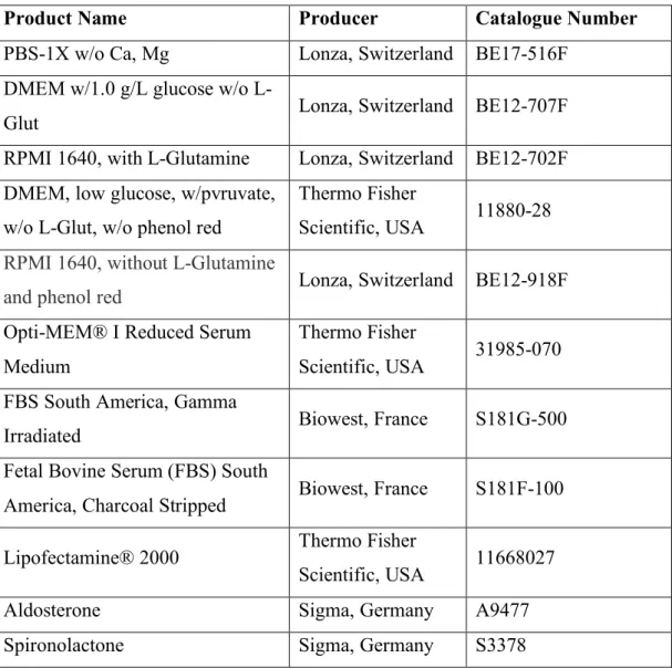
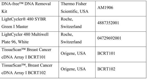
Table 1. 1 Numerical representation and statistical analysis of recovered cells of ALU-based detection system and laser scanning cytometry method-ALU-based detection. ... 16! Table 2. 1 The name of the attributes and their explanations are presented for the Data Table and PDX Details modules. ... 46! Table 2. 2 Laboratory materials, reagents and kits for used cell culture studies. ... 52! Table 2. 3 Laboratory materials, reagents and kits for RNA, protein and immunohistochemistry studies are listed. ... 52! Table 2. 4 List of primers used, their sequences and annealing temperatures. ... 53! Table 2. 5 The RNA integrity (RIN) values of RNA samples submitted for RNA-Seq. ... 58!
CHAPTER 1
INTRODUCTION
1.1. Xenotransplantation applications in zebrafish model organism
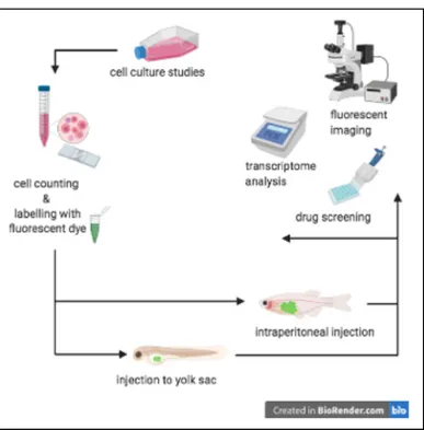
Cancer has been the disease of the 21st century due to increased rates of diagnosis and its therapy presents challenges and heterogeneity. Precision medicine is only possible if tumor material from patients are tested for series of drugs to find the one tailored for that patient. Today, tumor xenograft models are central for better understanding and evaluation of cancer biology research. In addition, they have become an important part of precision medicine. For decades, mouse models have been the gold-standard for executing xenograft studies. With the successful xenotransplantation of the human cells into zebrafish embryos in the beginning of this millennia (Lee et al., 2005; Nicoli et al., 2007; Nicoli & Presta, 2007) a whole new era has risen for xenotransplantation applications in cancer research. An example representation of a standard zebrafish xenotransplantation into embryos and adult fish is shown in Figure 1. 1 .
Figure 1. 1 Schematic representation of a standard zebrafish xenotransplantation procedure applied on zebrafish embryos and adult fish.
Zebrafish embryo renders an ideal host model organism for xenotransplantation studies due to several reasons, i.e., external fertilization, high fecundity, optic clarity of the larvae, short generation periods, yet most importantly lack of fully functional adaptive immune system development until 4-6 post week fertilization (Lam et al., 2004; Sullivan & Kim, 2008). Use of zebrafish embryos as xenograft hosts, therefore, could be a preferable alternative model for xenograft studies. Furthermore, humanization of zebrafish for generating immunocompromised strains has led to better assessment of xenotransplantation studies eliminating possible immune-rejection of the host organism. The immunodeficient strains of zebrafish used for xenotransplantation studies are as follows; recombination activation gene 2 (rag2) mutants (Q. Tang et al., 2014; Tenente et al., 2014), DNA-dependent protein kinase gene (pkrdc) mutants (I. H. Jung et al., 2016) and janus kinase 3 (jak3) gene (Moore et al., 2016) mutants. rag1 and rag2 are required for B and T lymphocyte differentiation (Willett et al., 1997) serve as adaptive immunity markers in zebrafish. rag2 mutant line (rag2(E450fs)), lacks mature T cells and possesses reduced numbers of functional B cells and is currently optimized for xenotransplantation studies in adult fish (Q. Tang et al., 2014; Tenente et al., 2014). Furthermore, pkrdc-null SCID zebrafish line functional B and T lymphocytes (I. H. Jung et al., 2016) has been established for adult fish xenotransplantation studies. In addition to rag2 and pkrdc mutant lines, jak3 mutant line (jak3P369fs) lacking both T lymphocytes and Natural
Killer (NK) cell immunity has been also generated for better mimicking of immune-deficiency in fish studies (Moore et al., 2016). Additionally, application of immunosuppressants such as dexamethasone (Eden et al., 2015; Stoletov et al., 2007) and Busulfan (Khan et al., 2019) have been also used to surpass any immunogenic response to engrafted cells or tissues. Although availability of these lines and drug exposure methods offer a broad range of immune-deficiency, still more research is required for fine-tuning both innate and adaptive immune systems for minimizing all kind of immune response to engrafted cells and/or tissues.
Use of zebrafish hosts for xenotransplantation studies could also overcome some disadvantages of mouse xenograft models specially by the terms of timing and cost efficiency. For decades, humanized mouse models have been used to prevent immune rejection of the grafted tumor tissue/cells and ongoing research focuses on fine-tuning
the real scenario of tumor-microenvironment interactions as in human physiology. Athymic nude mice lacking thymus (Flanagan, 1966; Pantelouris, 1968), SCID mice (G. C. Bosma et al., 1983; M. J. Bosma & Carroll, 1991), NOD-SCID mice (Prochazka et al., 1992), Rag2 knockout mice (Shinkai et al., 1992) are well-established immunocompromised host models. Although zebrafish and mouse models share equivalent immune-compromised models by the means of gene mutations, several other parameters such as cost, timing and possible tumor-microenvironment interactions are to be considered for the experimental design. Accordingly, most xenografting is performed in embryonic/larval zebrafish providing a platform relatively cost- and time-efficient.
Overall, the above-mentioned characteristics of the zebrafish organism with the feasibility of the current technologies for applying tumor cell injections and generation of ‘humanized’ fish strains paves the way for standardizing the use of zebrafish xenograft models in cancer research more efficiently.
1.1.1. The nature of existing zebrafish xenograft models
1.1.1.1. Immortalized cell line injections
First ever zebrafish xenograft study was performed by Lee and colleagues in 2005 where they had transplanted human metastatic melanoma cells (C8161) into blastula-stage zebrafish embryos and showed successful survival, division and migration of cancer cells (Lee et al., 2005). Herein, survival and motility of primary melanocytes and normal fibroblasts have also been investigated and these cells survived in zebrafish embryos yet exhibited less migratory effects (Lee et al., 2005). Engraftment of immortalized cell lines to zebrafish host model has been carried out since, and to date, cell line engraftments of different human cancer types including breast cancer cell lines; MDA-MB-231 (Qiong Wu et al., 2018; B. Yang et al., 2019; X. Zhao et al., 2019), MCF7 (Mercatali et al., 2016; Sun et al., 2015), T47D (Vazquez Rodriguez et al., 2018), ZR-75-1 (Rodriguez et al., 2017); glioma cell lines, U-87MG (Fan et al., 2019) and U-251MG (Gamble et al., 2018); leukemia cell lines, K562 (Dale P. Corkery et al., 2011); prostate cancer cell lines, PC3, DU-145 and LNCap cells (Bansal et al., 2014) and many other cell lines. In addition to immortalized cell-lines derived from
humans, murine (Weijts et al., 2018), rat (Sandquist et al., 2018), zebrafish (Mizgirev & Revskoy, 2010), dog (Timmermans-Sprang et al., 2019) cell line injections are also present.
Zebrafish, enabling successful transplantation of immortalized cell lines of numerous types derived from numerous organisms, serves as an ideal model for xenotransplantation studies by allowing feasible, efficient, cheap and high-throughput research in the cancer research community.
1.1.1.2. PDXs of zebrafish
Successful patient-derived xenograft engraftment to zebrafish host performed by two individual studies of Bagowski group, i.e., pancreas, ampulla vateri tissue engraftment (Weiss et al., 2009) and pancreas, colon, and stomach tissue engraftment (Marques et al., 2009), had opened a new era for easy application and evaluation of PDXs in zebrafish embryos for research community. Majority of studies using zebrafish PDXs (zPDXs) possesses glioblastoma (Banasavadi-Siddegowda et al., 2018), liver cancer (Lin et al., 2019) and colorectal cancer (Fior et al., 2017)-derived primary cell cultures and/or tissue engraftments. In addition to these cancer types, a broad range of zPDXs exist in the literature and gastric cancer (J. Q. Wu et al., 2017), prostate cancer (Bansal et al., 2014), pancreatic ductal adenocarcinoma (L. Wang et al., 2019), melanoma (Yan et al., 2019), adrenocortical carcinoma (Gianoncelli et al., 2019), breast cancer (Yan et al., 2019) and acute myeloid leukemia (Gacha-Garay et al., 2019) are among many. Most of the zPDX studies have established patient tissue derived primary cell line injections yet several studies exemplified direct tissue transplantation into the zebrafish yolk (Marques et al., 2009; Weiss et al., 2009). In addition to patient derived xenografts, primary cell lines derived from healthy donors have been also used as controls or as test subjects to assess different biological hypotheses raised by the research community. Overall, successful implementation of patient-derived tissue to zebrafish hosts idealizes standardized use of zPDXs in cancer research overcoming the disadvantages of mice model PDXs i.e. longer time-period for tumor establishment and assessment in mice models (J. Jung et al., 2018).
1.1.1.3. Stem cell and cancer stem cell injections
Zebrafish model has been also a useful and preferable model organism for studying stem-cell associated events through xenotransplantation of stem cells and cancer stem cell injections (Banasavadi-Siddegowda et al., 2018; Li et al., 2015; Orlova et al., 2014). One of the pioneer studies of stem-like cell population xenotransplantation in zebrafish embryos have been established by Eugiera and colleagues in 2011 wherein they tested tumorigenic and migratory capacity of the monolayer culture-derived control cell- and mammosphere culture-derived breast cancer cell- xenografts in zebrafish model organism (Eguiara et al., 2011). Herein, both tumor mass and tail migration capacity have significantly increased in mammosphere-derived cell injections when compared to monolayer-derived cell injections when the fish were incubated at 34°C. Additionally, a dietary polyphenol, curcumin, previously shown to modulate self-renewal of breast stem cells of normal and malignant origin, were applied to parental monolayer culture prior to injection and hereby a significant decline were detected in the tail migration and tumor mass in comparison to mock treated control bearing xenografts (Eguiara et al., 2011). This platform, hence, is ideal for pursuing rapid, feasible and inexpensive approach for testing novel drugs on self-renewal capacity of cancer stem cells. A similar approach has been also taken for studying different cancer stem cells such as glioma (X. jun Yang et al., 2013), glioblastoma (Lai et al., 2017; Welker et al., 2017), leukemia (Gacha-Garay et al., 2019; B. Zhang et al., 2015; B. Zhang, Shimada, Kuroyanagi, Umemoto, et al., 2014) and prostate (Nouri et al., 2017) as well as many other cell types.
Studies harboring injection of multiple cell lines are also present in the literature, study presented by Zhang and colleagues, for instance, highlights the use of leukemia and solid tumor-derived stem cell xenografts for high-throughput drug screening. Herein, K562-KOr cell derived leukemia stem cells (LSCs), DU-145-KOr cell line-derived prostate cancer stem cells (CSCs), HepG2-KOr cell line-line-derived liver cancer stem cells had been engrafted to both zebrafish embryos and adult-stage fish for cancer stem cell characterization and high-throughput therapy screening. In line with this hypothesis, cells sorted according to their aldehyde dehydrogenase (ALDH) expression, ALDH positive representing the cancer stem cell characteristic, ALDH+ and ALDH- cells had been transplanted to both zebrafish embryos and 20Gy-irradiated
adult fish. As a result, for K562-KOr-ALDH+(LSCs) and K562-KOr-ALDH-(Non-LSCs injected adult fish experimental groups; 7/10 LCS-injected group whereas 4/10 of Non-LSCs had established tumor in their trunks after 35 dpi.
Injection of prostate cancer stem cells and non-cancer stem cells, DU145-KOr-ALDH+ (CSCs) and DU145-KOr-ALDH-, respectively has been another experimental group of this study. Herein, tumor proliferation in irradiated adult fish have been assessed on 0dpi, 7dpi and 14dpi and DU145-KOr-ALDH+ (CSCs) had proliferated more significantly when compared to non-CSCs. Yet, another experimental group of this study has been transplantation of HepG2-KOr-ALDH+ and HepG2-KOr-ALDH- cells into irradiated fish and herein, tumor formation had been observed in both groups at 28 dpi with metastasis formation in HepG2-KOr-ALDH+ transplanted fish. Zebrafish embryo injection of DU145-KOr-ALDH+ and K562-BFP-ALDH+ and- ALDH- experimental groups into yolk sac of 48hpf fish; 1dpi images of fish have been captured and fish were exposed to drug treatment for additional 48 hours and before final images were captured. Docetaxel treated-DU145-KOr-ALDH+-injected zebrafish embryo proliferation were found to be significantly diminished when compared with untreated-DU145-KOr-ALDH+-injected fish. K562-BFP zebrafish xenograft model high-throughput drug screening for one chemical library has revealed the LSC-selective compound, B647, which later were used carry out its effect on the proliferation of K562-BFP-ALDH+ and K562-BFP-ALDH+ injected zebrafish embryos. LSCs have been shown to exhibit significantly increased proliferation compared to Non-LSCs and B647 treatment has been shown to reduce proliferation of LCSs when compared to control treated fish (B. Zhang, Shimada, Kuroyanagi, Nishimura, et al., 2014). This study demonstrates well that stem cell xenografting in zebrafish can lead to new drug discovery through high-throughput screening.
To summarize, successful zebrafish xenograft models of cancer stem cell applications, hematopoietic stem cells (Hamilton et al., 2018), mesenchymal stem cell (Han & Hsu, 2016; Patel et al., 2012) and induced pluripotent stem cell xenotransplantation (Chan et al., 2015) are also present and stem-cell zebrafish xenograft studies are now in rise.
1.1.2. Molecularly-modified cell injections
Zebrafish xenograft model organism also allows evaluation of immortalized cell lines, patient-derived primary cell lines or primary cell lines of any kind of tissue of origin which has been targeted to any kind of molecular/genetic modification. Thorough screening of the literature has facilitated identification of different kinds of molecularly-modified cell transplantation to zebrafish embryos including cells modified with tag-expression vectors for cell tracking purposes (Banasavadi-Siddegowda et al., 2018; Marranci et al., 2019), overexpression vectors (Jerónimo et al., 2017; Naber et al., 2013; von Mässenhausen et al., 2016), interfering RNAs (RNAi) (D. P. Corkery et al., 2018; Halasz et al., 2013; Leung et al., 2017), morpholinos (Topczewska et al., 2006; Weiss et al., 2009) as well as cells modified with Cre-LoxP (Karkampouna et al., 2018; Weijts et al., 2018) and Cripsr/Cas9 (Gaytan-Cervantes et al., 2017) technologies. Use of zebrafish xenograft models, therefore, enables testing gene specific-derived hypothesis employing different technologies altering gene expression levels and/or gene structures of interest of the cell lines to be injected.
1.1.2.1. Tag expression vectors
One of the earliest studies harboring such molecular modifications is performed by Topczewska and colleagues where they have injected aggressive melanoma cells tagged with GFP expression vector (C8161-GFP), a tag expression vector, and treated with anti-Nodal morpholinos (or scramble morpholinos) where they have been injected to blastula-stage zebrafish embryos in order to investigate the interactions among cancer cells and embryogenic progenitors and C8161 cells have been shown to form ectopic gene expression and had Nodal expression; and this phenotype had been reversed by the anti-Nodal morpholino treated C8161 injection in zebrafish embryos. In parallel, tumorgenicity of the anti-Nodal morpholino (or scramble morpholino)-treated C8161 cells had been examined on subcutaneously injected 5-week-old nude mice at different time intervals (day 3, 7, 14 and 17) and Nodal inhibition had diminished tumor volume significantly at these specified time intervals. This study therefore exemplifies an application of the same cell line transplantation both in zebrafish and mice models and bearing comparative results hence, solid evidence for compatible use of both xenograft models for a given hypothesis (Lee et
al., 2005). This study also highlights the use of GFP tag expression vector for visualizing cell behavior in vivo.
Use of tag expression vectors such as GFP (Lee et al., 2005) and mCherry (Marranci et al., 2019) are often preferred as cell tracking sources in fish and use of fluorescent protein tag enables assessment of tumor growth, migration, metastasis, dissemination and other fluorescence-based quantification assessments performed on fish.
1.1.2.2. Overexpression models
Another earliest example of the use of molecularly modified cell xenografts has been established by Nicoli and colleagues where they have developed a zebrafish xenograft angiogenesis model in which they have successfully examined the angiogenic capacity of tumorigenic FGF2-overexpressing mouse aortic endothelial cells (FGF2-T-MAE) (Nicoli et al., 2007). Applications of molecularly-modified cell line injections are not solely peculiar to immortalized cell lines, patient derived primary cell lines subjected to modifications have been also successfully engrafted into zebrafish embryos (Banasavadi-Siddegowda et al., 2018; L. Wang et al., 2019). Wang and colleagues have pursued zebrafish xenograft models of PDAC tissue derived primary cancer cells and fibroblasts which have been genetically modified to express anti-apoptotic BCL2L1 and, fluorescent proteins, eGFP and mKate2, respectively (L. Wang et al., 2019). Herein, modified PDAC primary cells subjected to fibroblast inhibitor treatment and modified fibroblasts have been injected to zebrafish embryo yolk sacs and have been treated with gemcitabine chemotherapy agent in the presence or absence of BCL2L1 inhibitor, Navitoclax. As a consequence, the gemcitabine treated cancer cells and fibroblasts without BCL2L1 inhibitor treatment showed significant fall in fluorescent intensity when compared to cancer cells and fibroblasts without BCL2L1 inhibitor and chemotherapeutic agent treatment, respectively. On the contrary, gemcitabine and BCL2L1 inhibitor-treated cancer cells and fibroblasts had not shown any significant alterations with counterparts that had only received gemcitabine treatment. This approach has enabled the examination of fluorescent intensity among differential groups aiming to reflect information on tumor viability upon genetic modifications and chemotherapeutic agent treatments while also tumor-microenvironment interaction had been also taken into consideration by co-injection
approach/strategy (L. Wang et al., 2019). However, it might be important to estimate the gene copy number of cells xenografted into the host and for this an accurate quantification method remains to be developed and tested.
1.1.2.3. RNA interference applications
RNA interference applications have been also widely used for generating molecularly-modified cell xenografts and assessments of gene-specifically driven physiological differences in tumor itself or the surrounding microenvironment. Silencing RNA (siRNA) treated cell transplantation to zebrafish embryos are of common use for cell proliferation, metastases, angiogenesis and drug sensitivity assessments in fish model. Wang and colleagues, for instance, had studied the association of COL3A1 mRNA and protein expression in colorectal cancer prognosis and further tested the functionality of the gene by in vitro and in vivo viability and proliferation assays, respectively. In fact, COL3A1-silenced colorectal cancer cells exhibited reduced growth in vitro (shRNA-treated) and xenografted COL3A1-silenced cells (siRNA-treated) exhibited decreased tumor proliferation on second day post injection when compared to initial injection time point (X. Q. Wang et al., 2016). Studies harboring several cell-line injections with different molecular modifications are also present and this way, comprehensive research could be performed to decipher the raised scientific question. For instance, a study led by Mukhopadhyay group studied the mechanistic model of extravasation and its association with neurophilin-2 (NRP-2), a semaphorin and vascular endothelial growth factor receptor, in clear cell renal carcinoma (RCC) and pancreatic adenocarcinoma (PDAC). Herein, NRP-2 overexpressing 786-O (an aggressive RCC cell line) and corresponding control cells were transplanted into pericardium of 72 hpf zebrafish embryo for examining extravasation. NRP-2 overexpressing 786-O cells has been shown to be extravasating from intersegmental vessels (ISVs) whereas controls had remained in the circulation after 24 hours. With a similar experimental approach, a highly metastatic cell line pancreatic cell line with high NRP-2- expression levels, ASPC-1, had been treated with short hairpin RNA (shRNA) targeting NRP-2 and ASPC-1 with reduced NRP-2 levels had been shown to reduce extravasation in zebrafish models (Y. Cao et al., 2013). Herein, different cell lines with different level of gene expression and modified with different molecular
modifications were used to comprehensively study of gene functionality in vivo using zebrafish models.
1.1.2.4. Mutations
In addition to gene expression reducing or overexpressing models of unmodified genes, expression of genes with mutations, has been also studied in zebrafish xenograft models. One of the few studies investigating effect of gene mutations in zebrafish xenograft model has aimed to identify metastatis associated events, i.e., invasion raised by differential phosphorylation of Na+/H+ exchanger regulatory factor 1, NHERF1, a scaffold protein, in breast cancer (Greco et al., 2019). An unidentified phosphorylated NHERF1 had been previously identified to be involved in B1 integrin-dependent invadopodia formation (Antelmi et al., 2013). In the study by Greco et al., non-phosphorylable form of S279 (S279A), S301(S301A) residues solely as well as S279A/S301A together as double mutants have been introduced to cells as well as corresponding controls, i.e., wild type-NHER1 and expression vector, and invasive behavior of these experimental groups were tested in zebrafish model. In fact, number of metastatic foci and invasiveness percentage were high in wild-type-NHERF1 over-expressing cells when compared to all of the mutation-introduced cell line xenografts (Greco et al., 2019). This shows that zebrafish host is amenable to test mutant form of proteins in tumor assessment, such as degree of invasiveness.
1.1.2.5. Gene editing tools
Application of Cre-LoxP system induced overexpression and/or knockout cell line zebrafish xenograft models are also present in the most up-to-date literature search for molecularly-modified cell line engraftment studies in zebrafish. One example is the use of CRIPTO-overexpressing HepG2 hepatocellular carcinoma cell line that has been generated using a lentiviral red-to-green pTOMO-CRIPTO construct wherein CRE recombinase activity leads to excision of the floxed RFP cassette, resulting in CRIPTO and GFP expression (Karkampouna et al., 2018). As CRIPTO expression had been associated with poor prognosis in HCC and behaved as a proto-oncogene in various cancer types, the migratory and metastatic capacity of the CRIPTO-overexpressing cells were tested in zebrafish xenograft model and increased tumor-foci formation was detected in CRIPTO-overexpression model when compared to
control model (Karkampouna et al., 2018). Another application of Cre-LoxP induced molecularly-modified cell xenotransplantation to zebrafish embryos is the engraftment of Cre/LoxP mediated EF7/EF8 double knockout mouse embryonic fibroblasts (MEFs) cell engraftment to zebrafish embryos. Herein, Myc/Ras transformed EF7/EF8 double knockout MEFs and Myc/Ras transformed control cells have been injected to perivitelline space to examine metastatic and angiogenic capacity of these cells. Myc/Ras transformed EF7/EF8 double knockout MEFs have been shown to be metastasizing in the trunk region as well as promoting the intra-tumoral blood vessel branching (Weijts et al., 2018). Molecular modifications introduced by Crispr/Cas9 technology are of common use in molecular cell biology, and zebrafish xenograft studies harboring Crispr/Cas9-modified cells are also present in the current literature (Gaytan-Cervantes et al., 2017). Gaytan-Cervantes and colleagues have studied the role of cis elements and trans factors which is involved in the formation of an anti-apoptotic survivin gene isoform which lacks exon 3, DEx3, whose expression is upregulated when compared to other survivin isoforms in different cancer types (Gaytan-Cervantes et al., 2017). Herein, zebrafish xenograft model has been used to examine the tumorgenicity of endogenous survivin exon 3 mutated-HeLa cells introduced by Cripspr/Cas9 system. Introduced mutation had aimed to affect the binding of Sam68 regulator protein (trans-acting) to examine its role in the alternative splicing of the surviving in vitro, and hence allowed to better reflect its physiological effects in vivo by assessing tumorgenicity in zebrafish xenograft models. In return, decreased DEx3 survivin through impaired Sam68 binding and hence alternative splicing, the mutant cell injections have exhibited increased tumor formation in vivo (Gaytan-Cervantes et al., 2017).
Overall, feasibility of immortalized cell line, stem cell and cancer stem cell injections, PDX-derived primary cell line injections as well as solid tissue engraftments with modifications into zebrafish embryos as well as adult fish encourages the standardization of zebrafish model organism for the use of transplantation studies and hence, reveals an alternative xenograft model organism to rodent studies.
1.1.3. Zebrafish xenograft host modifications
Zebrafish xenograft studies focusing on tumor growth, proliferation, apoptosis, tumor viability and similar assessments which is based on evaluation of tumor volume and/or, tumor cell numbers, dispersion in the fish embryo mostly uses casper mutant fish, compound roy-/-:nacre-/- homozygous double mutant, which is transparent and named after the friendly ghost casper (White et al., 2008). Alternatively, achieving transparency in wild-type fish is also available through PTU administration. In addition to casper mutants, transgenic fish possessing fluorescently labelled fish are also commonly used for assessing metastasis and/or angiogenesis. In fact, use of the transgenic fish, Tg(fli1:EGFP) (Lawson & Weinstein, 2002), that expresses GFP under the early-endothelial marker, fli1, is often preferred in zebrafish xenotransplantation studies. Similarly, another transgenic fish, Tg(flk1:EGFP)s843, which also drives GFP expression under another endothelial marker, flk1 (Jin et al., 2005) is also used for again assessing metastatic capacity and angiogenesis.
In addition to above-mentioned fish which is applicable for more-generalized use fish with desired features are also selected for specific assessment performed in xenograft models. One of the examples, is the use of ache mutants and effect of the tumor microenvironment in tumor development (Avci et al., 2018); this study i also a part of this thesis. Herein, homozygous recessive ache mutants led to excess accumulation of acetylcholine in fish and its effect on liver cancer was further tested. Another assessment-specific use of fish was the use of cloche mutants lacking functional circulation and vasculature and hence, screens the necessity of intact vasculature in tumorigenesis and metastasis (Marques et al., 2009; C. Zhao et al., 2016). Recently, a ‘humanized’ zebrafish transgenic strain has been also constructed to express human hematopoietic cytokines and has been used to as a host for human stem cell transplantation (Rajan et al., 2019).
In adult fish xenotransplantation studies hosts in use are often modified to lack functional immune system. Herein, immunosuppressant-induced, i.e., Busulphan induced immunosuppressed casper mutants (Khan et al., 2019), prkdc null mutant SCID fish lacking functional T and B cells (I. H. Jung et al., 2016), fish manipulated to possess cancer-specific immunotolerance without immunosuppression (B. Zhang et
al., 2016), nacre/rose/fli1:egfp zebrafish fish subjected to 20-Gy ionizing radiation 2 days prior to engraftment for immune suppression (B. Zhang, Shimada, Kuroyanagi, Nishimura, et al., 2014) are among adult fish used for tumorigenesis, proliferation and tumor growth assessments.
1.1.4. Biological assessment and validation methods in zebrafish
xenografts
Utilizing zebrafish model organism for transplantation studies has enabled assessment of numerous tumor-biology associated events. Tumor growth, proliferation, apoptosis, invasion, migration, metastasis, extravasation and angiogenesis are among many assessments which could be pursued using current zebrafish xenograft models. However, there is not a database existing in which this information is searchable. Availability of transgenic and/or mutant zebrafish lines of special features are the main reason for feasibility of biological assessments. Hence, host modifications, biological assessments and validation methods are crucial for establishing right zebrafish xenotransplantation model for desired experimentation. Evaluation of biological assessment together with the use of assessment-targeted modified host is therefore explained in detailed titles, namely, tumor growth, proliferation and apoptosis; invasion extravasation, dissemination and metastasis; angiogenesis.
.
1.1.4.1. Tumor growth, proliferation and apoptosis
Zebrafish xenograft models of tumorigenesis, tumor formation, viability, growth and/or proliferation have been applied to many tumor types including acute myeloid leukemia, hepatocellular carcinoma, melanoma, esophageal squamous cell carcinoma, mouse ovarian surface epithelial cells, colorectal carcinoma, hematopoietic stem and progenitor cells and to numerous cancer types along with other assessments run in parallel. Herein, studies or part of the studies only focusing on tumor growth and/or proliferation will be further exemplified and explained in detail to better portrait the picture of host modifications used, injection sites preferred and/or type of methods applied in order to quantify tumor growth, proliferation and apoptosis.
Assessment of proliferation in embryos and/or adult fish could be performed through several routes which have been extensively applied in majority of zebrafish xenograft
studies. Overall, fluorescence intensity, cell mass area, mean fluorescent area (Marranci et al., 2019) (Liu et al., 2019), fluorescent integrated density (Mazumder et al., 2018; X. Q. Wang et al., 2016; Z. Xie et al., 2018), tumor area (µm2) (Avci et al.,
2018), tumor growth ratio and cell spread (Martinez-Ordoñez et al., 2018) and number of fluorescent cells counted (D. P. Corkery et al., 2018; Dale P. Corkery et al., 2011) were used for assessing proliferation and tumor growth. However, there is a need for more high-throughput methods as used in mouse (Prigent et al., 2015) (please see section 1.1.4.1.1.). In addition, biochemical measurements on human cells xenografted are possible (see below).
In adult fish, for instance, fluorescently-labelled tumor cell injected prkdc-null SCID zebrafish have been assayed for proliferation markers, PCNA and Ki67, employing immunohistochemistry (IHC) on xenografted tissue for evaluating the proliferative activity of tumor cells in adult fish (I. H. Jung et al., 2016). Similarly, optically-clear prkdc and il2rga compound mutant fish, lacking both B, T and NK cells, xenografted with various type of cancer cells have been assessed for the presence of Ki67 staining by IHC and TUNEL for reflecting proliferation and apoptosis rates, respectively (Yan et al., 2019).
In addition to above-mentioned applications for proliferation assessment, several zebrafish-specific tools reflecting tumor burden have been developed. One of such tools is an open-source software tool, ZF-Mapper and it assesses tumor proliferation by quantifying fluorescence intensity of each pixel in zebrafish images for estimating/quantifying the implanted cell numbers (Yamamoto et al., 2019). ZF-tool offers another methodology that uses the number of GFP pixels and GFP intensity medium value in order to compare two different time points and compute proliferation index (Cabezas-Sainz et al., 2018).
Although majority of the studies used fluorescence-based tumor quantification methods, quantitative-PCR based tumor DNA quantification method was also initially used by our group for precise definition of tumor proliferation in xenograft models (Avci et al., 2018).
Availability of different techniques and tools, when applied in parallel, could help deliver high-precision data on proliferation and tumor growth assessments performed both in zebrafish embryos and adult fish. Moreover, development of novel tools or modifications of those already applied for mouse studies are still needed.
1.1.4.1.1. ALU-based tumor quantification studies
Quantitative detection of ALU repeats of human DNA employing qRT-PCR are in use for quantifying xenografted human cells in mouse models (McBride et al., 2003; Zijlstra et al., 2002) and chick embryo chorioallantoic membrane (Mira et al., 2002) since the early 2000s. Zijlstra and colleagues have established ALU-based detection method for identifying presence of human cancer cell DNA in metastatic organs of mouse xenografted with different cancer cell lines bearing different metastatic capacity. Employing this approach, the sensitivity of the method was approved as 1 human cell per 1.000.000 murine could be detected. The ALU-repeat quantification at the metastatic sites were correlative with histological markers and cells with higher metastatic capacity were projecting intensified ALU-repeat signals. Human cell transplanted xenografts when treated with anti-metastatic the ALU-repeat quantification declined (Zijlstra et al., 2002). Overall, this was an initial method to reflect the precision and quality of ALU-based DNA quantification in tumor quantification in different metastatic sites in vivo.
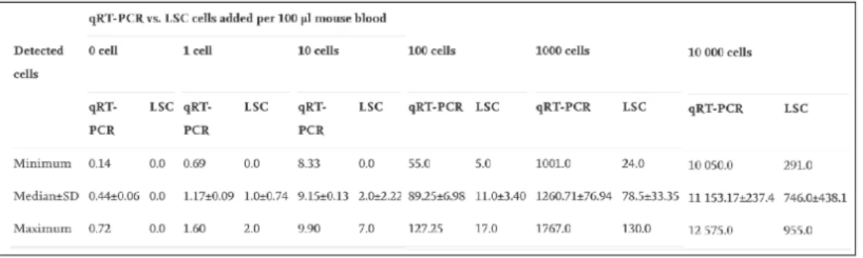
In 2009, study lead by Nehmann and collogues performed a comparative analysis among ALU-based quantification and fluorescent image-based analysis performed by laser scanning cytometry (Nehmann et al., 2010). Herein, they have performed human cancer cell detection by analyzing blood samples derived from immunodeficient mice (100 micromols) which was mixed with different number of colon cancer cells ranging from (1 cell to 10.000 cells) as treatment groups. Detection of presence of single cell has become possible employing either one of the techniques however, overall, ALU-based detection system was shown to be much more sensitive when compared to image based detection method laser scanning cytometry (Nehmann et al., 2010) (Figure 1. 2, Error! Reference source not found.).
Figure 1. 2 Comparison of ALU-based detection system and laser scanning cytometry system reflecting the number of cells present in colon cancer cell-spiked immune deficient mice blood. A) ALU-based detection system is determined
by comparing cell-added groups in 100µl mouse blood with calibration curve formed by known colon cancer tumor DNA concentration. B) Laser scanning cytometry-based system establishment by comparing cell-added groups in 100µl mouse blood with calibration curve formed by known colon cancer tumor DNA concentration.
(The image was reproduced by copy right permission and obtained from N ehmann, N., Wicklein, D., Schumacher, U., & Müller, R. (2010). Comparison of two techniques for the screening of human tumor cells in mouse blood: Quantitative real-time polymerase chain reaction (qRT-PCR) versus laser scanning cytometry (LSC). Acta Histochemica. https://doi.org/10.1016/j.acthis.2009.05.004)
Table 1. 1 Numerical representation and statistical analysis of recovered cells of ALU-based detection system and laser scanning cytometry method-based detection.
(The table was reproduced by copy right permission and obtained from N ehmann, N., Wicklein, D., Schumacher, U., & Müller, R. (2010). Comparison of two techniques for the screening of human tumor cells in mouse blood: Quantitative real-time polymerase chain reaction (qRT-PCR) versus laser scanning cytometry (LSC). Acta Histochemica. https://doi.org/10.1016/j.acthis.2009.05.004).
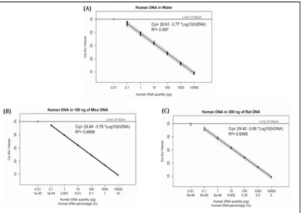
Another lead study of the field was established by Prigent and colleagues wherein they have studied ALU-based quantification method from a different perspective, instead of using the number of cells as base line they have optimized the technique by initially extracting the tumor cell DNA, quantifying it, performing dilutions, mixing them with a constant amount of mouse or rat DNA and measuring their Ct with ALU primers. This technique was shown to be very sensitive and precise as small as 0.01 pg human DNA could be detected in 100g of mouse DNA and 200g of rat DNA mix (Figure 1. 3). ALU-based detection system therefore enables precise detection of human DNA when also considered by the terms of added DNA quantity.
Figure 1. 3 Linear regression analysis of ALU-based detection method (AluYb8) performed across different species. A) Human DNA detection in water. B) Human DNA detection in 100ng mice DNA C) Human DNA detection in 200ng rat DNA. (The copy right permission was taken and this imae was taken from Prigent, J., Herrero, A., Ambroise, J., Smets, F., Deblandre, G. A., & Sokal, E. M. (2015). Human progenitor cell quantification after xenotransplantation in rat and mouse models by a sensitive qPCR assay. Cell Transplantation, 24(8), 1639–1652. https://doi.org/10.3727/096368914X681955).
1.1.4.2. Invasion, extravasation, dissemination and metastasis
Zebrafish xenograft models also revolutionized assessment of metastasis-related events in vivo. In fact, vast number of studies assesses presence of metastatic foci and/or micrometastasic foci in cancer cell-transplanted fish in parallel with evaluating presence of migration, invasion, dissemination and extravasation in these fish. A generalized approach has been adopted in most of these studies focusing on metastasis-related events assays wherein micrometastatic/metastatic foci forming at different sites, i.e., head, trunk, back, tail fin, are stated (Lara et al., 2011; Spender et al., 2016; C. Thomas et al., 2012). In several studies, step-wise approaches also have been adopted to demonstrate dissemination, extravasation and tissue invasion at one cell-resolution eventually demonstrating micrometastasis at distant foci (Shuning He et al., 2012).
Automated tools are also available for assessing metastasis-related events such as dissemination (Ghotra et al., 2012) and organ-specific distant foci formation (Annila et al., 2013). Ghotra and colleagues developed an automated whole animal bioimaging system for evaluating disseminating cells in zebrafish xenograft models. Herein, multiple parameters, i.e., total tumor cell foci, mean distance, mean intensity, mean area and cumulative distance of tumor burden, are initially evaluated for each embryo. Van der Ent and colleagues exemplify the use of this tool (Ghotra et al., 2012), where they performed automated quantitative image analysis of proliferation and migration. For tumor burden, number of tumor foci detected by its fluorescent emission were multiplied with the mean area of all fluorescence cell clusters. For migratory cells, the distance between site of injection and individual cell clusters are measured and averaged for calculating migration mean value or individual added up for reflecting cumulative/total migration value for each embryo (van der Ent et al., 2015). Similarly, Tan and colleagues used a method wherein metastatic tumor cell foci were initially identified by discarding single migrator cells by minimum area filtering criteria then, cumulative distance (CD) of metastatic tumor cell foci was calculated by summing up all identified tumor foci distances from the transplantation site in each embryo followed by use of a migration index (Tan et al., 2014). These studies show that quantification of migrating cells is important as quantifying the tumor size and volume.
A metastasis-quantifying image analysis tool, ZebIAT, was established and it provides a platform for automated cancer metastasis analyses per organ(Annila et al., 2013). The tool works with fish aging between 2dpf and 5dpf enabling registry of all major organs and upon injection, the mean fraction area of organs harboring cancer cells are examined to reflect the site/organ of metastasis (Annila et al., 2013)
Numerous methods and automated tools exist for assessing metastasis-related events in zebrafish xenograft models and extensive number of studies will give birth to novel assessment methods in time. However, there is still a need for a database through which differences in methodologies can be extracted systematically and over time.
1.1.4.3. Angiogenesis
One of the key and early studies to show successful cell transplantation to zebrafish involved establishing zebrafish xenograft angiogenesis model that was performed by Nicoli and colleagues in the beginning of 2000s where they assessed the angiogenic capacity of tumorigenic FGF2-overexpressing mouse aortic endothelial (FGF2-MAE) cells by performing three independent assays: i. alkaline phosphatase staining; ii. Whole mount in situ RNA hybridization of early endothelial markers including VE-cadherin, Fli-1 and VEGFR2/KDR; and iii. Employing fluorescence-based assessment of neovessels formation from subintestinal vessels migrating and infiltrating the FGF2-MAE cells in VEGFR2:G-RCFP zebrafish embryos (Nicoli et al., 2007). Similarly, majority of the study employed alkaline phosphatase staining for angiogenic assessment (Leali et al., 2010; Moshal et al., 2011) or fluorescence-based assessment of neo vessel formation stemming from the subintestinal vessel and/or intersegmental vessel formation in fluorescently-labelled endothelial marker expressing transgenic fish (Cheng et al., 2011; Hollenbach et al., 2013; Moshal et al., 2011; S. Zhang et al., 2011).
To conclude this section, zebrafish xenograft model provides a feasible platform for application of angiogenesis assessment and aid investigate the angiogenic capacity of xenografted cell lines and PDXs in fish. Nevertheless, there is not an online structured format by which researchers can extract knowledge in table format or visualize the