Therapeutic Nanomaterials, First Edition. Edited by Mustafa O. Guler and Ayse B. Tekinay.
© 2016 John Wiley & Sons, Inc. Published 2016 by John Wiley & Sons, Inc.
BIOSENSORS FOR EARLY DISEASE
DIAGNOSIS
Ahmet E. Topal, Alper Devrim Ozkan, Aykutlu Dana,
Ayse B. Tekinay, and Mustafa O. Guler
Institute of Materials Science and Nanotechnology, National Nanotechnology Research Center (UNAM), Bilkent University, Ankara, Turkey
10.1 INTRODUCTION
Early diagnosis is an important aspect of modern medicine and significantly improves the prognosis of a wide range of life‐threatening disorders (Ping et al., 1997; Wang, 2006). However, early diagnosis is often difficult or impossible without an appropriate means of detecting small differences in the patient’s physiology. As such, a great diversity of both general and spe cialized methods has been developed to identify the presence of genetic disorders, cancers, viral or bacterial infections, and other diseases prior to the point they present their pathological effects (Saravolatz et al., 2003; Wulfkuhle et al., 2003). These methods principally involve either medical imaging, in which the patient’s tissues are checked in situ with or without the assistance of a dye or contrast agent (see Chapter 9); histopathology, in which a sample of the patient’s tissues is removed by biopsy for visual inspection, usually with the help of specific cell and tissue stains; or a variety of diagnostic assays, in which the patient’s bodily fluids (or tissue samples) are biochemically analyzed for disease‐specific biomarkers.
236 BIOSENSORS FOR EARLY DISEASE DIAGNOSIS While powerful, current medical diagnostic techniques nonetheless suffer from clinical limitations. Histopathology is the gold standard for the diagnosis of many diseases but also requires an expert practitioner and introduces an element of subjectivity to disease diagnosis (Uckermann et al., 2014). Likewise, medical imaging is commonly used in disease diagnosis but cannot easily detect early‐stage disorders, which often affect tissues on scales too small to be resolved by the imaging technique. In contrast, biochemical assays combine low detection limits with objective criteria of quantification and are therefore more suitable for the diagnosis of hard‐to‐detect diseases. Standard biochemical assays are now employed for the diagnosis of tuberculosis, HIV, hepatitis, Escherichia coli enteritis, and many other pathological conditions.
Nonetheless, these assays frequently require a laboratory and a human practitioner, which may not be readily available in rural areas where infectious diseases are common (Peeling and Mabey, 2010). In addition, complex diseases often require testing of multiple biomarkers in tandem, and an automated system would be of considerable advantage for the parallel testing (or “multiplexing”) of a broad range of molecules. The amount of analytes and buffer solutions used in these assays can also be reduced if the protocols of these assays could be replicated on a small device, allowing the development of cheaper diagnostic methods. The benefits of such fast, repeatable “autoassay” devices are therefore obvious, and numerous attempts have been made to perform each step of a diagnostic assay on a small device that ultimately produces an output that can either be confirmed by the naked eye or quantified by a spectrometer or similar, commonly available equip ment. These devices are typically referred to as biosensors.
A biosensor is a compact analytical device that is capable of selectively identifying biological signals, such as proteins, nucleic acids, small mole cules, or secondary metabolites, which are collectively called analytes. The presence of a specific biological moiety can either be detected directly (“label‐free”) or through the assistance of a label. Label‐bearing biosen sors usually have a recognition element, which specifically interacts with the target analyte, and a signal transducer element, which transforms that interaction into an optical, electrochemical, or mechanical signal. Recog nition elements are called bioreceptors if they consist of biological mate rials with recognition capability, such as antibodies and complementary DNA or RNA sequences (due to their specificity, most biosensors employ bioreceptors as their recognition element). Transducers, in turn, convert the analyte/recognition element interaction into a measurable signal and are composed of one or more interface elements, which are device elements such as thin films and field‐effect transistor (FET) devices or nanomaterials
BIOSENSOR ELEMENTS 237
such as nanoparticles and nanowire arrays. The output of the biosensor is either confirmed visually (especially in the case of colorimetric biosensors) or quantified by a readout system (Medley et al., 2008).
The first biosensor, designed for the detection of glucose, was intro duced in 1962 by Clark and Lyons (Rapp et al., 2010). Also called an “enzyme electrode,” it was a biosensor of the amperometric type. Since then, the number and diversity of biosensors have grown enormously, and the design of biosensors has become an important field in medical diag nostics. As such, this chapter will focus on biosensor types, their detection limits, analysis times, and the diseases they are suitable for detecting. In addition, as nanomaterials are an effective means of producing small‐scale diagnostic devices, nanostructures have been commonly employed in bio sensor design. Consequently, a section will be devoted to the types of nanomaterials currently under use in biosensor design.
10.2 BIOSENSOR ELEMENTS
Biosensors can be classified according to their recognition element (e.g., enzymes, antibodies, nucleic acids), output type (e.g., optical, electrical, mechanical), detection principle (e.g., surface plasmon resonance (SPR) based, surface‐enhanced Raman spectroscopy (SERS) based, quartz crystal microbalance (QCM) based), or intended use (in vivo or ex vivo). These factors all play vital roles in determining the sensitivity and selectivity of a biosensor and will be considered separately.
10.2.1 Recognition Elements
Recognition elements are almost uniformly biological, since enzymes, anti bodies, and complementary nucleic acid sequences display specificities unparalleled by almost any nonbiological material (molecular imprinting, however, is also capable of creating highly specific binding sites for bio logical analytes and can be employed in sensor design). Biological sensing materials used for analyte recognition include enzymes, antibodies, and DNA or RNA constructs, which may either be used in purified form or expressed on the surface of bacteria or viruses (live‐cell detection) (Van Dorst et al., 2010).
Enzyme‐based biosensors typically detect the breakdown products of an enzymatic reaction between the analyte and the enzyme (i.e., catalytic recog nition) (Iles and Kallichurn, 2012). They are often employed for the detection of small molecules, as antibody‐ and nucleic acid‐based methods are either unfeasible (if the analyte isn’t recognized as an antigen) or unnecessary (if easy‐to‐produce enzymes are able to yield accurate measurements) for these
238 BIOSENSORS FOR EARLY DISEASE DIAGNOSIS materials. As enzyme targets are often found in both healthy and diseased tissues, diagnosis often relies on the concentration, rather than the presence, of the analyte. Enzymes have been used in the design of biosensors for glucose and other sugars, nerve gas agents, heavy metals, urea, ascorbic acid, acetylcholine, malate, and other small molecules (Mulchandani et al., 1999; Tsai and Doong, 2005; Wang, 2001). In addition to acting as the recognition element, enzymes can also be used as a means of visualizing the analyte– recognition element, such as by the use of HRP‐tagged secondary antibodies. Antibodies are often considered the gold standard for the detection of proteins and are commonly used in protein‐detecting biosensors. Antibodies may either be monoclonal or polyclonal; monoclonal antibodies target a specific recognition site, while polyclonal antibodies bind to different recog nition sites on the same antigen. As such, monoclonal antibodies are gener ally more specific, although they are costlier to produce and the recognition specificity of even polyclonal antibodies is considerable. Antibodies are also selective enough to quantify very small differences in the concentration of a specific protein, which is of considerable advantage in situations where small changes in expression patterns are indicative of early‐stage disease. In addition to antibodies, binding peptides with similarly high affinities (down to the picomolar range) to specific analytes can be used as detector elements (Sidhu et al., 2000). Antibody‐based biosensors have been designed for the detection of a wide range of proteins, varying from cancer markers to viral antigens and bacterial cell membrane components (El‐Sayed et al., 2005; Pathirana et al., 2000; Torrance et al., 2006).
The most prominent technique for antibody‐based detection is the enzyme‐linked immunosorbent assay (ELISA). Digital ELISA is capable of detecting analyte concentrations at as low as the femtomolar scale, for example, prostate‐specific antigen (PSA) could be detected at 14 fg/ml (0.4 fM) from patients who had undergone radical prostatectomy (Rissin et al., 2010). However, ELISA is relatively costly as a routine diagnostic technique and, in some cases, not sensitive enough for use in diagnosis, especially for the detection of early biomarkers for cancer (Tothill, 2009). As such, one main focus in biosensor development is on surpassing the detection limit of conventional ELISA (Park et al., 2009).
While antibodies can be raised against specific nucleic acid sequences, a complementary DNA or RNA strand can also be used for nucleic acid detection. These can be used not only for the detection of bacterial or viral nucleic acids but also to rapidly measure gene expression at the RNA level or to detect mutations in genomic DNA (Dell’Atti et al., 2006). However, DNA and RNA may face stability issues when exposed to serum or other biological media; as such, nucleic acids with alternative backbones, such as peptide
BIOSENSOR ELEMENTS 239
nucleic acids (PNAs), have been developed for use as recognition elements with increased stability (Ray and Norden, 2000). In addition, oligonucleotide aptamers are promising recognition agents that, much like antibodies, can be raised against specific protein targets (Du et al., 2013). Aptamer production does not require a cell line or animal to serve as a source, which is an advantage compared to antibodies (Iliuk et al., 2011). Nucleic acid‐based biosensor probes have been reported for the detection of mutations, human and animal viruses, and heavy metals, as well as for use in gene expression studies.
Recognition elements are typically of biological origin, as it is difficult to match the efficiency of detection mechanisms that have been continu ously improved by natural selection since the emergence of the earliest immune systems. However, polymers can also be etched to create highly specific binding sites using a technique called molecular imprinting, which has been employed in the detection of amino acids, sugars, antibiotics, and simple organic molecules (Kriz et al., 1997). Electronic noses, devices that contain no biological elements and instead rely on the differential binding of gas or solute molecules to the device surface, have also been used in the development of breath test biosensors for diabetes, pneumonia, fungal toxins, and blood in urine (Di Natale et al., 1999; Hanson and Thaler, 2005; Logrieco et al., 2005; Ping et al., 1997).
10.2.2 Output Type and Detection Techniques
The signal created by the binding of the analyte to the recognition element is transformed into a detectable form by a transducer. The transducer may detect either the binding event itself or, in the case of an enzymatic reaction, the products that are formed in the aftermath of catalytic activity. Alter natively, the recognition element itself might yield a detectable signal, such as a change in absorption properties, after binding to the analyte. No matter the case, the resulting output must be measured and quantified for the biosensor to function. This output may be optical, electrical, electro chemical, gravimetric/piezoelectric, mechanical, or magnetic and may be amplified and processed to increase signal quality prior to diagnostic assessment. Secondary equipment, such as spectrometers, is commonly used in data evaluation in this manner.
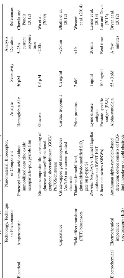
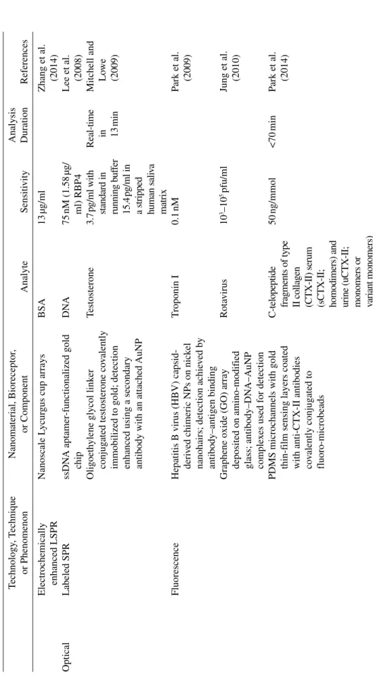
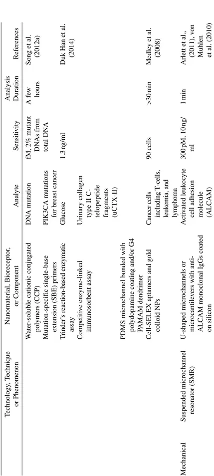
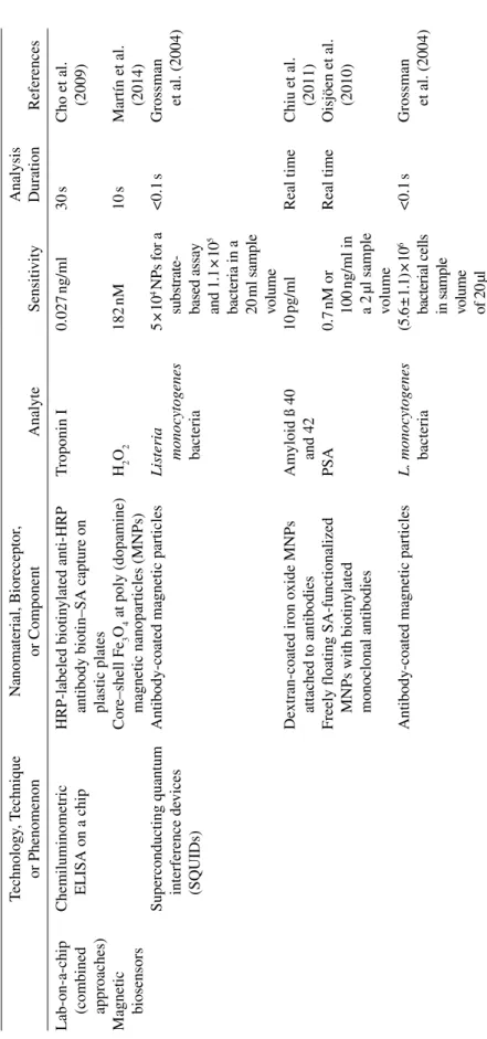
The output signal and the method used for its detection are important factors in determining the sensitivity of the biosensor. A table of biosensor output types, detection techniques, and the associated sensitivity and anal ysis time comparisons is provided in Table 10.1. It is worth noting that most biosensors provide optical, electrical/electrochemical, or mechanical output and other detection methods are relatively rare.
T
ABLE
10.1
Output
T
ypes, Recognition Elements, Sensiti
vities, and Analysis T imes f or Recent Biosensors Technology , T echnique or Phenomenon Nanomaterial, Bioreceptor , or Component Analyte Sensiti vity Analysis Duration References Electrical Amperometric
Fructosyl amino acid oxidase immobilized onto zinc oxide nanoparticle–polyp
yrrole f ilm Hemoglobin A1c 50 μM 5–25 s
current response time
Cha
wla and Pundir (2012)
Bionanocomposite f
ilm consisting of
glucose oxidase/Pt/functional graphene sheets/chitosan (GOD/ Pt/FGS/chitosan)
Glucose 0.6 μM ~200 s W u et al. (2009) Capacitance
Citrate‐capped gold nanoparticles (AuNPs) on a screen‐printed electrode
Cardiac troponin I 0.2 ng/ml ~25 min Bhalla et al. (2012) Field‐ef fect transistor (FET) biosensors Thiamine‐immobilized, glutaraldeh yde‐modif ied SiO 2 gate on p‐type Si Prion proteins 2 nM >1 h W ustoni et al. (2014) Borr elia b ur gdorferi (L yme) flagellar antibodies on SWNT FET
Lyme disease antigen
1 ng/ml 20 min Lerner et al. (2013) Silicon nano wires (SiNWs) Prostate‐specif ic antigen (PSA) 10 −6 ng/ml Real time Luo and Da vis (2013) Electrochemical
Electrochemical impedance spectroscop
y (EIS)
Antihuman alpha‐synuclein‐based detection; de
vice consists of PEG‐
thiol monolayer on gold electrode
Alpha‐synuclein 55 + 3 pM A fe w minutes Bryan et al. (2012)
Amperometric
Fructosyl amino acid oxidase (F
A
O)
immobilized on core–shell magnetic Fe–Si bionanoparticles (with chitosan); de
vice uses
modif
ied gold electrode
Glycosylated hemoglobin 0.1 mM <4 s Cha
wla and Pundir (2011)
Porous redox‐acti ve Cu 2 O–SiO 2 nanoparticles (NPs) Ferritin 0.4 ng/ml 30 min Y ang et al. (2009) Square w av e v oltammetry
CdSe QDs functionalized with strepta
vidin‐ (SA) labeled DN
A;
detection based on endonuclease acti
vity;
AuNPs used for signal
amplif ication Mycobacterium D NA 8.7 × 10 −15 M 10 s for pH = 2 Zhang et al. (2015)
Cyclic and dif
ferential
pulse v
oltammetry
Chitosan‐modif
ied glassy carbon
electrode (GCE) SCWL‐diseased plant ssDN A 4.709 ng/ µl >10 min W ongkae w and Poosittisak (2014) SiNW/AuNP‐modif
ied indium tin
oxide (IT
O)
DN
A related to dengue virus
3.5 ng/ml W ithin seconds Rashid et al. (2014) Electrochemiluminescence (ECL) Biotin‐anti‐cTnI‐luminol‐AuNPs and SA‐AuNPs
T roponin I 0.06 ng/ml A fe w minutes
(Li et al., 2013a)
Gold electrode labeled with hairpin DN
A incorporating a ruthenium comple x Tar get ssDN A 9 × 10 −11 M 2–4 s Bertoncello and F orster (2009) Electrochemical immunosensor Antitestosterone antibody on h ybrid gold NP/CNT–T eflon composite electrodes
Testosterone in human serum
85 ± 6 pg/ml Seraf ín et al. (2011)
Interdigitated electrodes (IDE) with nanoislands; de
vice functions
through protein immobilization to a parylene‐A surf
ace
Hepatitis B virus surf
ace antigen (HBsAg) <100 pg/ml Jung et al. (2014) (C ontinued )
Electrochemically enhanced LSPR
Nanoscale L
ycur
gus cup arrays
BSA 13 µg/ml Zhang et al. (2014) Optical Labeled SPR ssDN A aptamer‐functionalized gold chip D NA 75 nM (1.58 µg/ ml) RBP4 Lee et al. (2008) Oligoeth
ylene glycol link
er
conjug
ated testosterone co
valently
immobilized to gold; detection enhanced using a secondary antibody with an attached
AuNP Testosterone 3.7 pg/ml with standard in running b uf fer 15.4 pg/ml in
a stripped human sali
va matrix Real‐time in 13 min Mitchell and Lo we (2009) Fluorescence
Hepatitis B virus (HBV) capsid‐ deri
ved chimeric NPs on nick
el
nanohairs; detection achie
ved by antibody–antigen binding T roponin I 0.1 nM Park et al. (2009)
Graphene oxide (GO) array deposited on amino‐modif
ied
glass; antibody–DN
A–AuNP
comple
xes used for detection
Rota virus 10 3–10 5 pfu/ml Jung et al. (2010)
PDMS microchannels with gold thin‐f
ilm sensing layers coated
with anti‐CTX‐II antibodies covalently conjug
ated to
fluoro‐microbeads
C‐telopeptide fragments of type II collagen (CTX‐II) serum (sCTX‐II; homodimers) and urine (uCTX‐II; monomers or variant monomers)
50 ng/mmol <70 min Park et al. (2014) T ABLE 10.1 (Continued ) Technology , T echnique or Phenomenon Nanomaterial, Bioreceptor , or Component Analyte Sensiti vity Analysis Duration References
T ransmission spectroscop
y
Fluorescent Qdot‐antinitrotyrosine conjug
ate
Nitrated ceruloplasmin, a signif
icant biomark er for cardio vascular disease 1 ng/ml 10 min Li et al. (2010)
Plasmonic gold nanorods
Single nucleotide polymorphism (SNP) in DN
A 310 nm/RIU, 10 nM Dodson et al. (2015) 100 nm Au thin f
ilm with plasmonic
nanoholes
V
esicular stomatitis virus (VSV), pseudotyped Ebola (PT‐Ebola), and
V accinia virus 10 6 PFU/ml >90 min Y anik et al. (2010) SERS
Detection of antibody conjug
ated to
gold nanostar or spheres bound to malachite green isothioc
yanate
(MGITC) Raman signals is enhanced by a silica sandwich nanostructure Human immunoglob
ulin
G (human IgG), VEGF
7 fg/ml 10 s Li et al. (2013b) Iodide‐modif ied AgNPs in fluid Lysozyme, a vidin, bo vine serum alb umin (BSA),
cytochrome c, and hemoglobin
3 µ g/ml W ithin minutes Xu et al. (2014) Colorimetric, nak ed e ye
AuNP‐based plasmonic ELISA
PSA HIV‐1 capsid antigen p24
1
×
10
−18
g/ml
de la Rica and Ste
vens
(2012) (Continued
W
ater‐soluble cationic conjug
ated polymers (CCP) DN A mutation fM, 2% mutant DN As from total DN A A fe w hours
Song et al. (2012a)
Mutation‐specif
ic single‐base
extension (SBE) primers
PIK3CA mutations for breast cancer
T rinder’ s reaction‐based enzymatic assay Glucose 1.3 ng/ml
Duk Han et al. (2014)
Competiti
ve enzyme‐link
ed
immunosorbent assay
Urinary collagen type II C‐ telopeptide fragments (uCTX‐II)
PDMS microchannel bonded with polydopamine coating and/or G4 PAMAM dendrimer Cell‐SELEX aptamers and gold colloid NPs Cancer cells including T‐cells, leuk
emia, and lymphoma 90 cells >30 min Medle y et al. (2008) Mechanical
Suspended microchannel resonator (SMR) U‐shaped microchannels or microcantile
vers with anti‐
ALCAM monoclonal IgGs coated on silicon
Acti
vated leuk
oc
yte
cell adhesion molecule (ALCAM)
300 pM, 10 ng/ ml 1 min Arlett et al., (2011), v on Muhlen et al. (2010) T ABLE 10.1 (Continued ) Technology , T echnique or Phenomenon Nanomaterial, Bioreceptor , or Component Analyte Sensiti vity Analysis Duration References
Atomic force microscop
y
(AFM)
Functionalization with 3‐ aminoprop
yltriethoxysilane
(APTES) follo
wed by another
functionalization step with a layer of v
ery small peptides, which ha
ve a high af finity to the Fc re gion of the antibody Antibody–antigen comple xes 10 ng/ml Ierardi et al. (2013) Surf ace acoustic w av e (SA W) biosensor MUC1 aptamer‐modif ied gold
interdigital transducers for leak
y
SA
W on lithium tantalite (LiT
aO
3
)
surf
ace
MCF‐7 human breast cancer cells
32
cells/ml
Real time
Chang et al. (2014)
Quartz crystal microbalance (QCM) biosensor (acoustic‐ based biosensor)
ssDN
A cross‐link
ed polymeric
hydrogel immobilized on the gold surf
ace
A
vian influenza virus (AIV) H5N1
0.0128 HA U (HA unit) 30 min W ang and Li (2013) Biotin‐labeled probe DN A
immobilized on gold surf
ace after
immobilizing a
vidin on carboxyl
chip prior to biotin
Fish pathogenic viral hemorrhagic septicemia (VHS) virus
About 0.1 nM of real‐tar get viral RN A ~25 min Hong et al. (2010) Microcantile ver biosensor Biotin
ylated polyclonal antibody‐
functionalized silicon microcantile
ver arrays Interleukin‐6, interferon‐ γ, and alpha‐fetoprotein < 0.1 pg/ml ~1 h Joo et al. (2012) Multifunctional Fe 3 O4 at SiO 2 core Ti O2 shell magnetic– photocatalytic NPs
FhuA‐containing proteoliposomes immobilized on the chemically acti
vated gold‐coated surf
ace Bacterial virus T5 3 pM T5 virus >20 min Braun et al. (2009) (C ontinued )
Lab‐on‐a‐chip (combined approaches) Chemiluminometric ELISA on a chip
HRP‐labeled biotin
ylated anti‐HRP
antibody biotin–SA capture on plastic plates
T roponin I 0.027 ng/ml 30 s Cho et al. (2009) Magnetic biosensors Core–shell Fe 3 O4 at poly (dopamine) magnetic nanoparticles (MNPs) H2 O2 182 nM 10 s Martín et al. (2014)
Superconducting quantum interference de
vices
(SQ
UIDs)
Antibody‐coated magnetic particles
Listeria monocyto genes bacteria 5 × 10 4 NPs for a
substrate‐ based assay and
1.1 × 10 5 bacteria in a 20 ml sample volume <0.1 s Grossman et al. (2004) De
xtran‐coated iron oxide MNPs attached to antibodies Amyloid ß 40 and 42
10
pg/ml
Real time
Chiu et al. (2011)
Freely floating SA‐functionalized MNPs with biotin
ylated monoclonal antibodies PSA 0.7 nM or 100 ng/ml in a 2 µl sample volume Real time Oisjöen et al. (2010)
Antibody‐coated magnetic particles
L. monocyto genes bacteria (5.6 ± 1.1) × 10 6
bacterial cells in sample volume
of 20 µl <0.1 s Grossman et al. (2004) T ABLE 10.1 (Continued ) Technology , T echnique or Phenomenon Nanomaterial, Bioreceptor , or Component Analyte Sensiti vity Analysis Duration References
Whole cell Microbial cell‐based biosensor
Bacterium
Sphingomonas
sp. on the
inner epidermis of onion b
ulb scale p‐Nitrophenol (PNP) 4–80 μM of meth yl parathion >5 min
Michelini and Roda (2012)
Cell‐based biosensor
TurboRFP‐based DN
A damage
reporter cell line in NIH‐3T3 cells that fluoresce in response to stress
Genotoxic stress caused by a wide variety of agents, from chemical genotoxic agents to UV‐C radiation
>80% After 12 h of exposure to UV‐C Fendyur et al. (2015) Microfluidics
Paper‐based chemiluminescence biosensor
Paper‐based,
μPCAD, glucose
oxidase (GOx), urate oxidase (UOx) enzymes Glucose and uric acid
0.14
mmol/l
for
glucose and 0.52
mmol/l
for uric acid
40–50
s
Y
u et al. (2011)
Electrophoretic microfluidic immunoassay with magnetic‐beads detection
Biotin
ylated magnetic beads
combined with anti‐SA‐IgG antibody array
SA 20 aM or 2 × 10 −17 M W ithin 2–3 min Morozo v et al. (2007) Other types Nanopore‐based detection Bionanopore phi29 DN A‐packaging motor
Colon cancer antigen EpCAM antibody
1
nM
at
single‐ molecule level
Real time
W
248 BIOSENSORS FOR EARLY DISEASE DIAGNOSIS 10.2.3 Optical Biosensors
Optical biosensors yield outputs that can be confirmed by the naked eye, by changes in absorption, fluorescence peaks, or refractive index or by using other optical or spectroscopic techniques such as fluorescence, FTIR, and Raman spectroscopies. The widespread availability of optical detection equipment makes optical biosensor techniques attractive for the development of low‐cost lab‐on‐a‐chip devices for use in areas where rapid diagnosis of potential disease is essential and more sophisticated diagnostic methods are not readily available, such as during pathogen outbreaks in rural regions.
Colorimetric biosensors change color when exposed to their target analyte, and quantification is performed through changes in absorption at a specific wavelength. Despite their simplicity, these biosensors may have considerable sensitivity and selectivities: An absorption wavelength shift‐ based biosensor for vesicular stomatitis virus (VSV), pseudotyped Ebola (PT‐Ebola), and vaccinia virus was able to reach a sensitivity figure of merit (FOM) of 40, a detection limit of below 105 PFU/ml virus and a resolution of 0.05 nm (Yanik et al., 2010). Likewise, HIV RNA molecules could be color imetrically detected using PNAs bearing different numbers of cyclopentane chemical groups, with sufficient sensitivity to detect 20–30,000 copies/ml plasma of this virus (Zhao et al., 2014a).
Changes in chemical composition can also be detected through spec troscopic methods, such as Raman or FTIR spectroscopy. These methods quantify the absorbance, reflectance, or fluorescence of a material follow ing exposure to light at a specific wavelength or range of wavelengths and yield chemical information in the form of molecular interactions, ionic and covalent bonds, and vibrational and rotational motions. They are useful for label‐free biosensing efforts, as IR absorption and Raman scattering can be used to detect conformational changes of proteins and structural variations between materials and molecules, allowing the detailed analysis of chemical bonds without an intermediary reporter or dye. Alzheimer’s disease, for example, could be detected by quantifying amyloid β (Aβ) peptide titers using an attenuated total reflection Fourier transform infrared (ATR‐FTIR) spectroscopy biosensor (Kleiren et al., 2010).
Raman signals can be enhanced through a method called SERS, which utilizes the fact that the Raman signal becomes much more prominent (up to a factor of 1014, although typical values are in the 106–108 range) when the analyte molecules are situated between gold and silver surfaces set apart by distances around 20–30 µm (Kneipp et al., 1997). The interstruc tural distances required for SERS are usually created using nanoparticles
BIOSENSOR ELEMENTS 249
or nanopatterned surfaces, and gold and silver are often the materials of choice because their plasmon resonances (and, therefore, Raman enhance ments) are in the near‐infrared and visible spectral ranges, respectively. SERS‐active substrates for protein detection may also be designed to display a tunable resonance in the infrared range in order to produce a signal enhancement effect around the spectral positions of amide bands; or the samples may be labeled by a Raman‐active dye for easier detection (Han et al., 2009). A detection level of 7 fg/ml could be achieved in a cancer biomarker detection study from plasma using SERS with 3D hierarchical plasmonic nanoarchitectures, including Au nanospheres and Au stars, as well as with a Raman dye (malachite green isothiocyanate, MGITC) and silica nanoparticles (Li et al., 2013b).
Surface plasmons can be used for the enhancement of other spectro scopic detection methods, often using noble metal nanoparticles or metallic thin films. SPR reflectivity measurements in particular are useful for the detection of molecular interactions and may reach very high sensitivities. SPR‐based sensing systems measure the coupling of light with the plas mons (electron cloud oscillations) present at the surface of a nanoscale thin film of gold or silver. Recognition agents for SPR sensing and imaging are generally antibodies. Picogram per milliliter‐level sensitivities can be obtained using SPR; for example, testosterone detection limits in one SPR‐ based system was found to be 3.7 pg/ml in standard running buffer and 15.4 pg/ml in a human saliva matrix (Mitchell and Lowe, 2009). Biological assays, nonetheless, may prove superior in detection capacity: In another study, the detection capacity of SPR for retinol binding protein 4 (RBP4, a useful marker for type 2 diabetes) was found to be greater than ELISA but lower than Western blot (Lee et al., 2008).
A variant technique of SPR, called localized SPR (LSPR), uses metal nanoparticles (usually gold or silver) of specific sizes and geometries to further improve the detection limits of this technique (Swierczewska et al., 2012). LSPR nanostructures may be employed for biomolecule detection through the measurement of refractive index changes and colorimetric SPR imaging, and it has been shown in a theoretical study that the detection capacity of LSPR‐ based methods can exceed that of systems based on traditional Kretschmann geometry systems in terms of wavelength shift sensitivity outperforming the detection system with Kretschmann geometry (Kaya et al., 2014). LSPR‐ based detection of colon cancer was also achieved using Au nanorods, and the width‐ and length‐dependent changes in the plasmonic and photonic prop erties of nanorod antennae were investigated to identify optimal geometries for nanorod arrays for use in cancer biosensors (Dodson et al., 2015).
250 BIOSENSORS FOR EARLY DISEASE DIAGNOSIS 10.2.4 Electrical and Electrochemical Biosensors
Like optical signals, electrical signals are generally easy to detect and quantify. Electrical and electrochemical biosensors consequently see common use in both research and commercial purposes. Electrical bio sensors typically use an electrode as their transducer, and the recognition element (usually an enzyme) is immobilized onto this electrode. The enzyme–ligand interaction creates a change in the electrical properties of the electrode (this may be electrical potential, resistivity, impedance, conductivity, or capacitance, as well as the current running between the recognition element‐bound electrode and a reference electrode), which is measured and quantified to determine the concentration of the analyte. Cyclic voltammetry, chronoamperometry, chronopotentiometry, imped ance spectroscopy, and various FET‐based methods are some common tools that are used in the measurement of the output signal. More sensitive measurements may be performed with the use of nanomaterials such as nanowires, nanotubes, and nanoparticles.
As one of the most popular biosensor types, amperometric biosensors quantify changes in current, which is usually linear with the change in ana lyte concentration within the detection range (Wang, 1999). Amperometric biosensors typically display relatively modest detection capabilities; for example, a biosensor based on a multiwalled carbon nanotube (CNT)–gold nanoparticle composite yielded a limit of detection of 0.01 mM for uric acid (Chauhan and Pundir, 2011). Likewise, a detection range of 0.001 –5 mM was obtained for glucose detection using an early biosensor based on Prussian blue‐functionalized electrodes (Wu et al., 2012). In addition to gold, nanoparticles of other metals, such as zinc and platinum, can also be used in biosensor design (Chawla and Pundir, 2012; Wu et al., 2009).
The transducer electrode can be modified to produce amperometric sensors with much higher sensitivities. Sotiropoulou and Chaniotakis have reported dichlorvos detection limits at the picomolar level using a biosensor with a nanoporous carbon electrode, which enhances enzyme– ligand interactions by adsorbing the enzyme on its surface (Sotiropoulou and Chaniotakis, 2005). Salimi et al. also reported picomolar‐level detec tion capacity using a modified electrode; their system used a glassy carbon electrode modified by guanine and nickel oxide nanoparticles with the help of cyclic voltammetry (Salimi et al., 2008). Likewise, an electrical detection‐based FET biosensor was used to detect prion proteins through the use of thiamine molecules immobilized on a glutaraldehyde‐modified SiO2 gate surface on p‐Si (Wustoni et al., 2014). Another biosensor design, using an antibody‐immobilized single‐walled carbon nanotube (SWNT)
BIOSENSOR ELEMENTS 251
FET, was hypothesized to be able to detect the Lyme disease antigen at concentrations up to 1 ng/ml in buffer (Lerner et al., 2013).
Potentiometric biosensors measure changes in the electrical potential of the electrode and are less common than amperometric biosensors. None theless, potentiometry‐based biosensors using modified electrodes have been described for biomaterials such as urea and polyglycerides, with working ranges between 10−2 and 30 mM (Lakard et al., 2004; Saurina et al., 1998). Due to the relative rarity of this detection type, high‐sensitivity studies involving potentiometric biosensors are lacking, although a com bined SPR–potentiometric analysis method for nerve gas detection at nanomolar‐to‐picomolar detection ranges was reported (Taranekar et al., 2006). Biosensors that utilize changes in conductivity, capacitance, admit tance, and impedance have also been recorded in the literature, although nonamperometric output types are somewhat uncommon (Berggren et al., 2001; Gerard et al., 2002; Varshney and Li, 2007).
10.2.5 Mechanical Biosensors
The binding interaction between the recognition element and the ana lyte imposes an additional weight on the surface on which the former is immobilized. This change can be quantified using specialized devices and measurement methods, such as microcantilevers, QCM, microring resona tors (MRR), suspended microchannel resonators (SMR), or atomic force microscopy (AFM). Many of these methods have exceptionally high sen sitivities; QCM biosensors, for example, can detect femtomolar concentra tions of DNA. However, they also have long analysis times and require uncommon and highly sensitive equipment for analysis (Arlett et al., 2011). As such, these techniques are generally suitable for research purposes rather than point‐of‐care diagnostics.
Mechanical biosensors can broadly be divided into two types, those that experience a surface deflection following analyte binding (surface stress mechanical biosensors) and those that change their oscillation frequency in the presence of the analyte (dynamic‐mode mechanical biosensors) (Arlett et al., 2011). Cantilever biosensors can be of both types and have been used for sensing cancer biomarkers by using the change in cantilever frequency or deflection following antigen binding (Choi et al., 2010). Microcantilever array biosensors can be used with protein–ligand interactions to detect viruses at sensitivity levels down to subpicomolar concentrations (Braun et al., 2009). QCM biosensors can likewise reach picomolar sensitivities and have been developed for the detection of biomaterials such as human antibodies and bacterial toxins (Alfonta et al., 2001; Das et al., 2003; Yao et al., 2010).
252 BIOSENSORS FOR EARLY DISEASE DIAGNOSIS Other types of mechanical biosensors include nanoelectromechanical systems (NEMS), such as nanomechanical resonators, which may possess single‐molecule detection capacities. NEMS devices may use mass, elastic modulus, and surface stress changes as sensing parameters and can possess complex device designs that render them ideal for lab‐on‐a‐chip applica tions that require more sophisticated assay conditions than the detection of a recognition element–analyte binding interaction. In addition, while not generally employed for the analysis of biosensor outputs, AFM is an exceptionally sensitive material characterization and manipulation tool that has seen extensive use in medical proteomics and potential diagnosis of cancer and infectious disease (Archakov and Ivanov, 2007). AFM is not an ideal technique for commercial biosensors; however, its subatomic detection capacity is effective for mechanically characterizing the recogni tion element–analyte interactions that drive biosensor development, and the similarity of AFM probes to mechanical biosensor cantilevers allows this technique to yield potentially valuable information about the design of novel mechanical biosensors (Baselt et al., 1998).
10.2.6 Other Biosensor Types
In addition to optical, electrical, and mechanical detection methods, changes in the magnetic field of an analyte‐bound material can be quanti fied for biological detection. One example in the literature involves quan tification of magnetic responsivity with a high‐density sensor array that exhibits giant magnetoresistance (GMR), and the method was shown reach very high antigen–antibody binding sensitivities. A solute sensitivity of 20 zeptomoles, which is difficult to reach even with SPR and mechanical detection methods, was obtained using this strategy (Gaster et al., 2011).
Acoustic biosensors are another uncommon type and can be regarded as a subtype of mechanical biosensors. In these biosensors, changes in the properties of an acoustic wave are used to gain information about the binding interaction, usually by measuring the mass increase that results when the recognition element binds to the analyte. QCM and other types of acoustic resonators are employed in biosensors of this type and such devices have been shown to detect bacteria at sensitivities up to 0.4 cells/µl (Ferreira et al., 2009; Rocha‐Gaso et al., 2009).
Entire cells can also be used for biosensor applications, especially for the detection of pollutants and other chemicals. In these biosensors, the phy siological response of the cell is used as an indicator of analyte presence, and the complex sensory systems of cells are employed in place of advanced device design. The cells used may be bacteria, yeasts, and
THE IMPACT OF NANOTECHNOLOGY 253
fungi and are often genetically modified to yield an easily detectable signal in the presence of the analyte. These “one‐cell biosensors” may be exposed to a water or soil sample to yield a bioluminescent or fluorescent response that is subsequently quantified in an assay‐like process or the cells may be immobilized in an integrated system that truly qualifies as a biosensor (Belkin, 2003).
While analytes are typically presented in a solution, volatile organic compounds (VOCs) in gas form can also be quantified through mass spec troscopy methods for disease diagnosis. Gas chromatography–mass spec trometry (GC‐MS), for example, was used to detect VOCs in human breath for the diagnosis of lung cancer (Dragonieri et al., 2009). Sensors for VOCs typically display sensitivities in the ppm range (Adiguzel and Kulah, 2014).
10.3 THE IMPACT OF NANOTECHNOLOGY AND NANOMATERIALS IN BIOSENSOR DESIGN
Although the analytes and recognition elements involved are often in the nanoscale, nanomaterials in the strict sense are not required for the design of biosensors. Nonetheless, the large total surface areas associated with nanomaterials, as well as the small amounts of analytes and buffers required for nanoscale device‐based assays, render them attractive for biosensor applications. In addition, nanostructures may present ideal surface prop erties for analyte or recognition element immobilization or offer increased detection capacity through phenomena such as self‐assembly or plasmon enhancement. Nanospheres, nanorods, nanowires, graphene‐ and CNT‐ based structures, quantum dots, magnetic nanoparticles, NEMS systems, and other nanoscale materials are therefore often used in biosensor design.
Nanospheres are the most common nanoscale biosensor components and may be functionalized with surface groups such as thiols to better bind to an enzyme, antibody, or other recognition molecules. In addition, they may be organized into nanostructure arrays to better present their enzyme or antibody load (Xu and Han, 2004). The total surface area presented by nanoparticles is larger relative to the bulk material of equal volume; as such, nanospheres and similar nanostructures can immobilize a greater amount of recognition molecule per volume compared to their macroscale counterparts (Li et al., 2008). In addition, the size, composition, or material properties of the nanospheres might allow enhanced detection capability, as is the case with magnetic nanoparticles: Cross‐linked magnetic nanopar ticles (CLIOs), manganese‐doped magnetic nanoparticles, and core–shell structures such as elemental iron‐ferrite nanoparticles are among the
254 BIOSENSORS FOR EARLY DISEASE DIAGNOSIS magnetic materials used in biosensor applications (Haun et al., 2010). Quantum dots can also be used to improve the detection capacity of con ventional recognition elements, and QD sensors that react to a broad range of factors, such as pH change, protein or nucleic acid cleavage, or DNA synthesis, have been reported in the literature (Suzuki et al., 2008). A quantum dot–aptamer conjugate was also shown to possess a detection limit in the attomolar scale, while conventional aptamer‐based biosensors generally display nanomolar‐level sensitivity (Hansen et al., 2006).
While nanospheres are commonly used for their ease of production, nanorods and other nonspherical nanoparticles may present advantages over the nanosphere morphology. Thiol groups, for example, prefer to bind to the tips of gold nanorods, and this effect can be used to produce self‐assembled nanoparticle chains by attaching thiolated oligonucleotides to gold nanorods and allowing complementary sequences to link individual nanoparticles (Sepulveda et al., 2009). Nonspherical morphologies also alter the optical response of nanoparticles; both theoretical and experimental reports suggest that triangular silver nanoparticles enhance sensitivity in SPR‐based bio sensors (Haes and Van Duyne, 2002; Peng and Miller, 2011; Xu and Kall, 2002). Another study on a ZnO‐based biosensor found that biotin‐bound ZnO nanorods could detect streptavidin at concentrations lower than that previously reported for ZnO nanospheres (Kim et al., 2006).
Nanowires of silicon and noble metals can also be incorporated into sensor design. An Au nanowire waveguide, for example, was successfully used for plasmonic waveguide sensing in water and other liquids (Wang et al., 2014). Nanowires can also be organized into larger assemblies using techniques such as flow‐assisted, Langmuir–Blodgett, bubble‐blown, electric‐field, smearing‐transfer, roll‐printing, and PDMS‐transfer assem bly processes (Chen et al., 2011). In addition to metal or metal oxide nano wires, single‐ or multiwalled CNTs can also be used for biosensor applications. In these biosensors, the carbon structure is used to modify the electrode of an amperometric biosensor and may be functionalized to carry the recognition molecule of interest. Different configurations can be used for CNT‐based biosensors: CNTs may be coated onto the electrode or synthesized in aligned networks around it or the CNT itself may serve as the electrode. Submicromolar detection limits have been obtained in CNT‐based biosensors for glucose monitoring (Wang, 2005).
Nanoporous silica and similar materials have also been used in the design of biosensors. Light reflected off a thin, porous layer of silicon creates interference patterns (Fabry–Perot fringes) that change following the binding of the target analyte to a recognition element immobilized on the surface of the silicon film. The extent of this change depends on the
EARLY DIAGNOSIS AND BIOSENSOR‐BASED DISEASE DETECTION 255
change in the refractive index of the silicon layer and can be quantified to detect DNA, proteins, and other organic molecules at picomolar‐to‐ femtomolar scales (Lin et al., 1997). Molecular imprinting can also be employed to create polymer matrices with “holes” that serve as recogni tion sites for specific biomolecules and may reach detection levels of 0.1 μM for small organic molecules (Yano and Karube, 1999). Lipid mem branes can also be employed as biosensors, either serving as a platform to support an enzyme or antibody or acting as the recognition element itself; however, these sensors are more suited to membrane transport‐related research than clinical diagnosis (Nikolelis and Krull, 1992; Reimhult and Kumar, 2008). Pore‐bearing proteins that mimic the structure and selectivity of biological pore components can also be used in biosensor applications (Braha et al., 1997).
In addition to nanoparticle‐, thin film‐, and nanoporous matrix‐based detection methods, the techniques used in biosensor design and quanti fication are often deeply rooted in nanotechnology. As such, advances in nanomaterial‐based detection methods, such as QCM (the sensitivity of which depends on crystal thickness) and SERS, or nanofabrication methods, such as laser, AFM, or electron beam lithographies (which are used for the fabrication of cantilevers used in mechanical biosensors), will indirectly improve the sensitivity of current biosensors. However, while nanostructures offer several advantages over conventional biosensors, it must be kept in mind the in vivo use of nanoparticles in disease diagnosis is largely still in preclinical stages of development (Thakor and Gambhir, 2013). Further advances are no doubt necessary for the use of these devices in clinical settings.
10.4 EARLY DIAGNOSIS AND BIOSENSOR‐BASED DISEASE DETECTION
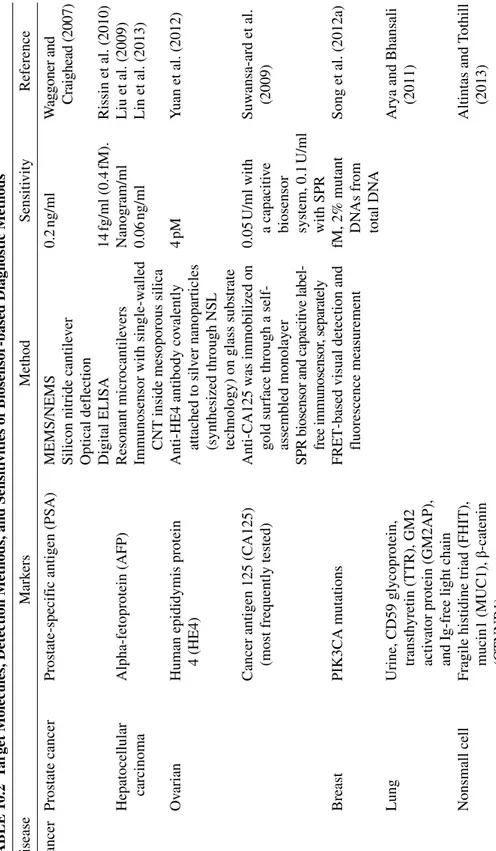
The greatest potential of biosensors lies in their medical applications. While the detection of a broad spectrum of biomaterials is feasible using biosensors, the sheer incidence rates, physiological diversity, and diffi culty of treatment that is characteristic of cancer, heart disease, and similar disorders ensure that a substantial amount of biosensor‐related research is directed toward their diagnosis. Likewise, the detection of widespread foodborne pathogens such as Salmonella, Klebsiella, and Staphylococcus or viruses such as HIV and dengue virus is vital for the control of the asso ciated diseases. As such, a list of diseases and target molecules that have been the subject of high‐sensitivity biosensor development efforts is pro vided in the present section (Table 10.2).
T
ABLE
10.2
T
ar
get Molecules, Detection Methods, and Sensiti
vities of Biosensor‐based Diagnostic Methods
Disease Mark ers Method Sensiti vity Reference Cancer Prostate cancer Prostate‐specif ic antigen (PSA) MEMS/NEMS 0.2 ng/ml W
aggoner and Craighead (2007)
Silicon nitride cantile
ver
Optical deflection Digital ELISA
14 fg/ml (0.4 fM). Rissin et al. (2010) Hepatocellular carcinoma Alpha‐fetoprotein (AFP) Resonant microcantile vers Nanogram/ml Liu et al. (2009)
Immunosensor with single‐w
alled
CNT inside mesoporous silica
0.06
ng/ml
Lin et al. (2013)
Ov
arian
Human epididymis protein 4 (HE4)
Anti‐HE4 antibody co
valently
attached to silv
er nanoparticles
(synthesized through NSL technology) on glass substrate
4
pM
Y
uan et al. (2012)
Cancer antigen 125 (CA125) (most frequently tested)
Anti‐CA125 w
as immobilized on
gold surf
ace through a self‐
assembled monolayer 0.05 U/ml with a capaciti ve biosensor system, 0.1 U/ml with SPR Suw ansa‐ard et al. (2009)
SPR biosensor and capaciti
ve label‐
free immunosensor
, separately
Breast
PIK3CA mutations
FRET‐based visual detection and fluorescence measurement fM, 2% mutant DN
As from
total DN
A
Song et al. (2012a)
Lung
Urine, CD59 glycoprotein, transth
yretin (TTR), GM2
acti
vator protein (GM2AP),
and Ig‐free light chain
Arya and Bhansali (2011)
Nonsmall cell
Fragile histidine triad (FHIT), mucin1 (MUC1),
β‐catenin (CTNNB1) Altintas and T othill (2013)
Mutations in the EGFR gene Circulating tumor cell detection using a microfluidic de
vice
Median of 74 cells per milliliter (mean, 133; range, 5–771)
Collura et al. (2008)
Cervical cancer
Electrochemical impedance spectroscop
y, impedimetric
biosensor based on PEGylated arginine‐functionalized magnetic nanoparticles
10
cells/ml
Chandra et al. (2011)
Squamous cell carcinoma antigen (SCCa)
LSPR biosensor 0.125 pM Zhao et al. (2014b) Myocardial inf arction T roponin I, T Steuer et al. (2009)
Fatty acid binding protein
Kak
oti and Gosw
ami (2013)
Rheumatoid arthritis (RA)
Rheumatoid f
actor (RF),
antic
yclic citrullinated peptide
antibody
Chandrashekara (2014)
Acti
vated CD8 cells
IL‐6, IL‐7, IL‐10 Antimutated citrullinated vimentin (anti‐MCV) antibody
Systemic lupus erythematosus (SLE)
Autoantibodies ag ainst C1q ELISA Andreje vic et al. (2013), V anheck e et al. (2012) Osteoarthritis
C‐telopeptide fragments of type II collagen (CTX‐II)
Fluorescence‐based biosensor
200
ng/mmol
Kim et al. (2013)
Cartilage oligomeric matrix protein (COMP)
ELISA with fluoro‐microbeads
0.8
ng/ml
258 BIOSENSORS FOR EARLY DISEASE DIAGNOSIS Cancer markers are some of the most common targets for biosensor design, as early detection is vital for successful treatment in many cancers. Early diagnosis of oral cancer, for example, increases survival rates from 50 to 80%, while pancreatic cancer, which is often diagnosed after the initial tumor has either spread locally or metastasized into another tissue, has a 5‐year survival rate of about 5% (Pannala et al., 2009; Silverman, 1988). Diagnosis of cervical cancer, likewise, must be performed at an early stage or “the cancer metastasizes to the rest of the uterus, bladder, rectum and abdominal wall and eventually reaches pelvic lymph nodes, thereby invading other organs and leading to death” (Chandra et al., 2011). As such, biosensors using protein markers, DNA sequences, membrane glycans, and cancer‐associated estrogen derivatives have been developed for the early detection of cancer, and the responses of cancer cells, such as drug resistance after taxane treatment in breast cancer cells, hydrogen per oxide production in human hepatoma cells, or formaldehyde presence in glioblastoma cells following treatment with a formaldehyde‐releasing drug, have been observed using various biosensor designs (Bareket et al., 2010; Braunhut et al., 2005; Cavalieri et al., 2006; Feng et al., 2006; Myung et al., 2011; Rui et al., 2010; Zhang et al., 2010).
Advancements in nanoscience have also created the prospect of devel oping small biosensors that are implanted to the body of a prospective patient and trigger at the onset of disease state or other changes in physiological conditions. This type of in vivo diagnostic method is useful for the detection of diseases that are nonsymptomatic in their early stages and for the monitoring of chronic conditions, such as diabetes, that require regular treatment. In addition to their direct healthcare benefits, these bio sensors may also serve as a means of collecting physiological data from a large set of patients, which would in turn enable the development of better treatment options. These sensors typically detect glucose and are designed for the management of diabetes; although biosensors that use fluorescein (introduced by the biosensor) or lactate for the detection of internal bleeding have also been developed (Kotanen et al., 2012; Mo and Smart, 2004; Ryou et al., 2011). Some glucose sensors of this type are approved for use by the FDA (Klonoff, 2007).
10.5 CONCLUSION AND FUTURE DIRECTIONS
Biosensor research combines the investigation of biochemical recognition processes, signal transduction systems, and output‐specific detection methods, which makes it a highly multidisciplinary research field. As
REFERENCES 259
such, the development of highly specific recognition elements and advancements in various signal detection techniques will indirectly result in the development of more accurate biosensors. Recent discoveries in gene regulation and biological signaling mechanisms have revealed the presence of various regulatory elements that are highly specific to their targets and may be used in biosensor applications. In addition, methods for the synthesis of proteins and nucleic acids incorporating nonstandard chemical groups have advanced greatly in the recent years; as such, combination systems incorporating signal detecting, amplifying, and transducing elements can now be produced with considerable ease.
As resolution limits are bypassed and more selective biomolecular rec ognition agents are discovered, the detection efficiency of future biosen sors will no doubt continue to increase. Furthermore, advances in nanotechnology and material fabrication methods may both allow novel detection techniques that employ nanoscale phenomena to be used in bio sensor design and decrease costs by lowering biosensor dimensions and the quantities of biological materials incorporated into the sensor struc ture. Consequently, it would appear that the multifaceted nature of bio sensor design, as well as the advantages it offers in terms of assay sensitivity, selectivity, and detection time and costs, will continue attract ing commercial and research efforts and may allow the early detection of diseases that currently are undetectable during periods for which their treatment would have been the most effective.
REFERENCES
Adiguzel, Y. and Kulah, H. (2014). Breath sensors for lung cancer diagnosis. Biosens. Bioelectron. 65, 121–138.
Alfonta, L., Willner, I., Throckmorton, D., and Singh, A. (2001). Electrochemical and quartz crystal microbalance detection of the cholera toxin employing horseradish peroxidase and GM1‐functionalized liposomes. Anal. Chem. 73, 5287–5295.
Altintas, Z. and Tothill, I. (2013). Biomarkers and biosensors for the early diagnosis of lung cancer. Sens. Actuators B Chem. 188, 988–998.
Andrejevic, S., Jeremic, I., Sefik‐Bukilica, M., Nikolic, M., Stojimirovic, B., and Bonaci‐Nikolic, B. (2013). Immunoserological parameters in SLE: High‐avidity anti‐dsDNA detected by ELISA are the most closely associated with the disease activity. Clin. Rheumatol. 32, 1619–1626.
Archakov, A.I. and Ivanov, Y.D. (2007). Analytical nanobiotechnology for medicine diagnostics. Mol. Biosyst. 3, 336–342.
260 BIOSENSORS FOR EARLY DISEASE DIAGNOSIS
Arlett, J., Myers, E., and Roukes, M. (2011). Comparative advantages of mechanical biosensors. Nat. Nanotechnol. 6, 203–215.
Arya, S.K. and Bhansali, S. (2011). Lung cancer and its early detection using biomarker‐based biosensors. Chem. Rev. 111, 6783–6809.
Bareket, L., Rephaeli, A., Berkovitch, G., Nudelman, A., and Rishpon, J. (2010). Carbon nanotubes based electrochemical biosensor for detection of formaldehyde released from a cancer cell line treated with formaldehyde‐releasing anticancer prodrugs. Bioelectrochemistry 77, 94–99.
Baselt, D., Lee, G., Natesan, M., Metzger, S., Sheehan, P., and Colton, R. (1998). A biosensor based on magnetoresistance technology. Biosens. Bioelectron. 13, 731–739.
Belkin, S. (2003). Microbial whole‐cell sensing systems of environmental pollutants. Curr. Opin. Microbiol. 6, 206–212.
Berggren, C., Bjarnason, B., and Johansson, G. (2001). Capacitive biosensors. Electroanalysis 13, 173–180.
Bertoncello, P. and Forster, R.J. (2009). Nanostructured materials for electroche miluminescence (ECL)‐based detection methods: Recent advances and future perspectives. Biosens. Bioelectron. 24, 3191–3200.
Bhalla, V., Carrara, S., Sharma, P., Nangia, Y., and Raman Suri, C. (2012). Gold nanoparticles mediated label‐free capacitance detection of cardiac troponin I. Sens. Actuators B Chem. 161, 761–768.
Braha, O., Walker, B., Cheley, S., Kasianowicz, J., Song, L., Gouaux, J., and Bayley, H. (1997). Designed protein pores as components for biosensors. Chem. Biol. 4, 497–505.
Braun, T., Ghatkesar, M.K., Backmann, N., Grange, W., Boulanger, P., Letellier, L., Lang, H.‐P., Bietsch, A., Gerber, C., and Hegner, M. (2009). Quantitative time‐resolved measurement of membrane protein‐ligand interactions using microcantilever array sensors. Nat. Nanotechnol. 4, 179–185.
Braunhut, S., McIntosh, D., Vorotnikova, E., Zhou, T., and Marx, K. (2005). Detection of apoptosis and drug resistance of human breast cancer cells to taxane treatments using quartz crystal microbalance biosensor technology. Assay Drug Dev. Technol. 3, 77–88.
Bryan, T., Luo, X., Forsgren, L., Morozova‐Roche, L.A., and Davis, J.J. (2012). The robust electrochemical detection of a Parkinson’s disease marker in whole blood sera. Chem. Sci. 3, 3468.
Cavalieri, E., Chakravarti, D., Guttenplan, J., Hart, E., Ingle, J., Jankowiak, R., Muti, P., Rogan, E., Russo, J., Santen, R., et al. (2006). Catechol estrogen quinones as initiators of breast and other human cancers: Implications for biomarkers of susceptibility and cancer prevention. Biochim. Biophys. Acta 1766, 63–78. Chandra, S., Barola, N., and Bahadur, D. (2011). Impedimetric biosensor for early
REFERENCES 261
Chandrashekara, S. (2014). Current studies of biomarkers for the early diagnosis of rheumatoid arthritis. Curr. Biomark. Find. 4, 107–119.
Chang, K., Pi, Y., Lu, W., Wang, F., Pan, F., Li, F., Jia, S., Shi, J., Deng, S., and Chen, M. (2014). Label‐free and high‐sensitive detection of human breast cancer cells by aptamer‐based leaky surface acoustic wave biosensor array. Biosens. Bioelectron. 60, 318–324.
Chauhan, N. and Pundir, C.S. (2011). An amperometric uric acid biosensor based on multiwalled carbon nanotube‐gold nanoparticle composite. Anal. Biochem.
413, 97–103.
Chawla, S. and Pundir, C.S. (2011). An electrochemical biosensor for fructosyl valine for glycosylated hemoglobin detection based on core‐shell magnetic bion anoparticles modified gold electrode. Biosens. Bioelectron. 26, 3438–3443. Chawla, S. and Pundir, C.S. (2012). An amperometric hemoglobin A1c biosensor
based on immobilization of fructosyl amino acid oxidase onto zinc oxide nanoparticles‐polypyrrole film. Anal. Biochem. 430, 156–162.
Chen, K.‐I., Li, B.‐R., and Chen, Y.‐T. (2011). Silicon nanowire field‐effect transistor‐based biosensors for biomedical diagnosis and cellular recording investigation. Nano Today 6, 131–154.
Chiu, M.J., Horng, H.E., Chieh, J.J., Liao, S.H., Chen, C.H., Shih, B.Y., Yang, C.C., Lee, C.L., Chen, T.F., Yang, S.Y., et al. (2011). Multi‐channel SQUID‐ based ultra‐high‐sensitivity in‐vitro detections for bio‐markers of Alzheimer’s disease via immunomagnetic reduction. IEEE Trans. Appl. Supercond. 21, 477–480.
Cho, I.‐H., Paek, E.‐H., Kim, Y.‐K., Kim, J.‐H., and Paek, S.‐H. (2009). Chemiluminometric enzyme‐linked immunosorbent assays (ELISA)‐on‐a‐chip biosensor based on cross‐flow chromatography. Anal. Chim. Acta 632, 247–255. Choi, Y.‐E., Kwak, J.‐W., and Park, J.W. (2010). Nanotechnology for early cancer
detection. Sensors (Basel) 10, 428–455.
Collura, C.V., Inserra, E., Diederichs, S., Ph, D., Iafrate, A.J., Bell, D.W., Digumarthy, S., Muzikansky, A., Irimia, D., Settleman, J., et al. (2008). Detection of mutations in EGFR in circulating lung‐cancer cells. N. Engl. J. Med.
359, 366–377.
Das, K., Penelle, J., and Rotello, V. (2003). Selective picomolar detection of hexachlorobenzene in water using a quartz crystal microbalance coated with a molecularly imprinted polymer thin film. Langmuir 19, 3921–3925.
De la Rica, R. and Stevens, M.M. (2012). Plasmonic ELISA for the ultrasensitive detection of disease biomarkers with the naked eye. Nat. Nanotechnol. 7, 821–824.
Dell’Atti, D., Tombelli, S., Minunni, M., and Mascini, M. (2006). Detection of clinically relevant point mutations by a novel piezoelectric biosensor. Biosens. Bioelectron. 21, 1876–1879.
262 BIOSENSORS FOR EARLY DISEASE DIAGNOSIS
Di Natale, C., Mantini, A., Macagnano, A., Antuzzi, D., Paolesse, R., and D’Amico, A. (1999). Electronic nose analysis of urine samples containing blood. Physiol. Meas. 20, 377–384.
Dodson, S.L., Cao, C., Zaribafzadeh, H., Li, S., and Xiong, Q. (2015). Engineering plasmonic nanorod arrays for colon cancer marker detection. Biosens. Bioelectron. 63, 472–477.
Dragonieri, S., Annema, J.T., Schot, R., van der Schee, M.P.C., Spanevello, A., Carratú, P., Resta, O., Rabe, K.F., and Sterk, P.J. (2009). An electronic nose in the discrimination of patients with non‐small cell lung cancer and COPD. Lung Cancer 64, 166–170.
Du, Y., Li, B., and Wang, E. (2013). “Fitting” makes “sensing” simple: Label‐free detection strategies based on nucleic acid aptamers. Acc. Chem. Res. 46, 203–213.
Duk Han, Y., Jin Chun, H., and Yoon, H.C. (2014). The transformation of common office supplies into a low‐cost optical biosensing platform. Biosens. Bioelectron. 59, 259–268.
El‐Sayed, I., Huang, X., and El‐Sayed, M. (2005). Surface plasmon resonance scattering and absorption of anti‐EGFR antibody conjugated gold nanoparti cles in cancer diagnostics: Applications in oral cancer. Nano Lett. 5, 829–834. Fendyur, A., Varma, S., Lo, C.T., and Voldman, J. (2015). Cell‐based biosensor to
report DNA damage in micro‐ and nanosystems. Anal. Chem. 86, 7598–7605. Feng, K., Yang, Y., Wang, Z., Jiang, J., Shen, G., and Yu, R. (2006). A nano‐ porous CeO2/chitosan composite film as the immobilization matrix for colorectal cancer DNA sequence‐selective electrochemical biosensor. Talanta
70, 561–565.
Ferreira, G., Da‐Silva, A., and Tome, B. (2009). Acoustic wave biosensors: Physical models and biological applications of quartz crystal microbalance. Trends Biotechnol. 27, 689–697.
Gaster, R.S., Xu, L., Han, S.‐J., Wilson, R.J., Hall, D.A., Osterfeld, S.J., Yu, H., and Wang, S.X. (2011). Quantification of protein interactions and solution transport using high‐density GMR sensor arrays. Nat. Nanotechnol. 6, 314–320.
Gerard, M., Chaubey, A., and Malhotra, B. (2002). Application of conducting polymers to biosensors. Biosens. Bioelectron. 17, 345–359.
Grossman, H.L., Myers, W.R., Vreeland, V.J., Bruehl, R., Alper, M.D., Bertozzi, C.R., and Clarke, J. (2004). Detection of bacteria in suspension by using a superconducting quantum interference device. Proc. Natl. Acad. Sci. U.S.A.
101, 1–6.
Haes, A. and Van Duyne, R. (2002). A nanoscale optical blosensor: Sensitivity and selectivity of an approach based on the localized surface plasmon reso nance spectroscopy of triangular silver nanoparticles. J. Am. Chem. Soc. 124, 10596–10604.
REFERENCES 263
Han, X.X., Zhao, B., and Ozaki, Y. (2009). Surface‐enhanced Raman scattering for protein detection. Anal. Bioanal. Chem. 394, 1719–1727.
Hansen, J., Wang, J., Kawde, A., Xiang, Y., Gothelf, K., and Collins, G. (2006). Quantum‐dot/aptamer‐based ultrasensitive multi‐analyte electrochemical biosensor. J. Am. Chem. Soc. 128, 2228–2229.
Hanson, C. and Thaler, E. (2005). Electronic nose prediction of a clinical pneumonia score: Biosensors and microbes. Anesthesiology 102, 63–68.
Haun, J., Yoon, T., Lee, H., and Weissleder, R. (2010). Magnetic nanoparticle biosensors. Wiley Interdiscip. Rev. Nanomed. Nanobiotechnol. 2, 291–304. Hong, S.‐R., Jeong, H.‐D., and Hong, S. (2010). QCM DNA biosensor for the
diagnosis of a fish pathogenic virus VHSV. Talanta 82, 899–903.
Ierardi, V., Ferrera, F., Millo, E., Damonte, G., Filaci, G., and Valbusa, U. (2013). Bioactive surfaces for antibody‐antigen complex detection by atomic force microscopy. J. Phys. Conf. Ser. 439, 012001.
Iles, R.K. and Kallichurn, H. (2012). What will be the future development of elec trochemical biosensors for the detection and quantification of biomarkers? J Bioeng. Biomed. Sci. 2, e110.
Iliuk, A.B., Hu, L., and Tao, W.A. (2011). Aptamer in bioanalytical applications. Anal. Chem. 83, 4440–4452.
Joo, J., Kwon, D., Yim, C., and Jeon, S. (2012). Highly sensitive diagnostic assay for the detection of protein biomarkers using microresonators and multifunc tional nanoparticles. ACS Nano 6, 4375–4381.
Jung, J., Cheon, D., Liu, F., Lee, K., and Seo, T. (2010). A graphene oxide based immuno‐biosensor for pathogen detection. Angew. Chem. Int. Ed. Engl. 49, 5708–5711.
Jung, H.‐W., Chang, Y.W., Lee, G., Cho, S., Kang, M.‐J., and Pyun, J.‐C. (2014). A capacitive biosensor based on an interdigitated electrode with nanoislands. Anal. Chim. Acta 844, 27–34.
Kakoti, A. and Goswami, P. (2013). Heart type fatty acid binding protein: Structure, function and biosensing applications for early detection of myocardial infarction. Biosens. Bioelectron. 43, 400–411.
Kaya, Y., Ayas, S., Topal, A.E., Guner, H., and Dana, A. (2014). Sensitivity comparison of localized plasmon resonance structures and prism coupler. Sens. Actuators B Chem. 191, 516–521.
Kim, J., Park, W.I., Lee, C., and Yi, G. (2006). ZnO nanorod biosensor for highly sensitive detection of specific protein binding. J. Kor. Phys. Soc. 49, 1635–1639.
Kim, S.J., Park, Y.M., Min, B.‐H., Lee, D.‐S., and Yoon, H.C. (2013). Optical immunosensor for quantifying C‐telopeptide fragments of type II collagen as an osteoarthritis biomarker in urine. BioChip J. 7, 399–407.
264 BIOSENSORS FOR EARLY DISEASE DIAGNOSIS
Kleiren, E., Ruysschaert, J., Goormaghtigh, E., and Raussens, V. (2010). Development of a quantitative and for Alzheimer’ s disease : The effect of deuteration on the detection of the A β peptide. Spectroscopy 24, 61–66. Klonoff, D.C. (2007). The benefits of implanted glucose sensors. J. Diabetes Sci.
Technol. 1, 797–800.
Kneipp, K., Wang, Y., Kneipp, H., Perelman, L., Itzkan, I., Dasari, R., and Feld, M. (1997). Single molecule detection using surface‐enhanced Raman scattering (SERS). Phys. Rev. Lett. 78, 1667–1670.
Kotanen, C., Moussy, F., Carrara, S., and Guiseppi‐Elie, A. (2012). Implantable enzyme amperometric biosensors. Biosens. Bioelectron. 35, 14–26.
Kriz, D., Ramstrom, O., and Mosbach, K. (1997). Molecular imprinting—New possibilities for sensor technology. Anal. Chem. 69, A345–A349.
Lakard, B., Herlem, G., Lakard, S., Antoniou, A., and Fahys, B. (2004). Urea potentiometric biosensor based on modified electrodes with urease immobi lized on polyethylenimine films. Biosens. Bioelectron. 19, 1641–1647. Lee, S.J., Youn, B.‐S., Park, J.W., Niazi, J.H., Kim, Y.S., and Gu, M.B. (2008).
ssDNA aptamer‐based surface plasmon resonance biosensor for the detection of retinol binding protein 4 for the early diagnosis of type 2 diabetes. Anal. Chem. 80, 2867–2873.
Lerner, M.B., Dailey, J., Goldsmith, B.R., Brisson, D., and Johnson, A.T.C. (2013). Detecting Lyme disease using antibody‐functionalized single‐walled carbon nanotube transistors. Biosens. Bioelectron. 45, 163–167.
Li, C., Liu, Y., Li, L., Du, Z., Xu, S., Zhang, M., Yin, X., and Wang, T. (2008). A novel amperometric biosensor based on NiO hollow nanospheres for bio sensing glucose. Talanta 77, 455–459.
Li, Z., Wang, Y., Wang, J., Tang, Z., Pounds, J.G., and Lin, Y. (2010). Rapid and sensitive detection of protein biomarker using a portable fluorescence bio sensor based on quantum dots and a lateral flow test strip. Anal. Chem. 82, 7008–7014.
Li, F., Yu, Y., Cui, H., Yang, D., and Bian, Z. (2013a). Label‐free electrochemilu minescence immunosensor for cardiac troponin I using luminol functionalized gold nanoparticles as a sensing platform. Analyst 138, 1844–1850.
Li, M., Cushing, S.K., Zhang, J., Suri, S., Evans, R., Petros, W.P., Gibson, L.F., Ma, D., Liu, Y., and Wu, N. (2013b). Three‐dimensional hierarchical plasmonic nano‐architecture enhanced surface‐enhanced Raman scattering immunosensor for cancer biomarker detection in blood plasma. ACS Nano
7, 4967–4976.
Lin, V., Motesharei, K., Dancil, K., Sailor, M., and Ghadiri, M. (1997). A porous silicon‐based optical interferometric biosensor. Science 278, 840–843.
Lin, J., Wei, Z., Zhang, H., and Shao, M. (2013). Sensitive immunosensor for the label‐free determination of tumor marker based on carbon nanotubes/