CYTOTOXIC ACTIVITY OF RESVERATROL IN DIFFERENT
CELL LINES EVALUATED BY MTT AND NRU ASSAYS
Hatice Gül GÖKTAŞ
1,2*, Merve BACANLI
1, Berkin KUTLUK
3, Arif
Ahmet BAŞARAN
4, Nurşen BAŞARAN
11
Hacettepe University, Faculty of Pharmacy, Department of Toxicology
06100 Ankara, TURKEY
2
Çukurova University, Faculty of Pharmacy, Department of Toxicology
01330 Adana, TURKEY
3
Bilkent University 06100 Ankara, TURKEY
4
Hacettepe University, Faculty of Pharmacy, Department of
Pharmacognosy 06100 Ankara, TURKEY
*Correspondence: E-mail: [email protected]; Tel: +90 312
305 21 78
ABSTRACT
Oxidative stress is the state of imbalance between the level of antioxidant defence system and production of reactive oxygen species (ROS) and is involded in the progression of several diseases such as inflammation, cancer, neurodegenerative disorders and cardiovascular diseases. It is suggested that plant polyphenols may act as antioxidants and therefore it has anti-cancer activities. Resveratrol (RV), is a naturally occuring polyphenolic compound which is found in many plant species including grapes, nuts, blueberries and raspberries. Data indicated that it has anti-oxidant, anti-inflamatory and anti-cancer activities. But there are also some studies reported that RV has not protective effects aganist cancer. In this study, the cytotoxicity of RV in human breast adenocarcinoma (MDA-MB 231), human cervical cancer (HeLa) and Chinese hamster lung fibroblast(V79) cells were evaluated by Neutral Red uptake assay (NRU) and MTT (3-[4,5-dimethylthiazol-2-yl]-2,5-diphenyltetrazolium bromide) assays after incubation at 24 h. We obtained more or the less same results by two cytotoxicity assays. In the concentrations between 2-400 µM, RV seemed not to induce a pronounced cytotoxicity in all cell types. Even at highest concentrations, it showed almost no cytotoxic effects. So the IC50 values were not calculated at the studied concentrations.
Key words: Resveratrol, cytotoxicity, breast cancer, cervical cancer, MTT assay, NRU assay.
Resveratrolün Farklı Hücre Hatlarında MTT ve NKA Yöntemleri ile Değerlendirilen Sitotoksik Etkileri
Oksidatif stres, reaktif oksijen bileşiklerinin üretimi ile antioksidan savunma sistemlerinin dengesinin bozulmasını tanımlayan bir terimdir. Oksidatif stres, inflamasyon kanser, nörodejeneratif bozukluklar ve kardiyovasküler hastalıklar gibi birçok hastalığın gelişmesinde rol oynamaktadır. Bitkisel fenolik bileşiklerin antioksidan etkili oldukları ve bu nedenle anti kanser etkileri oldukları öne sürülmektedir. Resveratrol (RV), üzüm, fındık, yaban mersini ve ahududu gibi pekçok bitkisel üründe bulunan, doğal olarak oluşan polifenolik bir bileşiktir. RV‟nin antioksidan, antiinflamatuvar ve antikanser etkileri olduğu gösteren çalışmalar bulunmaktadır. Ancak RV‟nin özellikle kanserin önlenmesinde koruyucu etkilerinin olmadığını da iddia edilmektedir. Bu çalışmada, insan meme adenokarsinoma (MDA-MB 231), insan servikal kanser (HeLa) ve Çin hamster akciğer fibroblast (V79) hücrelerinde RV‟nin sitotoksik etkileri, 24 saat inkübasyon sonrasında Nötral Kırmızı Alım (NKA) ve 3-(4,5-dimetiltiyazol-2-il)-2,5-difeniltetrazolyum bromür (MTT) yöntemleri ile değerlendirilmiştir. Her iki sitotoksisite yönteminde de benzer sonuçlar elde edilmiştir. 2-400 µM aralığında, RV‟ün tüm hücre tiplerinde belirgin bir sitotoksik etkisi olmadığı görülmüştür. Hatta en yüksek konsatrasyonlarda bile neredeyse hiç sitotoksik etkileri olmamıştır. Bu nedenle IC50 değerleri, bu konsantrasyonaralığında hesaplanamamıştır.
Anahtar kelimeler: Resveratrol, sitotoksisite, meme kanseri, servikal kanser, MTT yöntemi,
NKA yöntemi
INTRODUCTION
Cancer is a one of the principal cause of death worldwide with more than 3 million new cases and 1.7 million deaths each year. According to the recent International Agency for Research on Cancer report, breast cancer was the most common cancer diagnosed in women (151 countries worldwide) and the second most common cancer type was repoted to be cervix cancer (30 countries in worldwide) (1).
Oxidative stress is the state of imbalance between the level of antioxidant defence system and production of reactive oxygen species (ROS). ROS are continuously produced during normal physiologic events and can easily initiate the peroxidation of membrane lipids, leading to the accumulation of lipid peroxides. ROS have been implicated in more than 100 diseases including different cancer types (2). All aerobic organisms have antioxidant defenses, including antioxidant enzymes and antioxidant food constituents to remove or repair the damaged molecules (3). Natural products are widely being used as dietary supplements for health protective effects and epidemiological studies indicate that populations consuming high levels of plant derived foods have low incidence rates of various cancers (4).Resveratrol (RV); 3,40,5-trihydroxy-trans-stilbene, is a naturally occuring polyphenolic compound containing a stilbene structure similar to estrogen and produced by as a response to stress, injury, ultraviolet and fungal infection in many plant species including grapes, nuts and berries (Figure 1) (5, 6). It was identified in 1963 as the active constituent of the dried roots of Polygonum cuspidatum, a plant used in traditional Chinese and Japanese medicine against suppurative dermatitis, gonorrhea and hyperlipemia (7-9). In addition that, it was used in India for herbal preparation named „‟Darakchasava‟‟ since ancient times (10). However its old history, first real intrest in resveratrol came in 1992 when it was assumed to explain of cardio-protective effects of wine (11). It was suggested to be the solution to the „„French Paradox‟‟, a term used to describe the observation that the French population had a very low incidence of cardiovascular disease than other European countries despite a high consumption of saturated fat (12). Afterwards, it has become an interesting and attracting subject for the research. Data showed that RV has anti-oxidant, anti-inflamatory, anti-cancer activities (13-15). It has been shown that RV has cytotoxic potential in human breast adenocarcinoma (MCF-7), rat brain glioma (C6), human neuroblastoma (SH-SY5Y), African green monkey kidney fibroblast (CV1-P), mouse macrophage (RAW264.7), mouse fibroblast (3T6) and the human promyelocytic leukemia tumor (HL60) cells (16-19). But studies about RV‟s ctotoxicity using HeLa, V79 and MDA-MB 231 cell lines is limited and also there are some studies reporting that RV has not cytotoxic effects aganist cancer cells.Molecules which can inhibit cell growth in tumor cells, without significantly affecting the viability of normal cells, represent potential anticancer agents. Therefore, in this study, we evaluated the cytotoxic property of RV aganisttwo different cancer cell lines (human breast adenocarcinoma MDA-MB 231) and human cervical cancer (HeLa)) and one healthy cell line (Chinese hamster lung fibroblast (V79)) by Neutral Red Uptake (NRU) and 3-[4,5-dimethylthiazol-2-yl]-2,5-diphenyltetrazolium bromide (MTT) assays.Also another aim of this study was to compare the sensitivity of the two cytotoxicity assays.
Figure 1. Structure of RV
EXPERIMENTAL
Chemicals
The chemicals used in the experiments were purchased from the following suppliers: fetal calf serum (FCS), trypsin-EDTA, penicillin-streptomycin from Biological Industries (Kibbutz Beit-Haemek, Israel), minimum essential medium (MEM), dimethyl sulfoxide (DMSO),Dulbecco's
phosphate buffered saline (DPBS), ethanol, neutral red (NR), MTT and RV from Sigma (St Louis, USA), acetic acid from Merck (Darmstadt, Germany).
Cell Culture
MDA-MB 231, HeLa and V79 cells were seeded in 75 cm2 flasks in 20 ml MEM supplemented with 10% FCS and 1% penicillin-streptomycin and then grown for 1 day in an incubator at 37°C in a humidified atmosphere supplemented with 5% CO2.
NRU Assay
NRU assay was performed following the protocols described by Virgilio et al. (2004) and Saquib et al. (2012) (20, 21). After disaggregation of cells with trypsin/EDTA and resuspension of cells in medium, a total of 105 cells/well were plated in 96 well tissue-culture plates. After 24 h incubation, the different concentrations (2-400 µM) of RV in medium were added. The cells were incubated for 24 h (2 cell cycle) at 37°C in 5% CO2 in air, then the medium was aspirated.
The cells were washed twice with DPBS and incubated for an additional 3 hours in the medium supplemented with NR (50 µg/ml of stock in medium) was added (200 μl/well). After the medium was discarded, the cells were rinsed five times with warm DPBS (37°C) to remove the nonincorporated excess dye and 200 µl of “fixation solution” (50% ethanol, 1% acetic acid, and 49% distilled water) was added to each well to fix the cells and bring NR into solution. The plates were shaken for 20 min, and the absorbance of the solution in each well was measured in a microplate reader at 540 nm and compared with the wells containing untreated cells.
MTT Assay
MTT assay was performed by the method of Mosmann (1983) with the modifications Kuz´ma et al. (2012) (22, 23). Following disaggregation of cells with trypsin/EDTA and resuspension of cells in medium, a total of 105 cells/ well were plated in 96 well tissue-culture plates. After 24 h incubation, cells were exposed to the different concentrations of RV (2-400 μM) in medium for 24 h at 37 °C in 5% CO2 in air. After exposure, the medium was aspirated and MTT (5 mg/ml of stock in DPBS) was added (20 μl/well in 200 μl of cell suspension), and cells were incubated for an additional 4 h with MTT dye. At the end of incubation, the dye was carefully taken out and 100 μl of DMSO was added to each well. The plates were shaken for 5 min. The absorbance of the solution in each well was measured in a microplate reader at 540 nm.
Statistical Analysis
All experiments were performed three times in duplicate and cell viability was plotted as percent of control (assuming data obtained from the absence of RV as 100%). Data was statistically analysed with the independent student‟s t-test by SPSS for Windows 20.0 computer program.p value of less than 0.05 was considered as statistically significant.IC50 values represent the concentrations that reduced the mean absorbance of 50% of those in the untreated cells.
RESULTS
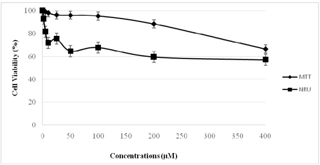
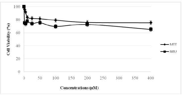
MDA-MB 231, HeLa and V79 cells were exposed to RV (2–400 µM) for 24 h and cytotoxicity was determined with the NRU and the MTT assays.We obtained more or the less same results by two cytotoxicity assays. In MDA-MB 231 cells, RV has almost no cytotoxic effects at the 2, 5, 10 and 25 µM concentrations. The cell viability seemed to decrease at higher concentrations than 50 µM. At the maximum concentration, cell viability was decreased to 55 %.(Figure 2). In HeLa cells, RV has almost no cytotoxic effects at the 2, 5 and 10 µM concentrations. The cell viability decreased slightly at higher concentrations than 25 µM and at the 400 µM, cell
viability was decreased to 57 % (Figure 3). No cytotoxic activity of RV was observed in healthy V79 cells at the studied concentrations. Even at the highest concentration, cell viability was higher than 60 % (Figure 4). Since RV has not strong cytotoxic activity against these cell lines, the IC50 values were not obtainedin the studied concentrations by the two assays for 24 h
incubation in these cell lines.
Fig. 2. Comparision of MTT and NRU assay in MDA-MB 231 cells after exposure to RV for 24
h; data presented as percentage of control ±SEM; SEM: standard error mean.
Fig. 3. Comparision of MTT and NRU assay in HeLa cells after exposure to RV for 24 h; data
Fig. 4. Comparision of MTT and NRU assay in V79 cells after exposure to RV for 24 h; data
presented as percentage of control ±SEM; SEM: standard error mean.
DISCUSSION
Cancer is a global problem and each year 14 million people are diagnosed with cancer and 8 million of them is resulted with death (24). Host and environmental factors such as diet and lifestyle, are effective factors in cancer development (25). In recents years, many studies have been focused on the cancer therapy with natural products. Current literature about anti-cancer properties of natural products is contradictory. There are some evidences on the effects of these products on cancer cells. But it is still unanswered argument whether it will help to the treatment of some cancer including breast and cervical cancers or not. Resveratrol (RV) is a natural polyphenolic compound and found in many plant species. It is suggested to show various biological activities such as cardioprotective, antiplatelet, anti-inflammatory, neuroprotective, antiviral and anti-cancer (26, 27). But it can also exhibit pro-oxidant activities depending on the concentration and cell type, especially in the presence of transition metal ions, that cause oxidative damage in cellular DNA (28). Cancer chemopreventive and anticancer effects is one of the most blazing activities of RV. These effects were first described by Jang et al. in 1997 and since then, there is a growing interest regarding clinical implications of RV on cancer treatment (29). RV is suggested to show anticancer effects by targeting the cell growth, inflamation, apoptosis, invasion and metastasis (30).
Most of the studies about RV‟s beneficial effects are based on in vitro studies. In a previous study, the cytotoxic effects of RV were increased in a dose depending manner on 3T6 and HL60 cells which was evaluated by MTT assay and Trypan Blue staining.(16).In a different study, SH-SY5Y and CV1-P cells were incubated with RV for 12 h and 24 h respectively and cytotoxicity was measured by MTT assay. It has been shown that RV significantly decreased the cell viability of CV1-P cells but had no significant cytotoxic effect on SH-SY5Y cells, although there was a slight decrease at 50 and 100 µM concentrations (17). The cytotoxic effect of RV was evaluated in C6 cells after incubation for 24 h and 72 h. The cell viability was decreased with the increasing concentrations of RV and the IC50 values of RV were found to be 85 and 15 μM respectively in this cell line(18). It was observed that RV reduced cell
viabilityapproximately 35% measured by MTT assay in RAW264.7 cells after 24 h treatment (19). In an another study, it is claimed that RV enhanced the anti-cancer effect induced by As2O3in vitro(31). It is suggested that RV‟s anticancer effects are based on its antioxidant
activities. On the other hand, a recent study showed that there was no difference in antioxidant activities between red wine and the red wine enriched 10-fold of resveratrol (32). In our study, we chosed MDA-MB 231 and HeLa cells as a cancer cell lines because breast and cervix cancer are the most common cancer types in women in the world. We found that RV seemed not to induce a pronounced cytotoxicity in all cell types at the studied concentrations after 24 h incubation. Recently Guisado et al. have showed that RV has cytotoxic effects in MDA-MB 231 cells but this cell line was almost insensitive to any concentrations of RV used for treatments shorter than 36 h(33). This result was consistent with our study. But inconsistent with the limited studies about cytotoxicity of RV in HeLa cells, we showed that it has not strong cytotoxic effect in this cell line (26). We chosed V79 cell as a healthy cell line. This cells are widely used in toxicity studies and they can keep basal cell functions in normal cell culture conditions since they do not have p450 enzyme system (34,35). Our results showed that the cytotoxic effects of RV were increased in a dose dependent manner but it has less cytotoxic effect at the higher concentrations.
Although, Gesher A. et al. mentioned that when RV added to diet of rodents, it impeded the development of cancer (36). However, they suggested the main issue of the clinical applications of RV in humans is its minimal oral bioavailability due to its rapid and extensive metabolism leading to the formation of various metabolite, mostly glucuronides and sulfates (37). In addition to that, many effects of RV does not followlinear but rather hermentic dose-response curves (38). Thus choising of the proper dosage is problematic (39).
In the present study, the results obtained from the cytotoxicity assays indicate that there are differences between the three cell lines concerning their sensitivity to RV. HeLa cells appear to be more sensitive as indicated by the NRU assay. This difference could be due to different uptake mechanisms of RV by these cell lines. When the two cytotoxicity assays, employed to assess cytotoxic effects of RV in vitro, we obtained more or the less same results with minor differences can be explained by the nature of each assay. The NRU assay is a colorimetric assay measuring the uptake of the dye by functional lysosomes whereas the MTT assay is mainly based on the enzymatic conversion of MTT in the mitochondria (40). When comparing the cytotoxicity assays, MTT assay appears to be more sensitive in detecting loss of viability than NRU assay.Increasing concentration resulted in increased cytotoxicity, which was observed more accurately with the MTT assay. The standard deviation was high in NRU assay. So more reliable and reproducible data can be obtained with MTT assay.
CONCLUSION
RV has suggested to have beneficial effects on some diseases related to oxidative damage based on its antioxidant properties. On the other hand, RV has show prooxidant properties depending on cell type and concentration. In our in vitro study, it seems that RV has no protective effects against human cervical and breast cancers since it is not cytotoxic in HeLa and MDA-MB 231 cells. Also it has almost no cytotoxic effects in healthy V79 cell line at the studied concentrations.Additional animal and human studies should be performed to confirm beneficial and toxic effects of RV in different disorders especially in different cancer types.
The authors declare that there are no conflicts of interest.
REFERENCES
1. http://globocan.iarc.fr/Pages/fact_sheets_cancer.aspx, Accessed: 01.07.2015
2. Halliwell B, Gutteridge J. Role of free radical and catalytic metal ions in human disease: an overview. Methods Enzymol 186(1), 1-85, 1990.
3. Halliwell B. Antioxidants in human health and disease. Annual review of nutrition 16(1), 33-50, 1996.
4. Balasundram N, Sundram K, Samman S. Phenolic compounds in plants and agri-industrial by-products: Antioxidant activity, occurrence, and potential uses. Food chemistry 99(1), 191-203, 2006.
5. Kramer MP, Wesierska-Gadek J. Monitoring of long-term effects of resveratrol on cell cycle progression of human HeLa cells after administration of a single dose. Annals of the New York Academy of Sciences 1171, 257-63, 2009.
6. Kasiotis KM, Pratsinis H, Kletsas D, Haroutounian SA. Resveratrol and related stilbenes: their anti-aging and anti-angiogenic properties. Food and Chemical Toxicology 61, 112-20, 2013.
7. Burns J, Yokota T, Ashihara H, Lean ME, Crozier A. Plant foods and herbal sources of resveratrol. Journal of Agricultural and Food Chemistry 50(11), 3337-40, 2002.
8. Nonomura S, Kanagawa H, Makimoto A. Chemical Constituents of Polygonaceous Plants. I. Studies on the Components of Ko-J O-Kon. Journal of the Pharmaceutical Society of Japan 83, 988-90, 1963.
9. Soleas GJ, Diamandis EP, Goldberg DM. Resveratrol: A molecule whose time has come? And gone? Clinical biochemistry 30(2), 91-113, 1997.
10. Singh CK, Kumar A, LaVoie HA, DiPette DJ, Singh US. Diabetic complications in pregnancy: is resveratrol a solution? Experimental Biology and Medicine 238(5), 482-90, 2013.
11. Siemann E, Creasy L. Concentration of the phytoalexin resveratrol in wine. American Journal of Enology and Viticulture 43(1), 49-52, 1992.
12. Renaud Sd, de Lorgeril M. Wine, alcohol, platelets, and the French paradox for coronary heart disease. The Lancet 339(8808), 1523-6, 1992.
13. Go J, Slowing KV, Thomas CF, Beecher CW, Fong HH, Farnsworth NR, Kinghorn AD, Mehta RG, Moon RC, Pezzuto JM. Cancer chemopreventive activity of resveratrol, a natural product derived from grapes. Science 275, 218-20, 1997.
14. Baur JA1, Pearson KJ, Price NL, Jamieson HA, Lerin C, Kalra A, Prabhu VV, Allard JS, Lopez-Lluch G, Lewis K, Pistell PJ, Poosala S, Becker KG, Boss O, Gwinn D, Wang M, Ramaswamy S, Fishbein KW, Spencer RG, Lakatta EG, Le Couteur D, Shaw RJ, Navas P, Puigserver P, Ingram DK, de Cabo R, Sinclair DA, Resveratrol improves health and survival of mice on a high-calorie diet. Nature 444(7117), 337-42, 2006.
15. Vang O, Ahmad N, Baile CA, Baur JA, Brown K, Csiszar A, Das DK, Delmas D, Gottfried C, Lin HY, Ma QY, Mukhopadhyay P, Nalini N, Pezzuto JM, Richard T, Shukla Y, Surh YJ, Szekeres T, Szkudelski T, Walle T, Wu JM. What is new for an old molecule? Systematic review and recommendations on the use of resveratrol. PLoS One 6(6), e19881, 2011.
16. Berardi V, Ricci F, Castelli M, Galati G, Risuleo G. Resveratrol exhibits a strong cytotoxic activity in cultured cells and has an antiviral action against polyomavirus: potential clinical use. Journal of experimental & clinical cancer research : CR. 28-96, 2009.
17. Lantto TA, Colucci M, Závadová V, Hiltunen R, Raasmaja A. Cytotoxicity of curcumin, resveratrol and plant extracts from basil, juniper, laurel and parsley in SH-SY5Y and CV1-P cells. Food Chemistry. 117(3), 405-11, 2009.
18. Ruweler M, Gulden M, Maser E, Murias M, Seibert H. Cytotoxic, cytoprotective and antioxidant activities of resveratrol and analogues in C6 astroglioma cells in vitro. Chemico-biological interactions. 182(2-3), 128-35, 2009.
19. Sang Kil J, Son Y, Cheong Y-K, Kim N-H, Jeong HJ, Don Kang S, et al. An anticancer/cytotoxic activity of resveratrol is not hampered by its ability to induce the expression of the antioxidant/cytoprotective heme oxygenase-1 in RAW264.7 cells. Biomedicine & Preventive Nutrition. 1(2), 146-52, 2011.
20. Di Virgilio AL, Iwami K, Wätjen W, Kahl R, Degen GH. Genotoxicity of the isoflavones genistein, daidzein and equol in V79 cells. Toxicology letters 151(1), 151-62, 2004. 21. Saquib Q, Al-Khedhairy AA, Siddiqui MA, Abou-Tarboush FM, Azam A, Musarrat J.
Titanium dioxide nanoparticles induced cytotoxicity, oxidative stress and DNA damage in human amnion epithelial (WISH) cells. Toxicology in vitro 26(2), 351-61, 2012. 22. Kuźma Ł, Wysokińska H, Różalski M, Krajewska U, Kisiel W. An unusual taxodione
derivative from hairy roots of Salvia austriaca. Fitoterapia 83(4), 770-3, 2012.
23. Mosmann T. Rapid colorimetric assay for cellular growth and survival: application to proliferation and cytotoxicity assays. Journal of immunological methods 65(1), 55-63, 1983.
24. Ferlay J, Soerjomataram I, Dikshit R, Eser S, Mathers C, Rebelo M, Parkin DM, Forman D, Bray F. Cancer incidence and mortality worldwide: sources, methods and major patterns in GLOBOCAN 2012. International journal of cancer Journal international du cancer 136(5), E359-86, 2015.
25. Boffetta P, Nyberg F. Contribution of environmental factors to cancer risk. British medical bulletin 68, 71-94, 2003.
26. García-Zepeda SP, García-Villa E, Díaz-Chávez J, Hernández-Pando R, Gariglio P. Resveratrol induces cell death in cervical cancer cells through apoptosis and autophagy. European Journal of Cancer Prevention 22(6), 577-84, 2013.
27. Gülçin İ. Antioxidant properties of resveratrol: A structure–activity insight. Innovative Food Science & Emerging Technologies 11(1), 210-8, 2010.
28. de la Lastra CA, Villegas I. Resveratrol as an antioxidant and pro-oxidant agent: mechanisms and clinical implications. Biochemical Society transactions 35(Pt 5), 1156-60, 2007.
29. Jang M, Cai L, Udeani GO, Slowing KV, Thomas CF, Beecher CW, Fong HH, Farnsworth NR, Kinghorn AD, Mehta RG, Moon RC, Pezzuto JM. Cancer chemopreventive activity of resveratrol, a natural product derived from grapes. Science 275(5297), 218-20, 1997.
30. Athar M, Back JH, Kopelovich L, Bickers DR, Kim AL. Multiple molecular targets of resveratrol: Anti-carcinogenic mechanisms. Archives of biochemistry and biophysics 486(2), 95-102, 2009.
31. Zhao X-Y, Yang S, Chen Y-R, Li P-C, Dou M-M, Zhang J. Resveratrol and arsenic trioxide act synergistically to kill tumor cells in vitro and in vivo. PloS one 9(6), e98925, 2014.
32. Xiang L, Xiao L, Wang Y, Li H, Huang Z, He X. Health benefits of wine: don't expect resveratrol too much. Food Chem 156, 258-63, 2014.
33. Pozo-Guisado E, Alvarez-Barrientos A, Mulero-Navarro S, Santiago-Josefat B, Fernandez-Salguero PM. The antiproliferative activity of resveratrol results in apoptosis in MCF-7 but not in MDA-MB-231 human breast cancer cells: cell-specific alteration of the cell cycle. Biochemical pharmacology. 64(9), 1375-86, 2002.
34. Rodriguez JA, Haun M. Cytotoxicity of trans-dehydrocrotonin from Croton cajucara on V79 cells and rat hepatocytes. Planta medica. 65(6), 522-6, 1999.
35. Cingi MR, De Angelis I, Fortunati E, Reggiani D, Bianchi V, Tiozzo R, Zucco F. Choice and standardization of test protocols in cytotoxicology: A multicentre approach. Toxicology in vitro : an international journal published in association with BIBRA. 5(2), 119-25, 1991.
36. Gescher A, Steward WP, Brown K. Resveratrol in the management of human cancer: how strong is the clinical evidence? Annals of the New York Academy of Sciences 1290, 12-20, 2013.
37. Wenzel E, Somoza V. Metabolism and bioavailability of trans-resveratrol. Mol Nutr Food Res. 49(5), 472-81, 2005.
38. Calabrese EJ, Mattson MP, Calabrese V. Resveratrol commonly displays hormesis: occurrence and biomedical significance. Human & experimental toxicology 29(12), 980-1015, 2010.
39. Scott E, Steward WP, Gescher AJ, Brown K. Resveratrol in human cancer chemoprevention--choosing the 'right' dose. Mol Nutr Food Res. 56(1), 7-13, 2012. 40. Fotakis G, Timbrell JA. In vitro cytotoxicity assays: Comparison of LDH, neutral red,
MTT and protein assay in hepatoma cell lines following exposure to cadmium chloride. Toxicology letters. 160(2), 171-7, 2006.