Selected serum biochemical parameters and acute phase
protein levels in a herd of Saanen goats showing signs
of pregnancy toxaemia
M.K. Albay
1, M.C. Karakurum
2, S. Sahinduran
1, K. Sezer
1, R. Yildiz
1,
T. Buyukoglu
11Faculty of Veterinary Medicine, University of Mehmet Akif Ersoy, Burdur, Turkey 2Faculty of Ceyhan Veterinary Medicine, University of Cukurova, Adana, Turkey
ABSTRACT: The purpose of this study was to examine selected serum biochemical parameters and acute phase
protein levels in a herd of Saanen goats showing signs of pregnancy toxaemia. Seventy five female goats were used and divided into three groups. Group 1 (n = 57) (blood serum glucose levels were within the physiological range), Group 2 (n = 11) (serum glucose values were low) and Group 3 (n = 7) (serum glucose values were high). Goats in Groups 2 and 3 were diagnosed with pregnancy toxaemia. Apart from serum glucose, β-hydroxybutyrate (BHB), triglycerides, blood pH, calcium (Ca), sodium (Na), potassium (K), aspartate aminotransferase (AST), alanine aminotransferase (ALT), haptoglobin (Hp), serum amyloid A (SAA) and tumour necrosis factor-α (TNF-α) were measured in all animals. In Group 3 average Hp and SAA values were found to be significantly (P < 0.001) higher than in Groups 1 and 2, and also higher in Group 2 than in Group 1. Acute phase proteins in goats with pregnancy toxaemia may be used in the course and the prognosis of the disease. The evaluation of acute phase proteins is useful and also quicker in cases of suspected pregnancy intoxication.
Keywords: goat; serum glucose; β-hydroxybutyrate; cytokines; enzymes; minerals; aciduria; ketonuria; acute
phase proteins
Supported by the Scientific Research Projects Commission of Mehmet Akif Ersoy University, Burdur, Turkey (Grant No. 0117-NAP-10).
Pregnancy toxaemia is a metabolic disorder of pregnant small ruminants, caused by an abnormal metabolism of carbohydrates and fats, which oc-curs at the final stage of pregnancy (Brozos et al. 2011). The condition is usually seen in females car-rying multiple foetuses and may result from their inability to consume enough energy (Mobini et al. 2002). Energy requirements for ewes and does car-rying twins or triplets are greatly increased during the final two months of gestation because 70–80% of foetal grow occurs during this time. The disease occurs in association with anorexia caused by other diseases or sudden stresses (Navarrei and Pugh 2002). Animals that are predisposed to the disease exhibit an ineffective gluconeogenic response to the continuous, preferential demands for glucose
by the growing foetuses resulting in hypoglycaemia, lipid mobilisation and the accumulation of ketone bodies. Acute phase proteins (APP) are a group of blood proteins that change in concentration in ani-mals subjected to external or internal challenges, such as infection, inflammation, surgical trauma or stress (Eckersall 2000). The acute phase response has been studied in ruminant species, and Hp and SAA are considered the most important and use-ful indicators of inflammatory processes in these animals (Gruys et al. 1994; Gonzalez et al. 2008; Eckersall and Bell 2010).
The purpose of this study was to examine selected serum biochemical parameters and acute phase protein levels in a herd of Saanen goats showing signs of pregnancy toxaemia.
MATERIAL AND METHODS
Animals. Goat breeding is, economically and
socially, important in the Burdur province which is situated in the south-west part of Turkey. This province harbours one of the highest concentrations of farm animals in the country. Although cattle are more numerous than small ruminants, a consider-able number of goats are reared in this area.
Saanen goats are one of the best known dairy goat breeds and have been successfully used to increase the milk yields of indigenous breeds of goats. They were introduced into Turkey in 1974 in order to upgrade the local breeds.
This study was conducted in March 2009. Six dead goats, from a group of 81 Saanen goats were brought to the Veterinary Medical Teaching Hospital for necropsy and diagnosis from a farm in Burdur province. According to the history taken from the owner, the dead goats had showed anorexia, depres-sion, some of them blindness, followed by recum-bency and coma before death. At necropsy, in two of six goats foetuses showed autolysis, in the other four goats there was severe fatty degeneration of the liver and abdominal cavity, dehydration, and the presence of more than one foetus. Ruminal fluid and urine samples were collected from these four dead goats. Ruminal fluids were acidic and pH values were 5.2, 5.3, 5.5 and 5.6, respectively. Urine sam-ples were analysed using a dipstick; all four samsam-ples showed aciduria and ketonuria. These findings led to a suspicion of pregnancy toxaemia in these ani-mals. The farm was then visited by the veterinarian of the Veterinary Teaching Hospital of the Faculty of Veterinary Medicine, University of Mehmet Akif Ersoy, and ultrasonography was used to confirm that all goats had a minimum of two foetuses. The farm had a history file of about two years. All pregnant goats (n = 75) were in the first or second gestation, 2–3 years old, weighed 40–50 kg, and were between days 100–140 of gestation. The goats were subjected to an intensive system of production and were fed with alfalfa hay, straw and 250 g of concentrate/ day at two months of gestation, increasing gradu-ally to 500 g/day per head for the last four weeks of pregnancy. The feed was provided twice a day. The animals had free access to water. All animals were vaccinated against foot and mouth disease, bru-cellosis, peste des petits ruminants, Mannheimia haemolytica, Mycoplasma agalactiae and goat pox.
Blood and urine samples were collected from all pregnant goats (n = 75) at the farm. Samples were
kept in a thermal box with ice during their trans-portation to the laboratory.
Clinical biochemistry. Blood samples were
collected in tubes without anticoagulant and were centrifuged at 3000 rpm, at 4 °C for 10 min. Serum samples were carefully harvested and stored at –20 °C until used. These sera were then used to establish the concentrations of glucose, Ca, ALT, AST and triglyceride levels using an auto analyser and commercial kits (VET TEST 8008, IDEXX Laboratories Inc., Westbrook, ME, USA).
Blood concentrations of D-3-hydroxybutyrate level (BHBA) were determined using a commer-cially available test kit spectrophotometrically ac-cording to the manufacturer’s instructions (Randox Laboratories Limited, Crumlin, UK).
Samples for determination of blood pH values were collected into vacuum tubes with heparin. Venous blood pH was analysed using an automated blood gas analyser (Roche OPTI CCA blood gas analyser, Roche, Mannheim, Germany).
Urine samples were obtained by voluntary mic-turition or induced by covering the nose and the mouth of the goat for a few seconds. Urine sam-ples were analysed using the Rothera test and Idexx UA Strips (urine test strips) for the diagnosis of ketosis (IDEXX UA Strips, IDEXX Laboratories, Westbrook, ME, USA).
Serum concentrations of haptoglobin, serum amyloid A and tumour necrosis factor were meas-ured using a commercially available enzyme-linked immunosorbent assay (ELISA) kits according to the manufacturer’s instructions (Goat Hpt/HP, SAA and TNF-α ELISA KIT, Cusabio Biotech Co., Suffolk, UK).
Statistical analyses. The one-way analysis of
variance test was used to statistically evaluate any differences between groups. For the determination of differences, Duncan’s test was used. Calculations were made using the SPSS 10.0 program pack. A P < 0.05 value was regarded as statistically significant. RESULTS
A total of 75 pregnant Saanen goats were included in this study. On the basis of serum glucose con-centrations goats were classified as having physi-ological, low or high glucose values (Tables 1 and 2). Fifty seven goats had glucose concentrations be-tween 3.04–4.68 mmol/l, and were included in the healthy control group (Group 1). Eleven goats had
glucose concentrations between 1.07–2.91 mmol/l; these animals were categorised as having sub-clinical pregnancy toxaemia (Group 2). Seven goats had serum glucose concentrations between 7.37–32.06 mmol/l; the animals in this group were categorised as having clinical pregnancy toxaemia (Group 3).
Goats in Group 1 (n = 57) were between days 100–110 of gestation. In this group there was no ketonuria or any clinical signs.
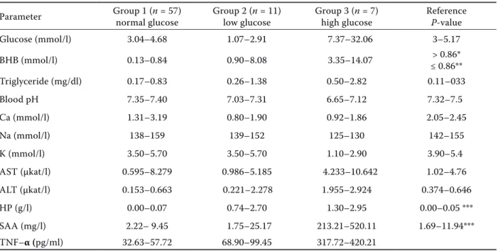
Goats in Group 2 (n = 11) were between days 110–130 of gestation. In these animals anorexia, grinding on the teeth, depression, in some of them blindness, and ketonuria (++) were seen. Three of Table1. Serum biochemical values in 75 pregnant goats with minimum and maximum values
Parameter Group 1 (n = 57) normal glucose Group 2 (n = 11) low glucose Group 3 (n = 7) high glucose Reference P-value
Glucose (mmol/l) 3.04–4.68 1.07–2.91 7.37–32.06 3–5.17 BHB (mmol/l) 0.13–0.84 0.90–8.08 3.35–14.07 ≤ 0.86**> 0.86* Triglyceride (mg/dl) 0.17–0.83 0.26–1.38 0.50–2.82 0.11–033 Blood pH 7.35–7.40 7.03–7.31 6.65–7.12 7.32–7.5 Ca (mmol/l) 1.31–3.19 0.80–1.90 0.92–1.86 2.05–2.45 Na (mmol/l) 138–159 139–152 125–130 142–155 K (mmol/l) 3.50–5.70 3.50–5.70 1.10–2.90 3.90–5.4 AST (µkat/l) 0.595–8.279 0.986–5.185 4.233–10.642 1.02–4.76 ALT (µkat/l) 0.153–0.663 0.221–2.278 1.955–2.924 0.374–0.646 HP (g/l) 0.00–0.07 0.74–2.70 1.30–2.95 0.00–0.05 *** SAA (mg/l) 2.22– 9.45 1.75–25.17 213.21–520.11 1.69–11.94*** TNF–α (pg/ml) 32.63–57.72 68.90–99.45 317.72–420.21
*subclinical pregnancy toxaemia, **healthy animals, ***limit of detection of the assay (Gonzalez et al. 2008)
Table 2. Mean blood biochemical values in healthy, subclinical and clinical pregnancy toxaemia goats
Parameter Group 1 (n = 57) normal glucose Group 2 (n = 11) low glucose Group 3 (n = 7) high glucose P-value
Glucose (mmol/l) 3.71 ± 0.44b 2.13 ± 0.69b 13.01 ± 9.04a < 0.001*** BHB (mmol/l) 0.47 ± 0.58c 2.34 ± 2.29b 6.63 ± 3.44a < 0.001*** Triglyceride (mg/dl) 0.53 ± 0.49c 0.70 ± 0.47b 1.60 ± 0.91a < 0.001*** Blood pH 7.36 ± 0.01c 7.16 ± 0.10b 6.80 ± 0.16a < 0.001*** Ca (mmol/l) 2.15 ± 0.43b 1.48 ± 0.38a 1.14 ± 0.46a < 0.001*** Na (mmol/l) 146.80 ± 5.52b 145.18 ± 4.44b 127.85 ± 1.77a < 0.001*** K (mmol/l) 4.56 ± 0.58b 4.32 ± 0.73b 2.21 ± 0.57a < 0.001*** AST (µkat/l) 1.47 ± 1.07b 2.46 ± 1.30b 7.10 ± 2.39a < 0.001*** ALT (µkat/l) 0.40 ± 0.12c 0.88 ± 0.78b 2.27 ± 0.37a < 0.001*** HP (g/l) 0.01 ± 0.01c 1.20 ± 0.67b 1.86 ± 0.69a < 0.001*** SAA (mg/l) 5.02 ± 2.19b 10.23 ± 6.35b 357.75 ± 105.50a < 0.001*** TNF–α (pg/ml) 46.68 ± 6.45c 84.75 ± 12.33b 352.79 ± 38.66a < 0.001*** a.b.cmean values marked with different superscripts in the same line are significantly different from each other (P < 0.001) ***statistically highly significant
them exhibited more severe clinical signs. Serum BHBA values in three goats were 3.60, 4.54 and 8.08 mmol/l. Caesarean sections were not per-formed on these animals because they were in the recumbent stage and their chance of survival was low. After injecting a combination of dexametha-sone (1 mg/10 kg b.w., i.m.), and dexcloprostenol (125 µl, i.m.)(Lima et al. 2012) two dead foetuses were delivered. Recovery was not seen in three goats. They died 48–72 h after treatment. Other goats in this group were treated with 5–7 g of glucose (i.v.) 6–8 times a day in conjunction with 30–40 units of zinc protamine insulin (i.m.) every other day for three days (Radostits et al. 2008).
Goats in Group 3 (n = 7) were between days 120–140 of gestation and they had shown recum-bency and coma. Ketonuria in these goats was se-vere (+++).
All goats in Group 3 (n = 7) died within 4 h after the farm was visited. In this group all animals car-ried two dead foetuses.
DISCUSSION
Pregnancy toxaemia has a significant economic impact on goat enterprises due to loss of foetuses, veterinary costs, and loss of the dams (Rook 2000). In severe cases, morbidity and mortality rates can reach up to 20% and 80%, respectively (Andrews 1997; Rook 2000).
The last six weeks of gestation in goats (and ewes) are a critical period for the pregnant animal be-cause approximately 80% of the foetal growth oc-curs during this period (Bergman 1993).
Pregnancy toxaemia in small ruminants occurs because of the competition for glucose between the pregnant animals and their foetuses, as the latter undergo intensive growth (Bulgin 2005).
Hypoglycaemia and hyperketonaemia are the primary metabolic disturbances in pregnancy tox-aemia (Radostits et al. 2008). However, hypergly-caemia may also develop. Blood levels of glucose in goats with pregnancy toxaemia vary dramatically and this gave rise to the idea that hypoglycaemia might indicate that the foetuses are alive and hy-perglycaemia that the foetuses are dead (Bulgin 2005; Lima et al. 2012). In this study all goats with high serum glucose concentrations and which were in the last four weeks of the gestation period (Group 3), had two dead foetuses and these results are similar to previous reports. Hyperglycaemia
occurs because foetal death removes the suppress-ing effect of the foetus on hepatic gluconeogenesis (Wastney et al. 1983). Compared with the control (Group 1), glucose values were decreased signifi-cantly (P < 0.001) in Group 2 and only three of them delivered dead foetuses.
The rate of hepatic ketone body production usu-ally increases 4–5 times in sheep during late gesta-tion and in early lactagesta-tion. Under condigesta-tions of an insufficient energy supply (starvation, spontaneous ketosis etc.), alimentary ketogenesis decreases and the rate of hepatic ketogenesis from NEFA dispro-portionately increases. The liver now becomes the main and eventually almost the sole ketogenic or-gan. Such metabolic changes are associated with marked increases in the rate of mobilisation of long chain fatty acids from adipose tissues and a marked rise in circulating concentrations of NEFA and ke-tone bodies. During hyperketonaemia keke-tone bod-ies are readily oxidised serving as fuels for energy production. Many peripheral tissues, like the heart, skeletal muscle, kidney, non-foetal uterine tissues and the lactating mammary gland can harvest large amounts of energy from the oxidation of ketone bodies (Harmeyer and Schlumbohm 2006).
In late gestation, the abdominal space is filled with accumulated fat and an ever-expanding uter-us. Because of the lack of rumen space, these fe-males have difficulty consuming enough feedstuff to satisfy their energy requirements (Pugh 2002). In our study, huge accumulations in fat were observed in the abdominal cavities at the necropsies.
In sheep and goats, pregnancy toxaemia is much more common in highly prolific breeds (Smith and Sherman 1994) such as the Saanen breed goats in this study.
Ewes and goats with BHB concentrations of 0.86–1.6 mmol/l are classified as having mild or subclinical pregnancy toxaemia (Ramin et al. 2005; Bani et al. 2008). According to the serum glucose and BHB concentrations in this study we classified goats as normal (Group 1), exhibiting subclinical pregnancy toxaemia (Group 2), or exhibiting clini-cal toxaemia (Group 3) (Table 1).
In this study all goats with high serum glucose and BHBA had two dead foetuses (Group 3); mean serum glucose and BHBA values were found to be 13.01 ± 9.04 and 6.63 ± 3.44,respectively. Glucose and BHB values were significantly (P < 0.001) higher in the subclinical (Group 2), and clinical (Group 3) pregnancy toxaemia groups than in the normal group.
In goats, it has been reported that ketonaemia leads to a metabolic acidosis and changes in acid base parameters could be used as indicators of early pregnancy toxaemia (Gonzalez et al. 2011). In this study in Group 1 serum BHB levels and blood pH values were within normal ranges; in Groups 2 and 3 serum BHB levels increased and blood pH values then gradually decreased.
Unlike previous findings in goats with subclini-cal pregnancy toxaemia (Bani et al. 2008), serum concentrations of triglycerides and serum activi-ties of AST in goats with subclinical and clinical pregnancy toxaemia were significantly (P < 0.001) increased compared with those of normal animals in this animals. However, the serum activity of AST significantly increased (P < 0.001) in clinical preg-nancy toxaemia goats compared with normal and subclinical pregnancy toxaemia animals.
In human patients with ketoacidosis and ke-tonuria, there is a marked loss of K+ in the urine leading to hypokalaemia (Rose and Post 2001). Hypokalaemia could be explained in part because the goats were not eating and, therefore, their di-etary K+ intake would have been reduced. These two mechanisms, acting together, resulted in hy-pokalaemia (Lima et al. 2012).
Serum potassium concentrations are decreased in ewes with pregnancy toxaemia (Halford and Sanson 1983); in this study in comparison to normal and subclinical pregnancy toxaemia goats, serum po-tassium concentrations progressively decreased in the clinical pregnancy toxaemia group. In ad-dition, serum sodium concentrations in normal and subclinical pregnancy toxaemic goats did not show significant differences, while a significant decrease in goats with clinical pregnancy toxae-mia was observed. Therefore, these parameteres are good indicators of clinical pregnancy toxaemia. On the other hand, serum calcium concentrations in both subclinical and clinic pregnancy toxaemic goats were found to be significantly altered.
The acute phase response (APR) is defined by the secretion of fibrinogen, serum amyloid A (SAA), haptoglobin (Hp) and α-1 acid glycoprotein prin-cipally by the liver (Vels et al. 2009).
It is clear that determination of animal APP values are not only useful for monitoring inflammatory processes for diagnostic and prognostic purposes but also for analysing various non-inflammatory conditions such as pregnancy, parturition, meta-bolic diseases and stress, which have previously been considered as not affecting APP values (Kent
1992). Haptoglobin and serum amyloid A proteins could be used as valuable indicators of inflamma-tion in goats (Gonzalez et al. 2008).
In ruminants the circulating levels of Hp are neg-ligible in normal animals, but increase over 100-fold on immune stimulation (Conner et al. 1998). Hp is induced in cows with fatty liver syndrome (Katoh et al. 2002). Increases in serum haptoglobin values can be a potential indicator of acidosis in goats (Gonzalez et al. 2010). In this study four of six dead goats exhibited acidic ruminal fluids at nec-ropsy. Further, haptoglobin concentrations were increased and blood pH values in goats with sub-clinical and sub-clinical pregnancy toxaemia were de-creased. Previous studies have reported that there is a significant correlation between Hp and BHB in subclinical pregnancy toxaemia in goats (Trevisi et al. 2005; Gonzalez et al. 2011).
SAA concentrations are not affected by the in-duction of pregnancy toxaemia (Gonzalez et al. 2011). In our study, serum concentrations of SAA in normal goats and in animals with subclinical pregnancy toxaemia were not significantly altered but in those with clinical pregnancy toxaemia val-ues were significantly increased (Table 2).
Of the proinflammatory cytokines, tumour ne-crosis factor-alpha (TNF-α) is one of the major me-diators of APP synthesis in the liver (Alsemgeest et al.1996; Yoshioka et al. 2002).
A previous study has shown that in early pregnant ewes, tumour necrosis factor-α and acute-phase pro-teins increased after challenge with peptidoglycan-polysaccharide (Dow et al. 2010). Similar findings were made in this study. A sharp increase was ob-served in serum TNF-α activity for the subclinical and especially clinical pregnancy toxaemia groups.
In conclusion, the blood levels of glucose in goats with pregnancy toxaemia can be a good indicator of the viability of the foetuses. Blood pH values and serum β-hydroxyl butyrate, triglyceride, calcium, ALT, haptoglobin and tumour necrosis factor-alpha values can be a good indicator of subclinical and clinical pregnancy toxaemia. On the other hand, so-dium, potassium, AST, and serum amyloid A values can be used for the diagnosis of clinical pregnancy toxaemia in goats.
REFERENCES
Alsemgeest SP, Van’t Klooster GA, Van Miert AS, Hul-skamp-Koch CK, Gruys E (1996): Primary bovine
hepatocytes in the study of cytokine induced acute-phase protein secretion in vitro. Veterinary Immunol-ogy and ImmunopatholImmunol-ogy 53, 179–184.
Andrews AH (1997): Pregnancy toxemia in the ewe. In Practice 19, 306–312.
Bani ZA, Al-Majali AM, Amireh F, Al-Rawashdeh OF (2008): Metabolic profiles in goat does in late preg-nancy with and without subclinical pregpreg-nancy tox-emia. Veterinary Clinical Pathology 37, 434–437. Bergman EN (1993): Disorders of carbohydrate and fat
metabolism. In: Swenson MJ, Reece WO (eds.): Dukes Physiology of Domestic Animals. Cornell University Press, Ithaca, New York. 492–516.
Brozos C, Mavrogianni VS, Fthenakis GC (2011): Treat-ment and control of peri-parturient metabolic diseases; pregnancy toxemia, hypocalemia, hypomagnesemia. Veterinary Clinics of North America Food Animal Prac-tice 27, 105–113.
Bulgin MS (2005): Pregnancy toxemia. In-depth Review. Wool and Wattles 33, 9–10.
Conner JG, Eckersall PD, Wiseman A, Aitchison TC, Douglas TA (1998): Bovine acute phase response fol-lowing turpentine injection. Research in Veterinary Science 44, 82–88.
Dow TL, Rogers-Nieman G, Holaskova I, Elsasser TH, Dailey RA (2010): Tumor necrosis factor-α and acute-phase proteins in early pregnant ewes after challenge with peptidoglycan-polysaccharide. Domestic Animal Endocrinology 39, 147–154.
Eckersall PD (2000): Recent advances and future pros-pects for the use of acute phase proteins as markers of disease in animals. Revue de Medecine Veterinaire 151, 577–584.
Eckersall PD, Bell R (2010): Acute phase proteins: Bio-markers of infection and inflammation in veterinary medicine. Veterinary Journal 185, 23–27.
Gonzalez FHD, Tacles F, Martinez-Subiela S, Tvarijona-viciute A, Soler L, Ceron JJ (2008): Acute phase protein response in goats. Journal of Veterinary Diagnostic Investigation 20, 580–584.
Gonzalez FHD, Ruiperez FH, Sanchez JM, Souza JC, Martinez-Subiela S, Ceron JJ (2010): Haptoglobin and serum amyloid A in subacute ruminal acidosis in goats. Revista de la Facultad de Medicina Veterinaria y de Zootecnia 57, 159–167.
Gonzalez FHD, Hernandez F, Madrid J, Martinez-Subiela S, Tvarijonaviciute A, Ceron JJ, Tecles F (2011): Acute phase proteins in experimentally induced pregnancy toxemia in goats. Journal of Veterinary Diagnostic Investigation 23, 57–62.
Gruys E, Obwolo MJ, Toussaint MJ (1994): Diagnostic significance of the major acute phase proteins in
vet-erinary clinical chemistry: a review. Vetvet-erinary Bul-letin 64, 1009–1018.
Halford DM, Sanson DW (1983): Serum profiles deter-mined during ovine pregnancy toxemia. Agri-Practice 4, 27–33.
Harmeyer J, Schlumbohm C (2006): Pregnancy impairs ketone body disposal in late gestating ewes: Implica-tions for cytoxemia. Research in Veterinary Science 81, 252–264.
Katoh N, Oikawa S, Oohashi T, Takahashi Y, Itoh F (2002): Decreases of apolipoprotein B-100 and A-1 concentrations and induction of haptoglobin and se-rum amyloid A in nonfed calves. Journal of Veterinary Medical Science 64, 51–55.
Kent JE (1992): Acute phase proteins: their use in vet-erinary diagnosis. British Vetvet-erinary Journal 148, 279–282.
Lima MS, Pascoal RA, Stilwell GT (2012): Glycaemia as a sign of the viability of the foetuses in the last days of gestation in dairy goats with pregnancy toxemia. Irish Veterinary Journal 65, 1. DOI:10.1186/2046-0481-65-1.
Mobini S, Heath AM, Pugh DG (2002): Theriogenology of sheep and goats. In: Pugh DG (ed.): Sheep and Goat Medicine. WB Saunders. An Imprint of Elsevier, Phil-adelphia, Pennsylvania. 129–186.
Navarrei CB, Pugh DG (2002): Diseases of the gastroin-testinal system. In: Pugh DG (ed.): Sheep and Goat Medicine. WB Saunders Company Ltd., London, UK. 69–105.
Pugh DG (2002): Diseases of the gastrointestinal system. In: Pugh DG (ed.), Sheep and goat medicine, WB Saun-ders Company Ltd. London, UK. 129–186.
Radostits OM, Gay CC, Hinchcliff KW, Constable PD (2008): Metabolic diseases. In: Veterinary Medicine: A Textbook of Diseases of Cattle, Sheep, Pigs, Goats and Horses. 10th ed. WB Saunders Company Ltd.,
Lon-don, UK. 1613–1690.
Ramin AG, Asri S, Majdani R (2005): Correlations among serum glucose, beta-hydroxybutyrate and urea con-centrations in non-pregnant ewes. Small Ruminant Research 57, 256–269.
Rook JS (2000): Pregnancy toxemia of ewes, does, and beef cows. Veterinary Clinics of North America Food Animal Practice 16, 293–317.
Rose BT, Post TW (2001): Hypokalemia. In: Clinical Physiology of Acid-Base And Electrolyte Disorders. 5th ed. McGraw-Hill, Medical Publishing Division,
New York. 836–887.
Smith MC, Sherman DM (1994): Nutrition and meta-bolic diseases. In: Goat Medicine. Lea and Febiger,
Trevisi E, D’Angelo A, Gaviraghi A, Noe L, Bertoni G (2005): Blood inflammatory indices in goats around kidding. Italian Journal of Animal Science 4, 404. Vels L, Rontved CM, Bjerring M, Ingvartsen KL (2009):
Cytokine and acute phase protein gene expression in repeated liver biopsies of dairy cows a lipopolysaccha-ride-induced mastitis. Journal of Dairy Science 92, 922–934.
Wastney ME, Wolf JR, Bickerstafe R (1983): Glucose turnover and hepatocyte glucose production of starved
and toxaemic pregnant sheep. Australian Journal of Biological Sciences 36, 271–284.
Yoshioka M, Watanabe A, Shimada N, Murata H, Yok-omizo Y, Nakajima Y (2002): Regulation of haptoglobin secretion by recombinant bovine cytokines in primary cultured bovine hepatocytes. Domestic Animal Endo-crinology 23, 425–433.
Received: 2014–04–14 Accepted after corrections: 2014–08–14
Corresponding Author:
Metin Koray Albay, University of Mehmet Akif Ersoy, Faculty of Veterinary Medicine, Department of Internal Medicine, Istiklal Yerleskesi, 15030 Burdur, Turkey