ORIGINAL ARTICLE
The Feasibility of Hepatic Resections Using a Bipolar
Radiofrequency Device (Habib®)
Osman Civil1&Metin Kement1&Nuri Okkabaz1&
Mustafa Haksal1&Cem Gezen1&Mustafa Oncel1,2
Received: 18 December 2014 / Accepted: 26 May 2015 / Published online: 5 June 2015 # Association of Surgeons of India 2015
Abstract The bipolar radiofrequency device (Habib®) has been recently introduced in order to reduce intraoperative bleeding for a safe hepatic resection as an alternative to the conventional tools. However, indications, perioperative find-ings, and outcome of the device for hepatic resections remain and deserve to be analyzed. The current study aims to analyze the feasibility of the bipolar radiofrequency device (Habib®) for hepatic resections. Information of the patients that underwent hepatic resection using with the Habib® device between 2007 and 2011 was abstracted. Patient, disease, and operation-related findings and perioperative data were inves-tigated. A total of 71 cases (38 [53.5 %] males, mean age was 56.8±11.9) were analyzed. Metastatic disease (n=55; 77.5 %) was the leading indication followed by primary liver and bil-iary malignancies (n=7; 9.9 %), hemangioma (n=5; 7 %), hydatid disease (n=3; 2.8 %), and hepatic gunshot trauma (n=1; 1.4 %). Metastasectomy was the most commonly per-formed procedure (n=31; 56.3 %), but in 24 (77.4 %) cases, it was performed in addition to extended resections. Other pro-cedures in the study patients include segmentectomy in 17, bisegmentectomy in 19, trisegmentectomy in 17, right or left hepatectomy in 8, and extended right/left hepatectomy in 3. The mean (±SD) operation time was 241.7±78.2 min. The median amount of bleeding was 300 cc (range 25–2500),
and 23 (32.4 %) cases required perioperative transfusion. The median hospitalization period was 5 days (range 1–47). Lengthened drainage (n=9, 12.7 %) and intraabdominal ab-scess (n=8, 11.23 %) were the most common problems. He-patic resections using the Habib® device seem to be feasible in cases with primary and metastatic hepatic lesions and benign liver masses and even those with hepatic trauma. It may lessen the amount of intraoperative hemorrhage, although length-ened drainage and intraabdominal abscess were the major postoperative problems in these cases.
Keywords Habib . Radiofrequency device . Hepatectomy . Liver surgery . Blood loss
Introduction
Hepatic resections have been performed more commonly for over the last two decades not only because it has been shown that the outcomes are better after the removal of the tumors but also since more sophisticated devices have been introduced in order to make the hepatic operations safer and more feasible [1–5]. These devices have been mostly generated for helping the surgeon dealing with bleeding, which is probably the most demanding complication during hepatic resections, since it is sometimes challenging to manage hemorrhage occurring from a solid and well-vascularized organ such as liver [4–7]. In addi-tion, more amount of blood loss during resection has been shown to lead to increased morbidity during the postoperative period and even worse survival rates in the long term of follow-up [8,9]. The bipolar radiofrequency device (Habib®) has been re-cently introduced in order to reduce intraoperative bleeding for a safe hepatic resection as an alternative to the convention-al tools such as cavitron ultrasonic aspirator (CUSA), harmon-ic scalpel, bipolar scalpels, and ligature diathermy.
This paper has been participated as a podium presentation to 18th National General Surgery Congress at May, 2012, in Izmir, Turkey * Osman Civil
1
Department of General Surgery, Kartal Education and Research Hospital, Istanbul, Turkey
2
Department of General Surgery, Medipol University Hospital, Istanbul, Turkey
Radiofrequency energy had been initially used in order to damage parenchymal lesions where the resection was not practical. Using the same technology, the Habib® device is advocated to provide a bloodless procedure while not requir-ing additional tyrequir-ing or clipprequir-ing of the large vessels or biliary channels [10]. Besides, others have stated that Habib® leaves a large amount of devascularized tissue within the resection plane, which may lead to necrosis and further septic compli-cations [10–13]. Thus, the indications, perioperative findings, and outcome of the device for hepatic resections remain and deserve to be analyzed. Consequently, the current study aims to evaluate the feasibility of the Habib® device for the resec-tion of various hepatic lesions.
Methodology
Patients that received a liver surgery using a Habib device at our institution between 2007 and 2011 were documented at a prospectively designed database. Institutional Review Board approved the content and design of the study prior to data evaluation (Approval number: B104İSM4340029/1009/14).
Patients were evaluated considering at least two of the fol-lowing diagnostic tools: ultrasonography (USG), computed tomography (CT), and magnetic resonance imaging (MRI). Positron emission tomography (PET) was used in recent years in patients with metastatic tumors. The indication of resection and type of the procedure were generally decided in a multi-disciplinary council, and the operations were performed or supervised by a senior surgeon (MO). After laparotomy was initiated, which was a subcostal incision with an extension to the left side and to the xiphoid process in most cases, the liver was totally mobilized and hepatic hilum was prepared for a possible Pringle maneuver. The same incision was also pre-ferred if a synchronous metastasectomy was aimed in addition
to the removal of the primary tumor, since colonic resections were laparoscopically performed in our department by the same team. In those cases, the colon was extracted via subcostal incision. A midline incision was used in the trauma patient. Complete mobilization of the liver was performed; however, no pedicle dissection and inflow or outflow ligations were necessitated. Hepatic examination using an intraopera-tive ultrasound, which was performed by a radiologist, was the routine practice in patients with metastasis.
Operative Technique The device included a generator (Habib® 4×, Generator 1500×, RITA Medical Systems, Inc., California, USA) creating radiofrequency power, and a hand applicator with four steel needles for transferring the energy directly to the tissue was used. The resection border was marked and the device was introduced into the liver abutting the transection line. The generator was programmed to pro-duce an alert signal when energy delivery had been automat-ically stopped. Then, the probe was gently moved out for a few centimeters, but not taken out of the liver, and energy was re-introduced at the same location. The device was step-by-step introduced through the resection line without leaving a skip area. Then, the resection was initiated with electrocautery through the ablated tissue. Suture ligations were also used for the visible bile ducts or vessels even if they were totally co-agulated (Fig.1). After the resection was completed, the re-gion was routinely drained. These data were prospectively abstracted using a special computer-based program, and the data were presented as percentages, means, and standard de-viations (SDs) or medians and ranges: patient-related informa-tion (demographics, American Society of Anesthesiology [ASA] scores, and associated comorbidities), disease- and procedure-specific data (indication, number, and location of the lesions; type of resections; operation time; amount of bleeding; hospitalization period; and postoperative
Fig. 1 The device was introduced into the liver through the transection line without leaving a skip area (a). Then, the resection was completed with electrocautery through the ablated tissue (b). The specimen (c)
morbidity), and pathological results. Postoperative complica-tions were classified according to the contracted form of Clavien-Dindo Classification. Grades of this classification range from any deviation from the normal postoperative course without the need for any treatment (grade 1) to death (grade 5) [14]. Major liver resection was defined as resection of three or more liver segments.
Results
There were a total of 71 patients (38 [53.5 %] males, mean [±SD] age was 56.8±11.9). The ASA scores were 2, 3, and 4 in 36 (50.7 %), 33 (46.5 %), and 2 (2.8 %) cases, respectively, and hypertension, type 2 diabetes mellitus, and chronic ob-structive pulmonary disease were the associated comorbidities in 23 (32.4 %), 13 (18.3 %), and 5 (7 %) patients, respectively. Twenty-seven (38 %) cases had received chemotherapy be-cause of their primary diseases during the last 6 months. These were the indications for resections: metastatic disease (n=55; 77.5 %), primary hepatic or biliary malignancies (n=7; 9.9 %) (gallbladder cancer [n=3; 4.2 %], hepatocellular carcinoma [n=2; 2.8 %], cholangiocarcinoma [n=1; 1.4 %], and hemangioendothelioma [n=1; 1.4 %]), hemangioma (n=5; 7 %), hydatid disease (n=3; 4.2 %), and hepatic gunshot trau-ma (n=1; 1.4 %) (Table1).
A total of 169 metastases were removed in 55 patients. The metastatic diseases were secondary to colorectal cancer (n= 48; 87.3 %), and breast, ovary, endometrial, renal, and gastric cancers; retroperitoneal sarcoma; and gastrointestinal stromal tumor (n=1 for each, 1.8 %). Intraoperative ultrasound was performed in all metastatic cases. The numbers of metastasis were 1, 2, 3, and 4 or more in 23 (41.8 %), 10 (18.2 %), 12 (21.8 %), and 10 (18.2 %) patients, respectively, reaching an average (±SD) quantity of 2.5±1.8 metastases in each patient. Segment VII was the most common location (n=30), followed by segments VIII (n=29), VI (n=28), and V (n=26). There were also 19, 18, 13, and 6 metastases at segments IV, II, III, and I, respectively. Metastasectomy was the most commonly performed procedure (n=31; 56.3 %), but in 24 (77.4 %) cases, the technique was performed in addition to an extended resection (segmentectomy [n=8], bisegmentectomy [n=6], trisegmentectomy [n=5], right/left hepatectomy [n=3], and extended right/left hepatectomy [n=2]) leaving the isolated metastasectomy operation in only 7 patients. These were the other resection procedures in patients with metastases: bisegmentectomy (n = 14), trisegmentectomy (n = 14), segmentectomy (n=12), right/left hepatectomy (n=5), and ex-tended right/left hepatectomy (n=3). In 23 (32.4 %) patients, synchronous resections of primary tumor were performed, and these were the locations of the resections: rectum (n=9; 39.1 %), sigmoid/left colon (n=6; 26 %), cecum/right colon (n=5; 21.7 %), transverse colon (n=1; 4.3 %), gastric cancer
(n=1; 4.3 %), and retroperitoneal sarcoma (n=1; 4.3 %) (Table2).
There were seven patients with primary biliary malignant tumors. In three cases with gallbladder cancer, two of whom had had a previous laparoscopic cholecystectomy; segments IV and V were resected. Right/left hepatectomy (n=3) or segmentectomy was necessitated in the remaining four pa-tients with other primary biliary malignant tumors. There were two patients (2.8 %) with primary hepatocellular carcinoma. Child-Pugh and MELD scores were 5–5 and 8–9 in these patients, respectively. Signs of portal hypertension were ab-sent in both of them. In those two patients with primary hepa-tocellular carcinoma, right and left hepatectomy was per-formed, respectively. There were five and three patients with hemangioma or hydatid disease, respectively. In these cases, bisegmentectomy (n=3) or segmentectomy (n=3) was neces-sitated. In a blunt trauma patient with severe bleeding from segment III, segmentectomy was performed.
The mean (±SD) operation time was 241.7±78.2 min. The median amount of bleeding was 300 cc (ranged from 25 to 2500), and 23 (32.4 %) cases required perioperative transfu-sion. After the exclusion of 23 cases, who had synchronous metastasectomy with primary tumor resection, the parameters dropped to 215.5±59.9 min, 250 cc (ranged from 25 to 1600), and 9 (18.8 %) cases. Although all patients were prepared for a
Table 1 Demographics and disease characteristics of the study cohort
Characteristics Findings
Demographics
Age (years) 56.8±11.9
Gender (male) (%) 38 (53.5)
Chemotherapy before surgery (%) 27 (38)
Diagnosis (%)
Metastatic disease 55 (77.5)
Colorectal 48 (67.6)
Gynecologic 2 (2.8)
Gastric carcinoma 1 (1.4)
Gastric stromal tumor 1 (1.4)
Renal 1 (1.4)
Breast 1 (1.4)
Malignant mesenchymal tumor 1 (1.4)
Benign diseases 8 (11.3)
Hemangioma 5 (7.0)
Hydatid disease 3 (4.2)
Primary hepatobiliary malignancies 7 (9.9)
Gall bladder cancer 3 (4.2)
Hepatocellular carcinoma 2 (2.8)
Cholangiocellular carcinoma 1 (1.4)
Hemangioendothelioma 1 (1.4)
Pringle maneuver, it was necessitated only in six (8.5 %) cases; all those patients underwent major liver resections. The examination of the specimens revealed that the mean (±SD) volume of the resection materials was 435.4 ± 293.3 cm3. The median hospitalization period was 5 days (ranged from 1 to 47) (Table2).
A total of 37 complications happened in 21 (29.6 %) cases, and re-hospitalization was necessitated in 20 (28.2 %) patients 20.8 ± 13.5 and 13.2 ± 12.0 days after the operation and discharge from the hospital, respective-ly. Lengthened drainage was the most common problem (n=9, 12.7 %) and generally characterized with a bile drainage continuing for a few days and then transforming to a sterile flow of necrosis. The drainage was spontane-ously decreased and stopped in weeks, and further inter-vention was not necessitated in any cases during the ini-tial hospitalization. However, re-hospitalization and endo-scopic sphincterotomy was required in three cases due to lengthened fistula. Intraabdominal abscess was another common complication, observed in eight (11.3 %), and mostly remained asymptomatic or resolved spontaneously. But, re-hospitalization and percutaneous drainage were required in two cases. Wound infection, hepatic failure, and anastomotic leak were observed in five (7.0 %), three (4.2 %), and two (2.8 %) patients, respectively, and all were treated with medical intervention and conservative
approach. These were other complications and all were observed in single cases (1.4 % for each): ileus, gastrotestinal hemorrhage, pulmonary emboli, myocardial in-farction, evisceration, sepsis secondary to catheter infec-tion, acute renal failure, eviscerainfec-tion, and pleural effusion. The mean (±SD) re-hospitalization period was 18.1 ± 15.6 days (Table 3). In our study, 28 (39.4 %) patients underwent major liver resection (Table 2); in these pa-tients, there was no significant difference between the patients who did not receive chemotherapy and those who received chemotherapy with respect to Clavien-Dindo Classification (Table 4). As known, RFA in the central location of the liver may increase injury to biliary tree. In our study, central liver resection was performed in eight (14.5 %) patients. Biliary leakage was seen in two (25 %) of these patients. Even though biliary leakage rate in these patients was higher than other patients (25 vs. 14.9 %), the difference was not significant (p=0.475).
Of the 27 patients who had received preoperative chemo-therapy, 15 (55.6 %) patients received FOLFIRI-bevacizumab 97.1 ± 55.0 days ago; 4 (14.8 %) patients received only bevacizumab 46.7 ± 2.9 days ago; 3 (11.1 %) FOLFOX 110.0±62.4 days ago; 2 (7.4 %) FUVA 90±4.4 days ago; and 1 (3.7 %) FOLFIRI 120 days ago, sunitinib 60 days ago, or 5-FU-taxane 180 days ago. Three (11.1 %) patients who had received FOLFIRI-Bevacizumab experienced lengthened drainage and abscess/collection (n=1) anastomot-ic leak (n=1) and mortality due to heart failure (n=1). Two (7.4 %) patients who received only bevacizumab experienced lengthened drainage (n=1) and mortality due to hepatic failure (n=1), and the patient who received only FOLFIRI had mor-tality due to hepatic failure (n=1). The other patients who had
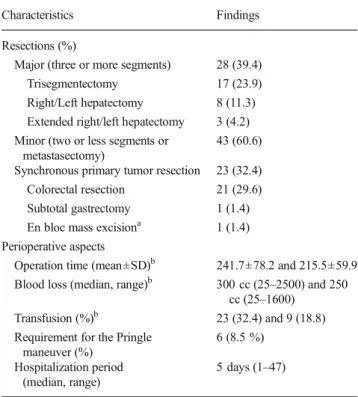
Table 2 Operative characteristics
Characteristics Findings
Resections (%)
Major (three or more segments) 28 (39.4)
Trisegmentectomy 17 (23.9)
Right/Left hepatectomy 8 (11.3)
Extended right/left hepatectomy 3 (4.2)
Minor (two or less segments or metastasectomy)
43 (60.6)
Synchronous primary tumor resection 23 (32.4)
Colorectal resection 21 (29.6)
Subtotal gastrectomy 1 (1.4)
En bloc mass excisiona 1 (1.4)
Perioperative aspects
Operation time (mean±SD)b 241.7±78.2 and 215.5±59.9
Blood loss (median, range)b 300 cc (25–2500) and 250
cc (25–1600)
Transfusion (%)b 23 (32.4) and 9 (18.8)
Requirement for the Pringle maneuver (%) 6 (8.5 %) Hospitalization period (median, range) 5 days (1–47) a
For mesenchymal tumor,bdata include the outcomes obtained from all
patients and those after the exclusion of the cases received synchronous primary tumor resections (n=23)
Table 3 Complications Complications (%) 21 (29.6) Lengthened drainagea 9 (12.7) Abscess/Collection 8 (11.3) Wound infection 5 (7) Hepatic failure 3 (4.2) Anastomotic leak 2 (2.8) Othersb 9 (12.7) Re-hospitalization (%) 20 (28.2) Additional intervention (%) 5 (7) Endoscopic sphincterotomy 3 (4.2) Percutaneous drainage 2 (2.8)
aUsually with biliary leakage,bother
com-plications include ileus, gastrointestinal hemorrhage, pulmonary emboli, myocar-dial infarction, evisceration, sepsis second-ary to catheter infection, acute renal fail-ure, evisceration, and pleural effusion (one case for each event)
received preoperative chemotherapy had not had any complications.
Thirty-day mortality in our study was six (8.5 %) and in-hospital mortality was seven (9.9 %). Of those, two died of postresection liver failure and both were following major re-section in postchemo patients. Three cases, two of which underwent major resection, one after chemotherapy, died due to sepsis related to catheter infection (n=1), leakage from synchronous ileocolic anastomosis (n=1), and intraabdominal abscess (n=1). One patient who underwent major resection for complicated, giant hydatid cyst died on postoperative day 1, subsequent to major intraoperative bleeding that re-quired liver packing. Pulmonary embolism was the cause of mortality in a patient following minor resection for metastasis from previous gynecological malignancy.
Discussion
Hemorrhage is a challenging problem at the time of hepatic resections, and the bipolar radiofrequency device (Habib®) has been used as a possible safe procedure for a decade. The current study evaluates the indication for the use of the Habib® device. Similar to the series of 604 hepatic resections by Habib et al., who was the designer of the device, metastases were the most common indication for the resection, followed by malignant or benign hepatic lesions in the current data [15]. In our opinion, the Habib® device may be used as an assisting tool in any indication for hepatic resection. Besides, to the best to our knowledge, the trauma patient in the current series was the only case presented in the literature for which the Habib® device was used at the time of resection. In our opinion, the device may also be safe and feasible in emergent situations in order to control hemorrhage, where active bleeding continues during the use of the apparatus. Thus, this occasional indica-tion may be well investigated in further analyses.
Patients’ characteristics, extent of resections, and synchro-nous procedures were evaluated in the current study. Twenty-seven (38 %) of patients received chemotherapy prior to the hepatic resection. In addition, almost 40 % (n=28) of cases in this series that received an extended resection may be defined
by at least removal of three segments. Consequently, the av-erage number of 2.5 metastases was resected in metastatic patients, and the mean volume of resection material reached 435.4±293.3 cm3of the liver in all study cases. Finally, one third (n=23) of procedures were synchronous resections with the primary tumors. These characteristics represent a model of non-selected cases in a general or hepatobiliary surgery de-partment, which is likely to make our results more adaptable to routine clinical practice.
The principle advantage using the Habib® device seems to be the reduction in the amount of bleeding. Conventional practices such as finger dissection and clamp crush techniques are believed to be safe during hepatic resections; however, they may be complicated with severe hemorrhage. In a retro-spective study on 1220 cases, the median amount of bleeding was reported to be 1450 and 850 cc in patients operated with the finger dissection technique and ultrasonic dissector (CUSA), respectively [3]. Likewise, in another analysis on 1269 patients, who had resections with the clamp crash tech-nique, the amount of intraoperative hemorrhage was 655 ml [16]. Similar to the previous studies evaluating the outcomes of the Habib® device in hepatic resections that stated that the average bleeding varied between 15 and 487 ml, the median hemorrhage was 300 cc in our cases [15,17,18]. In addition, the Pringle maneuver was necessitated in only 8.5 % of all cases in the current study. This is also compatible to the infor-mation in the literature, which varies between 0 and 26 % among the patients who received Habib®-assisted hepatic re-sections [17,19,20]. Consequently, the current study has sup-ported the hypothesis that the need for transfusion may be decreased with Habib® use. A recent analysis questioning the place for laparoscopic hepatic resections has exposed that more than 25 % of cases require transfusion after conventional hepatic resections [21]. Another series has advocated that al-most half of the cases required transfusion during the periop-erative period [22]. Finally, previous data evaluating the infor-mation regarding Habib® use and a recent review on seven published series analyzing the same issue have revealed that transfusion was necessitated in less than 14 % of cases [15,17,
18]. This is somehow more than the rates in the current study, which were 32.4 and 18.8 % for patients who had synchro-nous primary tumor resections and those who had only hepatic resections, respectively. Although our results are similar to the reports evaluating the outcomes of conventional surgery, they may be considered to be worse than those assessing the con-sequences of Habib® use [21, 22]. Besides, the transfusion rate in our study was similar to one that included conventional hepatic resections as mentioned by a National Surgical Qual-ity Improvement Program [23]. Since the amount of bleeding was reasonable in our patients, this unexpected result even in those who did not receive a synchronous primary tumor re-section was probably related to the general condition of cases in the current study, 38 % of whom had had chemotherapy
Table 4 Complications after major resections in patients who received
chemotherapy and those who did not receive Clavien-Dindo Classification Chemotherapy received (n=13) Chemotherapy not received (n=15) p value I 0 0 0.454 II 2 (15.4) 0 III 0 2 (13.3) IV 1 (7.7) 1 (6.7) V 3 (23.1) 2 (13.3)
prior to the surgical intervention. Thus, we still believe that these results may show that the Habib® device may lessen the amount of bleeding and consequent requirement for the Pringle maneuver.
Operation period may be a significant parameter in patients undergoing hepatic resections. Although some may claim that the use of the Habib® device may lengthen the operation time, since each application lasts approximately 20 s depending on the surrounding vascularity of the tissue, previous informa-tion, as well as the current study, has shown that Habib® use does not have an impact on operation time [18,22].
Hepatic procedures may be associated with several num-bers of problems, and these complications may commonly occur reaching up to 36.5 % of patients as mentioned in a large-volume nationally based study [23]. Although a recent study has advocated that Habib®-assisted resections may be less likely to cause surgical morbidity, we believe that the complication rate after Habib® use is similar to that in con-ventional surgery, as we have reported a complication rate of 29.6 % in the current series. Besides, the characteristics and severity of the complications may vary, probably because most surgery-related problems occur due to the necrotic tissue at the border of resection line that the device has left. Conse-quently, lengthened drainage of pus or bile and abscess occur as the most common problems. Bile leak has been reported to happen between 1.2 and 9.2 % in large series consisting of patients operated with clamp crash and/or ultrasonic dissector [24,25]. Similarly, intraabdominal abscess has been reported to happen up to 12.5 % even in large volume series [2,16,22,
23]. Besides, a recent analysis evaluating 604 patients that received Habib®-assisted hepatectomy exposed that the bile leak and collection/abscess rates were 4.1 and 11.4 %, respec-tively [15]. In these series, 10 and 4 patients necessitated en-doscopic retrograde cholangiography with stenting and percu-taneous transhepatic biliary drainage, respectively [15]. A lengthened drainage was observed in 12.7 % in the current study; however, it is generally characterized with a bile drain-age continuing for a few days, then transforming to a sterile flow of necrosis. In our opinion, this kind of sequence seems to occur due to the necrotic tissue at the border of resection, which is generated after the use of the device. The drainage generally stopped in weeks, and an emergent intervention has never been necessitated during the initial hospitalization in our practice. However, re-hospitalization and endoscopic sphincterotomy were required in three cases, since the fistulas d i d n o t r e s o l v e w i t h c o n s e r v a t i v e m a n a g e m e n t . Intraabdominal abscess was another problem in our cases, and eight (11.3 %) patients suffered from this complication. In contrast to the data by Pai et al., in which most cases re-quired a percutaneous drainage (63 out of 69 subjects), no further intervention was required in our patients [15]. We be-lieve that the higher rates of complications in our series com-pared to the data evaluating Habib® use in the literature may
be due to the policy of our department, which follows exten-sive examinations and diagnostic investigations, and re-hospitalization in case of any uncertainty. Consequently, most cases, which were reported to have a complication in this study, were asymptomatic, and therefore, further interventions were rarely necessitated. Thus, we believe that the higher complication rates in the current study compared to the liter-ature may be secondary to our precise criteria in documenting the complications even in asymptomatic patients. In addition, in our opinion, it may be logical to use the drains at the time of surgery and leave them for a while after Habib®-assisted he-patic resections in order to reduce the rate of symptomatic abscesses and consequent necessity of percutaneous drainage. Finally, we believe that the re-hospitalization rates in the cur-rent study are also high, most likely to be related to elevated complication risks.
The current study has some limitations, mostly occurring from its design. The study had information of patients operat-ed with the Habib® device, and there was no comparison group. Although abstracted prospectively, the data included cases with different diseases making the information highly heterogeneous. Consequently, it was difficult to reach to a reliable conclusion after the analysis of the data. However, since the primary aim of the study was to evaluate the feasi-bility of the device, we believe that the data included informa-tive pieces of evidence regarding the indications and periop-erative outcomes.
It should convey that based on our individual series, Habib’s technique of liver resection in a predominantly non-cirrhotic patient cohort, although technically feasible, con-tinues to be associated with mortality and modest rates of postoperative morbidity with a higher need for re-admission for bile leaks and infective complications. Further prospective/ comparative studies would be required to establish its current place in the surgical armamentarium for liver resection.
Conflict of Interest Osman Civil, Metin Kement, Nuri Okkabaz,
Mustafa Haksal, Cem Gezen, and Mustafa Oncel declare that they have no conflict of interest.
References
1. Belghiti J, Hiramatsu K, Benoist S, Massault P, Sauvanet A, Farges
O (2000) Seven hundred forty-seven hepatectomies in the 1990s: an update to evaluate the actual risk of liver resection. J Am Coll Surg
191:38–46
2. Jarnagin WR, Gonen M, Fong Y, DeMatteo RP, Ben-Porat L, Little
S et al (2002) Improvement in perioperative outcome after hepatic resection: analysis of 1,803 consecutive cases over the past decade. Ann Surg 236:397–406
3. Poon RT, Fan ST, Lo CM, Liu CL, Lam CM, Yuen WK et al (2004)
Improving perioperative outcome expands the role of hepatectomy in management of benign and malignant hepatobiliary diseases:
analysis of 1222 consecutive patients from a prospective database.
Ann Surg 240:698–708
4. Cunningham JD, Fong Y, Shriver C, Melendez J, Marx WL,
Blumgart LH (1994) One hundred consecutive hepatic resections. Blood loss, transfusion, and operative technique. Arch Surg 129:
1050–1056
5. Arnoletti JP, Brodsky J (1999) Reduction of transfusion
require-ments during major hepatic resection for metastatic disease.
Surgery 125:166–171
6. Gozzetti G, Mazziotti A, Grazi GL, Jovine E, Gallucci A,
Gruttadauria S et al (1995) Liver resection without blood
transfu-sion. Br J Surg 82:1105–1110
7. Kooby DA, Stockman J, Ben-Porat L, Gonen M, Jarnagin WR,
Dematteo RP et al (2003) Influence of transfusions on perioperative and long term outcome in patients following hepatic resection for colorectal metastases. Ann Surg 237:860–869
8. Sitzmann JV, Greene PS (1994) Perioperative predictors of
morbid-ity following hepatic resection for neoplasm. A multivariate analy-sis of a single surgeon experience with 105 patients. Ann Surg 219: 13–17
9. Makuuchi M, Takayama T, Gunven P, Kosuge T, Yamazaki S,
Hasegawa H (1989) Restrictive versus liberal blood transfusion policy for hepatectomies in cirrhotic patients. World J Surg 13: 644–648
10. Weber JC, Navarra G, Jiao LR, Nicholls JP, Jensen SL, Habib NA
(2002) New technique for liver resection using heat coagulative necrosis. Ann Surg 236:560–563
11. Navarra G, Ayav A, Weber JC, Jensen SL, Smadga C, Nicholls JP
et al (2005) Short and long term results of intraoperative radiofre-quency ablation of liver metastases. Int J Color Dis 20:521–528
12. Ayav A, Jiao LR, Habib NA (2007) Bloodless liver resection using
radiofrequency energy. Dig Surg 24:314–317
13. Ayav A, Bachellier P, Habib NA, Pellicci R, Tierris J, Milicevic M
et al (2007) Impact of radiofrequency assisted hepatectomy for
reduction of transfusion requirements. Am J Surg 193:143–148
14. Dindo D, Demartines N, Clavien PA (2004) Classification of
sur-gical complications: a new proposal with evaluation in a cohort of
6336 patients and results of a survey. Ann Surg 240(2):205–213
15. Pai M, Frampton AE, Mikhail S, Resende V, Kornasiewicz O,
Spalding DR, Jiao LR, Habib NA (2012) Radiofrequency assisted liver resection: analysis of 604 consecutive cases. Eur J Surg Oncol 38(3):274.80
16. Kyoden Y, Imamura H, Sano K, Beck Y, Sugawara Y, Kokudo N,
Makuuchi M (2010) Value of prophylactic abdominal drainage in 1269 consecutive cases of elective liver resection. J Hepatobiliary
Pancreat Sci 17(2):186–192
17. Wagman LD, Lee B, Castillo E, El-Bayar H, Lai L (2009) Liver
resection using a four-prong radiofrequency transection device. Am Surg 75:991–994
18. Pai M, Spalding D, Jiao L, Habib N (2012) Use of bipolar
radio-frequency in parenchymal transection of liver, pancreas and kidney.
Dig Surg 29:43–47
19. Akyildiz HY, Morris-Stiff G, Aucejo F, Fung J, Berber E (2011)
Techniques of radiofrequency assisted precoagulation in laparo-scopic liver resection. Surg Endosc 25(4):1143–1147
20. Curro G, Bartolotta M, Barbera A, Jiao L, Habib N, Navarra G
(2009) Ultrasound-guided radiofrequency-assisted segmental liver
resection: a new technique. Ann Surg 250:229–233
21. Rao A, Rao G, Ahmed I (2012) Laparoscopic vs open liver
resec-tion for malignant liver disease. Syst Rev Surg 10:194–201
22. Huang ZQ, Xu LN, Yang T, Zhang WZ, Huang XQ, Cai SW et al
(2009) Hepatic resection: an analysis of the impact of operative and perioperative factors on morbidity and mortality rates in 2008
con-secutive hepatectomy cases. Chin Med J 122:2268–2277
23. Worni M, Mantyh CR, Akushevich I, Pietrobon R, Clary BM
(2012) Is there a role for simultaneous hepatic and colorectal resec-tions? A contemporary view from NSQIP. J Gastrointest Surg
16(11):2074–2085
24. Imamura H, Seyama Y, Kokudo N, Maema A, Sugawara Y, Sano K
et al (2003) One thousand fifty-six hepatectomies without mortality
in 8 years. Arch Surg 138:1198–1206
25. Feng ZQ, Huang ZQ, Xu LN, Liu R, Zhang AQ, Huang XQ et al
(2008) Liver resection for benign hepatic lesions: a retrospective analysis of 827 consecutive cases. World J Gastroenterol 14:7247– 7251