DOI: 10.5455/annalsmedres.2020.02.171 2020;27(3):711-6
Evaluation of the expression and proliferation of
degenerative markers in primary cell cultures obtained
from human intervertebral disc tissue
Yasin Emre Kaya1, Hande Akalan2, Ibrahim Yilmaz3, Numan Karaarslan4, Duygu Yasar Sirin5, Hanefi Ozbek6
1Abant Izzet Baysal University, Faculty of Medicine, Department of Orthopaedic and Traumatology, Bolu, Turkey 2Namik Kemal University, Faculty of Arts and Sciences, Department of Molecular Biology and Genetics, Tekirdag, Turkey 3Istanbul MedIpol University, Faculty of Medicine, Department of Medical Pharmacology, Istanbul, Turkey
4Namik Kemal University, Faculty of Medicine, Department of Neurosurgery, Tekirdag, Turkey
5Namik Kemal University, Faculty of Arts and Sciences, Department of Molecular Biology and Genetics, Tekirdag, Turkey 6Istanbul MedIpol University, Faculty of Medicine, Department of Medical Pharmacology, Istanbul, Turkey
Copyright © 2020 by authors and Annals of Medical Research Publishing Inc. Abstract
Aim: A major cause of low back pain is disc degeneration. Nevertheless, no specific and reliable markers of the degeneration of the
nucleus pulposus (NP) are available. This presented study aimed to examine changes in the expressions of genes in primary cell cultures isolated from intact and degenerated tissues to give insights into the biopathogenesis of intervertebral disc (IVD) tissue.
Material and Methods: Tissues of eight patients (n = 8; average age: 41.74 ± 9.86 years) were resected through microdiscectomy,
and primary cell cultures were prepared using degenerated disc tissue. The cultured degenerated tissues served as the study group. The samples in the control group comprised the intact tissues of patients (n = 8; average age: 38.68 ± 7.91 years) resected following a trauma. Morphology of the cell surface were evaluated using an inverted light/fluorescent microscopy at 0 and 24 h on days 10 and 21. The expressions of the chondroadherin (CHAD), cartilage oligomeric matrix protein (COMP), interleukin-1 beta (IL-1 beta), and matrix metalloproteinase (MMP)-7 and MMP-19 genes were evaluated using the reverse transcription-quantitative polymerase chain reaction (RT-qPCR). The data obtained were statistically analyzed.
Results: The four genes investigated, except COMP (P > 0.05), changed significantly in primary cell cultures isolated from degenerative
IVD tissues. This result was statistically significant (P < 0.05). The gene expressions in the samples derived from intact IVD tissues changed markedly and these changes were associated with proliferation (P < 0.05).
Conclusion: Analyzing the changes in gene expression levels associated with IVD should contribute to future studies on the
prevention and treatment of such pathologies. The data obtained from the present study will shed light on cellular-based personal targeted therapies through which genetic information can be transmitted to cells.
Keywords: CHAD; COMP; degenerative markers; IL-1β; intervertebral disc; MMP
Received: 02.03.2020 Accepted: 10.03.2020 Available online: 10.03.2020
Corresponding Author: Numan Karaarslan, Namik Kemal University, Faculty of Medicine, Department of Neurosurgery, Tekirdag,
Turkey E-mail: [email protected]
INTRODUCTION
The intervertebral disc (IVD) provides the mobility, load-bearing capacity, and flexibility of the spine. The IVD may undergo morphological changes with a person’s age or if it gets damaged owing to trauma (1,2).
The IVD, is the largest avascular structure in the human body and known to have low cell content. IVD cells experience a significant level of mechanical stress and must therefore adapt to these novel conditions (3). Mechanoproteins regulates this adaptation by converting mechanical signals into cellular response. Therefore, these proteins modify the gene expression of cells (4).
Modalities based on synthetic materials or composite implants are widely used in the treatment of IVD, but unfortunately these materials do not interact with the biological components of the disc necessary for a complete recovery. Cell-integrated materials or biological substances allowing cell-material interactions and remodeling have also been used in the treatment of IVD (5,6). Together with the current conservative and surgical treatment methods, the use of bioengineering-based therapy, a promising alternative for the clinical treatment of IVD, has markedly increased. However, satisfying results have not yet been achieved (5). Discectomy and surgical fusion are mainly palliative and may not regenerate the
baseline motion and mechanical load-bearing features of the IVD, especially if fused (6). Artificial replacements for the NP generate complications owing to the swelling or migration of the implants that are loosely integrated with the adjacent structures (5-7). The anatomical changes in the IVD result in nerve root compression, narrowing of the spinal canal, and facet joint compression that may cause painful symptoms and neurological deficits (7). Unsatisfying outcomes markedly decrease the quality of life of patients and impose a significant economic burden on the healthcare system.
To slow down or reverse the progression of IVD degeneration, researchers have focused on cellular studies that aim to provide understandings into the molecular mechanisms of the disc. The molecular mechanism of the autoimmune reaction induced by the NP exposed after a disc herniation remains uncertain. Whether certain key cytokines trigger the inflammatory cascade and support angiogenesis in the degenerative process of the IVD remains unknown and needs to be elucidated (8,9).
The present study aimed to evaluate the gene expressions of markers, that is, chondroadherin gene (CHAD), cartilage oligo matrix protein (COMP), interleukin-1 beta (IL-1β), matrix metalloproteinase (MMP)-7, and MMP-19, in primary cell cultures prepared from degenerated and intact IVD tissues.
MATERIAL and METHODS
Approval of Local Ethics CommitteeBefore starting this presented research approval of the local ethics committee (Istanbul Medipol University), and consent forms were achieved from the participants. Study Design
NP samples were collected from two groups of patients: a degenerated disc group (n = 8) undergoing microdiscectomy and fusion with significant signs of disc degeneration (10) and a trauma control group (n = 8) undergoing anterior vertebral body and disc excision and fusion without signs of disc degeneration (11). Lumbar spine magnetic resonance imaging of NP tissues was performed to assess the severity of disc degeneration in all samples. Degenerated samples were also divided into groups according to the Pfirrmann grading system to elucidate the correlation between the severity of degeneration (12) and gene transcriptional levels.
The correlation between the mRNA levels of these genes and cell proliferation was also tested. Reverse transcription-quantitative polymerase chain reaction (RT-qPCR) was performed to determine the mRNA expression levels of these genes as well as those of CHAD, COMP, IL-1β, MMP-7, and MMP-19.
Preparation of Primary Cell Cultures
Tissues obtained from two different anatomic regions were placed into Falcon tubes containing 5% penicillin-streptomycin/amphotericin and Dulbecco’s modified eagle
medium under appropriate conditions (13). The tissues in the flow cabinet were irrigated by a sterile phosphate buffer saline solution, and red blood cells were cleaned (13). The samples were degraded mechanically and then incubated overnight after the addition of Clostridium
histolyticum based collagenase type I (475 µg/mL) and
type II (125 µg/mL) enzymes, which were solubilized in Hank’s balanced salt solution (13). The degraded tissues were centrifuged at 4°C and 1,300 rpm for 10 min (13). Cell pellets were resuspended using the Dulbecco’s modified Eagle’s culture medium (13). Obtained cell pellets were transferred to flasks and incubated for 72 h (13). The degenerated and healthy samples of NP/AF cells obtained from humans were transferred into wells and fed for 21 days. Molecular analyses of primary cell cultures were performed at 0 h and 24 h on days 10 and 21.
Molecular Analyses
To determine cell viability, acridine orange (AO)/propidium iodide (PI) dyes were used (14). AO stains all nucleated cells whether they are alive or dead and can be observed green under fluorescent microscope, PI stains only dead nucleated cells with poor membrane integrity and produces red fluorescence. A fluorescent microscope was used for AO/PI staining.
Cell viability, toxicity, and proliferation analyses were performed using 3-(4,5-dimethylimidazole-2-yl)-2,5-diphenyltetrazolium bromide (MTT) and enzyme-linked immunosorbent assay (ELISA). MTT assays were performed using a commercial kit (Vybrant MTT Cell Proliferation Assay, Cat. no. V-13154; Cell Biolabs, USA) (15).
RNA isolation was carried out, and the amount of RNA obtained from all cultures was measured. Reverse transcription-polymerase chain reaction (RT-PCR), a procedure that integrates the reverse transcription of RNA into DNA and the amplification of specific DNA targets using PCR, was performed (16). To obtain cDNA, 50 ng of RNA was reverse transcribed with a High-Capacity cDNA Reverse Transcription Kit (Thermo Fisher Scientific, Cat#4368814) using a thermal cycler (ProFlex, Thermo Fisher Scientific).
Statistical Analyses
Time-dependent changes in gene expression were presented as a fold change. An analysis of variance (ANOVA) test was performed to test for significant differences across the group means. Nonparametric data were evaluated with a Kruskal-Wallis H test. The correlation between the proliferation and the gene expression was evaluated with Pearson’s correlation coefficient. Descriptive statistics were presented as the mean ± standard deviation (M ± SD). The alpha significance value was accepted as <0.05.
RESULTS
Healthy and viable cells were observed in cultures isolated from IVD tissues during microscopical analyses (Figure 1).
MTT and RT-qPCR assays revealed significant changes in the four genes investigated, except COMP, in the primary cell cultures prepared from degenerative IVD tissues (P < 0.05) (Table 1).
Figure 1. Cell surface morphologies of primary cell cultures
prepared from intact and degenerative IVD tissues were evaluated by an inverted light/fluorescent microscopy. Viable and dead cells produced green and red light, respectively, on day 21 The changes in CHAD (r = -0.346; P = 0.161), IL-1β (r = 0.749; P = 0.000), MMP-7 (r = -0.483; P = 0.049), and MMP-19 (r = 0.872; P = 0.000) gene expressions and proliferation were statistically significant in the cell cultures isolated from degenerative tissues. However, the correlation between COMP (r = -0.490; P = 0.061) expression and proliferation was not statistically significant. The correlation between the expressions and proliferations of the five genes analyzed was statistically significant (P < 0.05).
DISCUSSION
Many studies conducted in the field of genetics and biotechnology have provided alternative methods for the diagnosis and treatment of diseases (5,13,17). These studies, which investigated the degeneration of cells and surrounding microenvironmental structures using cell cultures isolated from IVD tissue, suggested that the transmission of preventive genetic information into the cell may be a promising method for the treatment of degeneration (5).
The investigation of differently expressed genes constitutes a pivotal step to realize targeted drug discovery through functional genomics and pharmacogenomics. The differences in the expression levels of genes between normal and diseased tissues provide considerable clues to detect disease pathogenesis. Analyzing changes in gene expression levels associated with the disease may contribute to the discovery of further treatment and diagnosis methods (5).
Despite conservative and surgical treatment modalities, IVD disorders still induce back and neck pain; as a result, they have adverse effects on individuals and healthcare costs. The present study aimed to evaluate the changes in some markers as well as the proliferation in the primary cell cultures prepared from intact and degenerative IVD tissues. The main goal was to elucidate the physiopathology of IVD disorders that leads to a significant decrease in quality of life and provide comprehensive insights for future treatment strategies.
Cytokines are small secreted proteins that play a crucial role in natural and adaptive immunity and provide a cell-mediated immunity (8,9,12-16,18). The relevant proteins currently find clinical use as biological response modifiers in the treatment of various disorders (19). ILs are a family of secreted cytokines that act as mediators between leukocytes. IL-1 can be produced by many cell types, including monocytes, macrophages, fibroblasts, epithelial
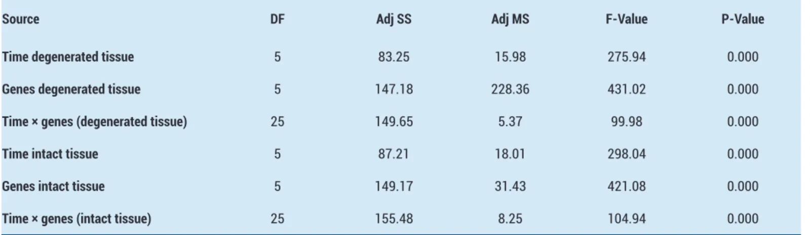
Table 1. Analysis of variance was performed in a 95% confidence interval between degenerated and intact groups
Source DF Adj SS Adj MS F-Value P-Value Time degenerated tissue 5 83.25 15.98 275.94 0.000
Genes degenerated tissue 5 147.18 228.36 431.02 0.000
Time × genes (degenerated tissue) 25 149.65 5.37 99.98 0.000
Time intact tissue 5 87.21 18.01 298.04 0.000
Genes intact tissue 5 149.17 31.43 421.08 0.000
Time × genes (intact tissue) 25 155.48 8.25 104.94 0.000
cells, endothelial cells, endothelin, and astrocytes (8,9,12-16,18). IL-1β is a local inflammatory mediator and is expressed at low concentrations. IL-1β, an inflammatory cytokine, also stimulates an acute inflammation by acting on endothelial cells and leukocytes (19) and plays a pivotal role in the healing process after IVD injury (8,9).
The expressions of hypoxia-inducible factor 1-alpha (HIF-1α) and MMP-3 upregulate IVD cells under hypoxic conditions. IL-1β upregulates the expression of pro-inflammatory cytokines and MMP-3, a disc degradation factor, in IVD cells. The application of antibodies as a treatment against IL-1β decreases the expression of VEGF and MMP-3. Studies have suggested that inflammation, chemotaxis, matrix degradation, and angiogenesis after disc herniation are affected by hypoxic conditions and regulated by some molecules, including IL-1β (8,9). The COMP gene encodes the cartilage oligomeric matrix protein present in the extracellular matrix (ECM) of the cells that make up ligaments and tendons and the near cartilage-forming cells. Ishii et al. studied the expression and distribution features of COMP using rat IVD. They reported that the mutation of COMP was known to result in skeletal dysplasia with characteristic platyspondyly; still, they did not clarify the expression and distribution of COMP in the spine and IVD. This study concluded that COMP is expressed at both the protein and mRNA levels in both the AF and NP of the lumbar spine and tail IVD (20). Another study conducted in the same year suggested that the remodeling of the matrix occurs in response to several factors and that the breakdown of aggrecan by the members of the ADAMTS (a disintegrin-like and metalloprotease with thrombospondin motifs) constitutes an early stage in a disease, and the degradation of molecules is important to secure the collagen network (21). COMP plays a significant role in cell growth and proliferation as well as in the regulation of cell movement and attachment (22). It may also be involved in the pathogenesis of osteoarthritis and serve as a potent suppressor of apoptosis in primary chondrocytes, intervertebral disc cells, and transformed cells (22).
COMP, which is present in the ECM as an integral part of ligaments and tendons, is known to play a crucial role in cellular proliferation and apoptosis as well as in the regulation of cell movement and attachment. COMP is highly expressed in AF cells (23). Osteoarthritis (OA) is a clinical syndrome caused by the degeneration of cartilage (24). COMP is believed to be found in the cartilage during articular degeneration or OA. However, studies have not yet fully clarified the involvement of COMP in IVD disorders (24). A study reported that COMP was not expressed in AF and NF cells (25). COMP is known to function in cross-linking with extracellular matrix elements (14,15). MMP-19 is an enzyme that cleaves COMP (14). Kaplan et al. (15) suggested that COMP is mainly found in the cartilage as an ECM protein and that it is a new biomarker involved in spine disc space narrowing, osteophytes, scoliosis, and joint metabolism.
CHAD, a constantly expressed NP-specific marker, is known to be associated with the development of the spinal cord and dorsal column (26,27). Studies comparing the expression of CHAD, COMP and MMP-19 examined that CHAD expression was associated with inadequately developing microenvironment (26,27). CHAD and COMP secreted from NP/AF cells plays a role in organization of ECM and contribute to the formation of a healthy microenvironment in the IVD tissue (26,27).
The primary role of cleaved/activated MMP is to degrade the ECM proteins, including casein, gelatin, fibronectin, and proteoglycan (12). Moreover, the changes in the gene expressions of MMP-13 and MMP-19, which play significant roles in many biological processes, including embryogenesis, tissue restructuring, wound healing, and angiogenesis, were examined (15).
Inflammatory cytokine IL-1β expression increases in degenerate IVD tissue. The increase of IL-1β expression results in MMP-7 and MMP-13 enhancement. Consequently, we have considered the gene expressions of MMP-7 and MMP-13 as well as IL-1β, even if they are related to the degenerated cell morphology which may be indicative of IVD degeneration (14).
Baptista et al. (28) reported that the sole presence of the studied molecules, including IL-1β and MMP-1, -2, and -3, in the IVD might not be accepted as a pathological condition. This study revealed that the expressions of remodeling enzymes and inflammatory mediators were almost the same in different vertebral segments and disc regions that generate a common degenerated pattern, whereas neurotrophins had a slightly higher expression in cervical discs. The authors suggested that the disc remodeling in distinct segments took a similar pathway that can be mediated to prevent structural failure.
In the present study, the four genes investigated, except COMP (P > 0.05), changed significantly in primary cell cultures isolated from degenerative IVD tissues. This result was statistically significant (P < 0.05). The changes in CHAD, IL-1β, MMP-7, and MMP-19 gene expressions were associated with proliferation in cell cultures isolated from intact IVD tissues (P < 0.05). This result suggests that changes in the four relevant genes’ expressions may be a risk factor for degenerative disc diseases.
This presented study has some limitations. Analyses were performed in vitro. Therefore, the results obtained may not be directly applicable in clinical settings. Further, the cell cultures were obtained from a small number of patients who were the entire same race.
CONCLUSION
The involvement of five NP markers in degenerative disc diseases was investigated. Of the analyzed markers, the results obtained from four were statistically significant. The expressions of CHAD, IL-1β, MMP-7, and MMP-19 may provide insights into the severity of disc degeneration.
The findings obtained may contribute to the clinical application of cell-based technology in IVD treatment. Despite the common belief that the IVD shows little to no capacity for recovery following injury, the IVD can be regenerated and restored by better understanding the cell biology and degeneration mechanisms.
Competing interests: The authors declare that they have no competing interest.
Financial Disclosure: There are no financial supports.
Ethical approval: This study was performed with the approval of the local ethics committee (Istanbul Medipol University).
Yasin Emre Kaya ORCID: 0000-0002-5412-8355 Hande Akalan ORCID: 0000-0002-5922-2498 Ibrahim Yilmaz ORCID: 0000-0003-2003-6337 Numan Karaarslan ORCID: 0000-0001-5590-0637 Duygu Yasar Sirin ORCID: 0000-0002-1224-442X Hanefi Ozbek ORCID: 0000-0002-8084-7855
REFERENCES
1. Patil P, Niedernhofer LJ, Robbins PD, et al. Cellular senescence in intervertebral disc aging and degeneration. Curr Mol Biol Rep 2018;4:180-90.
2. Dudli S, Ferguson SJ, Haschtmann D. Severity and pattern of post-traumatic intervertebral disc degeneration depend on the type of injury. Spine J 2014;14:1256-64.
3. Jiang L, Yuan F, Yin X, et al. Responses and adaptations of intervertebral disc cells to microenvironmental stress: a possible central role of autophagy in the adaptive mechanism. Connect Tissue Res 2014;55:311-21.
4. González Martínez E, García-Cosamalón J, Cosamalón-Gan I, et al. Biology and mechanobiology of the intervertebral disc. Neurocirugia (Astur) 2017;28:135-40.
5. Karaarslan N, Sirin DY, Yilmaz I, et al. Promising molecular developments in spinal surgery. Van Medical Journal 2018;25:496-501.
6. Bowles RD, Setton LA. Biomaterials for intervertebral disc regeneration and repair. Biomaterials 2017;129:54-67.
7. Kaplan N, Kasim HBF, Ozger O, et al. Complications of 200 cervical anterior surgery cases and the management of these complications in light of the literature. Ann Med Res 2019;26:1890-5.
8. Hsu YH, Lin RM, Chiu YS, et al. Effects of IL-1β, IL-20, and BMP-2 on intervertebral disc inflammation under hypoxia. J Clin Med 2020;9:140.
9. Huang KY, Hsu YH, Chen WY, et al. The roles of IL-19 and IL-20 in the inflammation of degenerative lumbar spondylolisthesis. J Inflamm (Lond) 2018;15:19.
10. Somay H, Karaarslan N. Sequestrectomy or microdiscectomy in patients with lumbar disc herniation. Ann Med Res 2019;26:753-8.
11. Akyuva Y, Kaplan N, Yilmaz I, et al. Delivering growth factors through a polymeric scaffold to cell cultures containing both nucleus pulposus and annulus fibrosus. Turk Neurosurg 2019;29:180-93.
12. Kaya YE, Karaarslan N, Sirin DY, et al. Investigation of the effects of methylphenidate, an amphetamine derivative, on intervertebral disc tissue cell cultures and matrix structures. Turk Neurosurg 2019;29:734-42.
13. Karaarslan N, Yilmaz I, Ozbek H, et al. Are specific gene expressions of extracellular matrix and nucleus pulposus affected by primary cell cultures prepared from intact or degenerative intervertebral disc tissues? Turk Neurosurg 2019;29:43-52.
14. Akgun FS, Sirin DY, Yilmaz I, et al. Investigation of the effect of dipyrone on cells isolated from intervertebral disc tissue. Exp Ther Med 2019;18:216-24.
15. Kaplan N, Karaarslan N, Yilmaz I, et al. Are intervertebral disc tissue cells damaged when attempting to prevent thrombus formation using dabigatran, a new oral anticoagulant? Turk Neurosurg 2019;29:470-7.
16. Karaarslan N, Yilmaz I, Ozbek H, et al. Are radio-contrast agents commonly used in discography toxic to the intact intervertebral disc tissue cells? Basic Clin Pharmacol Toxicol 2019;124:181-9.
17. Harrison RG. Observations on the living developing nerve fibers. Proc Soc Exp Biol Med 1907;4:140-3. 18. Gorth DJ, Shapiro IM, Risbud MV. Transgenic mice
overexpressing human TNF-α experience early onset spontaneous intervertebral disc herniation in the absence of overt degeneration. Cell Death Dis 2018;10:7.
19. Caliskan T, Sirin DY, Karaarslan N, et al. Effects of etanercept, a tumor necrosis factor receptor fusion protein, on primary cell cultures prepared from intact human intervertebral disc tissue. Exp Ther Med 2019;18:69-76.
20. Ishii Y, Thomas AO, Guo XE, et al. Localization and distribution of cartilage oligomeric matrix protein in the rat intervertebral disc. Spine (Phila Pa 1976) 2006;31:1539-46.
21. Feng H, Danfelter M, Strömqvist B, et al. Extracellular matrix in disc degeneration. J Bone Joint Surg Am 2006;88:25-9.
22. Karaarslan N, Yilmaz I, Sirin DY, et al. Do we damage nucleus pulposus tissue while treating cerebrovascular ischemic neurological deficits with nimodipine? Ann Med Res 2018;25:266-73.
23. Rutges J, Creemers LB, Dhert W, et al. Variations in gene and protein expression in human nucleus pulposus in comparison with annulus fibrosus and cartilage cells: potential associations with aging and degeneration. Osteoarthritis Cartilage 2010;18:416-23.
24. Yee A, Lam MP, Tam V, et al. Fibrotic-like changes in degenerate human intervertebral discs revealed by quantitative proteomic analysis. Osteoarthritis Cartilage 2016;24:503-13.
25. Van den Akker GG, Surtel DA, Cremers A, et al. Novel immortal cell lines support cellular heterogeneity in the human annulus fibrosus. PLoS One 2016;11:0144497. 26. Karaarslan N, Yilmaz I, Sirin DY, et al. Does pregabalin
used in the treatment of neuropathic pain damage intervertebral disc tissue? Exp Ther Med 2018;16: 1259-65.
27. Lv FJ, Peng Y, Lim FL, et al. Matrix metalloproteinase
12 is an indicator of intervertebral disc degeneration co-expressed with fibrotic markers. Osteoarthritis Cartilage 2016;24:1826-36.
28. Baptista JS, Traynelis VC, Liberti EA, et al. Expression of degenerative markers in intervertebral discs of young and elderly asymptomatic individuals. PLoS One 2020;15:0228155.