Selection signatures in goats reveal copy
number variants underlying breed-defining
coat color phenotypes
Jan HenkelID1,2, Rashid Saif1,3, Vidhya Jagannathan1,2, Corinne Schmocker1,
Flurina Zeindler4, Erika Bangerter5, Ursula Herren5, Dimitris Posantzis6, Zafer BulutID7, Philippe Ammann8, Cord Dro¨ gemu¨ llerID1,2, Christine Flury4, Tosso LeebID1,2*
1 Institute of Genetics, Vetsuisse Faculty, University of Bern, Bern, Switzerland, 2 DermFocus, University of Bern, Bern, Switzerland, 3 Institute of Biotechnology, Gulab Devi Educational Complex, Lahore, Pakistan, 4 School of Agricultural, Forest and Food Sciences, Bern University of Applied Sciences, Zollikofen, Switzerland, 5 Swiss Goat Breeding Association, Zollikofen, Switzerland, 6 Attica Zoological Park, Spata, Greece, 7 Department of Biochemistry, Faculty of Veterinary Medicine, Selcuk University, Konya, Turkey, 8 ProSpecieRara, Basel, Switzerland
Abstract
Domestication and human selection have formed diverse goat breeds with characteristic phenotypes. This process correlated with the fixation of causative genetic variants control-ling breed-specific traits within regions of reduced genetic diversity, so called selection sig-natures or selective sweeps. Using whole genome sequencing of DNA pools (pool-seq) from 20 genetically diverse modern goat breeds and bezoars, we identified 2,239 putative selection signatures. In two Pakistani goat breeds, Pak Angora and Barbari, we found selec-tion signatures in a region harboring KIT, a gene involved in melanoblast development, migration, and survival. The search for candidate causative variants responsible for these selective sweeps revealed two different copy number variants (CNVs) downstream of KIT that were exclusively present in white Pak Angora and white-spotted Barbari goats. Several Swiss goat breeds selected for specific coat colors showed selection signatures at the ASIP locus encoding the agouti signaling protein. Analysis of these selective sweeps revealed four different CNVs associated with the white or tan (AWt), Swiss markings (Asm), badger-face (Ab), and the newly proposed peacock (Apc) allele. RNA-seq analyses on skin samples from goats with the different CNV alleles suggest that the identified structural variants lead to an altered expression of ASIP between eumelanistic and pheomelanistic body areas. Our study yields novel insights into the genetic control of pigmentation by identifying six function-ally relevant CNVs. It illustrates how structural changes of the genome have contributed to phenotypic evolution in domestic goats.
Author summary
Domestic animals have been selected for hundreds or sometimes even thousands of years for traits that were appreciated by their human owners. This process correlated with the a1111111111 a1111111111 a1111111111 a1111111111 a1111111111 OPEN ACCESS
Citation: Henkel J, Saif R, Jagannathan V, Schmocker C, Zeindler F, Bangerter E, et al. (2019) Selection signatures in goats reveal copy number variants underlying breed-defining coat color phenotypes. PLoS Genet 15(12): e1008536.
https://doi.org/10.1371/journal.pgen.1008536
Editor: Gregory P. Copenhaver, The University of North Carolina at Chapel Hill, UNITED STATES
Received: August 12, 2019 Accepted: November 23, 2019 Published: December 16, 2019
Peer Review History: PLOS recognizes the benefits of transparency in the peer review process; therefore, we enable the publication of all of the content of peer review and author responses alongside final, published articles. The editorial history of this article is available here:
https://doi.org/10.1371/journal.pgen.1008536
Copyright:© 2019 Henkel et al. This is an open access article distributed under the terms of the
Creative Commons Attribution License, which permits unrestricted use, distribution, and reproduction in any medium, provided the original author and source are credited.
Data Availability Statement: All relevant data are within the manuscript and its Supporting Information files.
fixation of causative genetic variants controlling breed-specific traits within regions of reduced genetic diversity, so called selection signatures or selective sweeps. We conducted a comprehensive screen for selection signatures in 20 phenotypically and genetically diverse modern goat breeds and identified a total of 2,239 putative selection signatures in our dataset. Follow-up experiments on selection signatures harboring known candidate genes for coat color revealed six different copy number variants (CNVs). Two of these CNVs were located in the 3’-flanking region ofKIT and associated with a completely
white coat color phenotype in Pak Angora goats and a white-spotted coat color phenotype in Barbari goats, respectively. The other four CNVs were located at theASIP locus. They
were associated with four different types of coat color patterning in seven Swiss goat breeds. Their functional effect is mediated by region-specific quantitative changes inASIP
mRNA expression. Our study illustrates how structural changes of the genome have con-tributed to phenotypic evolution in domestic goats.
Introduction
Goat domestication started around 10,000 years ago in the fertile crescent and is believed to be one of the earliest domestication events of livestock animals [1,2]. Bezoars, the wild ancestors of domestic goats are an extant species with a distribution in Western Asia from Turkey to Pakistan. Since domestication, goats followed the human migration [3] and played an eco-nomically important role for their owners by providing various products like milk, meat or fibers. These economical values were further increased by production-orientated breeding, which led to more than 600 diverse goat breeds at present time [4–6].
Artificial selection of domesticated goats not only resulted in specialized elite breeds for milk, meat or fibers, but also in breeds with unique coat color phenotypes [4,7]. Due to their striking appearance, these goat breeds are of special value to their owners, selected for uniform coat color, and kept in closed populations. Coat color phenotypes are one of the most inten-sively studied traits in goats [8–12]. They include solid colored animals of different color, ani-mals with symmetrical color patterns, and aniani-mals with white markings, white spotting phenotypes or completely white animals.
White markings, white spotting and completely white phenotypes typically result from a lack of melanocytes in the skin and hair follicles. This group of phenotypes is also termed leu-cism or piebaldism and characterized by defects in melanoblast development or migration [13–17].
Very light coat colors resembling white are also seen in animals that have a normal set of melanocytes synthesizing a very pale pheomelanin [18]. Melanocytes produce two types of ments, the brown to black eumelanin and the red to yellow pheomelanin. The so-called pig-ment type switching, an intensively studied signaling process, governs whether a given melanocyte produces eumelanin or pheomelanin [19]. Eumelanin is produced, if MC1R is activated by its ligandα-melanocyte stimulating hormone (α-MSH), while pheomelanin is produced ifα-MSH is absent and/or outcompeted by binding of the competitive antagonist ASIP to MC1R [20–25]. Different alternative promoters of theASIP gene enable spatially and
temporally regulatedASIP expression, which results in characteristic patterns of eumelanin
and pheomelanin synthesis [25–28].
Domestication and artificial selection correlated with the fixation of causative genetic vari-ants controlling breed-specific traits within regions of reduced genetic diversity, so called selection signatures or selective sweeps [29–31]. A method detecting regions of low
Funding: This study was funded by a grant from the Swiss National Science Foundation
(31003A_172964). R.S. was supported by a Swiss Government Excellence Scholarship and a supplementary grant from the Hans Sigrist Foundation. The funders had no role in study design, data collection and analysis, decision to publish, or preparation of the manuscript.
Competing interests: The authors have declared that no competing interests exist.
heterozygosity from sequence data of pooled individuals (pool-seq) was developed and used to identify loci under selection in chicken, pigs, rabbits and the Atlantic herring [32–35].
Recently, pooled heterozygosity scores were also applied to monitor loci under selection in goats [36].
In the present study, we aimed to gain a better understanding of the genetic variants deter-mining breed-specific coat color phenotypes in goats. We therefore performed a comprehen-sive screen for selection signatures in bezoars and 20 breeds of domesticated goats.
Results
Selection signature analysis
For the present study, we selected 8–12 animals each from 20 phenotypically diverse domesti-cated goat breeds and their wild ancestor, the bezoar. We isolated genomic DNA from these animals and prepared equimolar DNA pools for sequencing (Table 1).
We obtained 2x150 bp paired-end sequence data corresponding to 30x genome coverage per pool, called high confidence SNVs and calculated pooled heterozygosity scores (−ZHp)
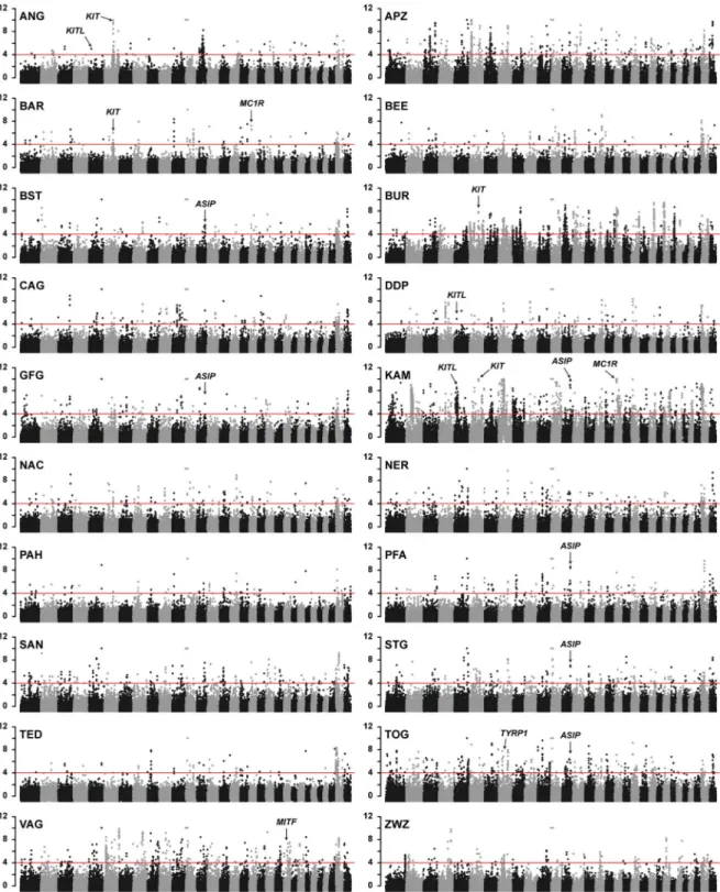
in 150 kb sliding windows with 75 kb step size (S1 Fig;S1andS2Tables). The significance threshold was conservatively set at−ZHp� 4, which identified 5,220 windows with extremely
reduced heterozygosity (0.8% of all windows). Overlapping windows were further merged into 2,239 selection signatures (1.1% of total genomic length). This corresponded to 112 selection signatures per breed pool on average (median = 81;S3 Table).
To evaluate the validity of the pool-seq approach, we compared the results from pool-seq data to individual whole genome sequence data of 120 goats from five different Swiss breeds (S1 Table). We called SNVs from the individual sequence data and calculated Hpand−ZHp
Table 1. Breeds collected for pool sequencing (pool-seq).
Breed Abbreviation Breed origin Animals per pool
Pak Angora ANG Pakistan 10
Appenzell goat APZ Switzerland 12
Barbari BAR Pakistan 12
Beetal BEE Pakistan 12
Bezoar (Capra aegagrus) BEZ wild ancestor 8
Grisons Striped goat BST Switzerland 12
Boer goat BUR Africa 12
Capra Grigia CAG Switzerland 12
Dera Din Panah DDP Pakistan 12
Chamois Colored goat GFG Switzerland 12
Kamori KAM Pakistan 12
Nachi NAC Pakistan 12
Nera Verzasca NER Switzerland 12
Pahari PAH Pakistan 12
Peacock goat PFA Switzerland 12
Saanen goat SAN Switzerland 12
St. Gallen Booted goat STG Switzerland 10
Teddy TED Pakistan 12
Toggenburg goat TOG Switzerland 12
Valais Blackneck goat VAG Switzerland 12
African Dwarf goat ZWZ Africa 12
scores respectively (S2 Table). The pool-seq dataset and the dataset with individual sequences yielded similar results (S2 Fig).
As a validation of the significance threshold, we inspected our data for selection signatures near known causative variants for breed-defining coat color traits. The characteristic brown coat color of the Toggenburg goat is caused by a missense SNV in theTYRP1 gene,
p.Gly496Asp [10]. Toggenburg goats showed the expected selection signature harboring the
TYRP1 gene with a −ZHpvalue of 4.88 (Fig 1;S3 Table).
In addition to the search for reduced heterozygosity, we calculated FSTvalues for each
breed pool in a pairwise comparison with bezoars. The FSTanalysis identified 847 selection
sig-natures or 0.4% of the total genomic length (S1andS3Figs,S4 Table).
CNVs at the
KIT locus in two Pakistani goat breeds
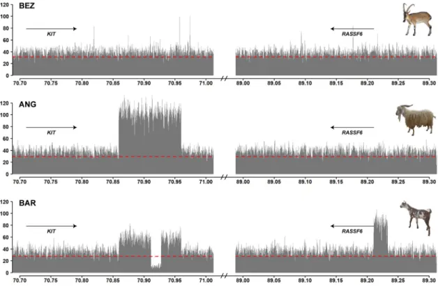
The completely white Pak Angora and the white spotted Barbari breeds showed strong selec-tion signatures harboring theKIT gene on chromosome 6 with −ZHpvalues of 7.20 and 4.56
(Fig 1;S3 Table). We searched for candidate causative variants within the signatures, but did not detect any coding variants in theKIT gene. However, visual inspection of the short read
alignments revealed two different copy number variants (CNVs) downstream of theKIT gene
in the Pak Angora and Barbari breeds (Fig 2).
Both CNVs started ~63 kb downstream ofKIT and covered ~100 kb of the genome
refer-ence sequrefer-ence without known coding DNA. The short read-aligments of read-pairs spanning the amplification breakpoints confirmed that the individual copies of the CNV were arranged in tandem in a head to tail orientation (S4 Fig). The Pak Angora allele consisted of a triplica-tion of the 100 kb region. The Barbari allele represented a duplicatriplica-tion of the same ~100 kb region with an additional 16,280 bp deletion in its central part. The read-pair information at the breakpoints indicated that the deleted part was replaced by a 22,702 bp genomic fragment from the 5’-flanking sequence of theRASSF6 gene, which is located 19 Mb further downstream
on the same chromosome (S4 Fig;S5 Table). The shared breakpoints of the two different CNV alleles suggested a common origin of these alleles.
CNVs at the
ASIP locus in Swiss goat breeds
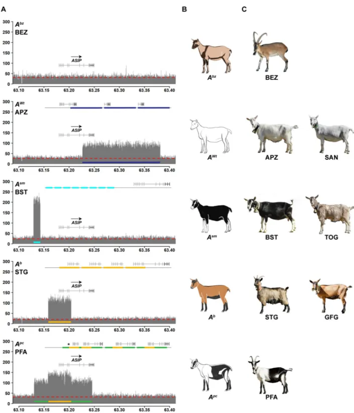
Five Swiss goat breeds with different coat color patterns had a selection signature with −ZHp � 4 in the region of the ASIP locus on chromosome 13 (Fig 1;S3 Table). We did not find anyASIP coding variation in the breeds with ASIP selection signatures.
Based on the segregation of coat color patterns in a large breeding experiment, the existence of up to 11 different caprineASIP alleles has been postulated [8]. The most dominant of this allelic series, termed “white or tan” (AWt), is responsible for white coat color in goats [8]. Fur-thermore, it has been shown that the white coat color in Saanen goats is caused by a triplication of theASIP gene [9].
Inspection of the short-read alignments of our own sequence data from Saanen goats at the
ASIP locus confirmed the previously reported triplication and revealed the exact boundaries of
the triplication. It spans 154,677 bp of the reference genome sequence and comprises the entire coding sequence of theASIP, AHCY and ITCH genes. The individual copies are arranged in
tandem in a head to tail orientation. Appenzell goats, another white goat breed, had the same CNV allele as the Saanen goats (Fig 3,S4 Fig,S5 Table).
We next investigated the coverage plots of Grisons Striped goats and Toggenburg goats. These two breeds show a characteristic color pattern, which has been postulated to be caused by anASIP allele termed “Swiss markings” (Asm) [8]. They are fixed for anASIP allele with 8
Fig 1. Manhattan plots showing−ZHp values from 20 diverse goat breeds. The red horizontal line indicates the chosen significance threshold of−ZHp = 4. Each dot represents a 150 kb window. Each plot contains 29 autosomes and two unplaced scaffolds representing the X chromosome. Selection signatures co-localizing with known coat color genes are marked with arrows.
tandem copies of a 13,433 bp sequence from the 5’-flanking region ofASIP (Fig 3,S4 Fig,S5 Table).
The Chamois Colored goat and the St. Gallen Booted goat are characterized by a color pat-tern and anASIP allele termed “badgerface” (Ab) [8]. Our pool-seq data revealed a five-fold amplification of 45,680 bp located ~61 kb downstream of theAsmamplification (Fig 3,S4 Fig,
S5 Table).
The Peacock goat is a rare Swiss goat breed with a unique and striking coat color pattern that has not been investigated previously. Pool-seq data from Peacock goats indicated a selec-tion signature at theASIP locus. The ASIP allele in Peacock goats, which we propose to term
“peacock” (Apc), has a quadruplication of the same ~45 kb region having five copies in theAb
allele. It is additionally flanked by triplicated segments of 27,996 bp and 41,807 bp on the left and right side of the quadruplicated sequence (Fig 3,S4 Fig,S5 Table). The central part of the
Apcallele has exactly the same breakpoints as theAballele suggesting a common origin ofAb
andApc.
The goat genome reference sequence is derived from a San Clemente goat, which has a sim-ilar coat color pattern as bezoars. The genome reference therefore supposedly represents the wildtype allele at theASIP locus, termed “bezoar” (Abz) [8]. Bezoars and all other Swiss goat breeds did not show any CNVs at theASIP locus. In the remaining goat breeds the AWt,Asm
andAballeles were segregating at low frequencies. These breeds are either not specifically
Fig 2. CNVs at theKIT locus. The coverage plot of bezoars (BEZ) does not show any copy number variation and represents the
wildtype allele. In the Pak Angora breed (ANG), the coverage plot shows a triplication of ~100 kb downstream of theKIT gene. In the Barbari breed (BAR), the same region is duplicated. The Barbari allele shows a complex rearrangement involving the insertion of a ~23 kb genome segment originating at 89.2 Mb into the duplicated sequence at ~70.9 Mb with the simultaneous deletion of ~16 kb ofKIT sequence. Please note that the coverage at ~89.2 Mb corresponds to three times the average. One genome equivalent corresponds to the wildtype sequence at ~89.2 Mb. Read-pair information indicated that the other two genome equivalents are inserted into the duplicated sequence at ~70.9 Mb (S4 Fig). The dashed red line indicates the average coverage across the whole genome of each pool-seq dataset.
Fig 3. CNVs at theASIP locus. A Coverage plots of the ASIP locus in different goat breeds reveal four different CNVs. The bezoar (BEZ) coverage
plot shows uniform coverage and is characteristic for the wildtype allele (Abz). Underneath, four different mutantASIP alleles associated with different CNVs are illustrated. The line on top of each plot schematically indicates the most likely configuration of these mutant alleles derived from the available short-read sequence information (S4 Fig). The dashed red line indicates the average coverage across the whole genome of each breed. B Schematic drawings and C representative photographs illustrating the coat color phenotypes of the studied breeds. The photo of the bezoar was obtained during summer, when the dark stripes at the collar and the belly are much less pronounced than in the winter coat. Note that some of the patterns show an exactly inverse distribution of eumelanin and pheomelanin. For example, goats with theAsmallele have white (pheomelanistic)
facial stripes and legs, while goats with theAborApcalleles have black (eumelanistic) facial stripes and legs.
selected for coat color (e.g. African dwarf goat, Beetal) or the effect ofASIP is phenotypically
not visible due to epistatic effects of other genes that lead to white spotting phenotypes (e.g. Boer goat, Pak Angora, Barbari).
Quantitative analysis of
ASIP mRNA expression
The different CNVs in putative regulatory regions of theASIP gene prompted us to
hypothe-size that quantitative differences inASIP expression may cause the different color patterns. We
therefore obtained whole skin samples from five goats carrying different CNV alleles. We iso-lated RNA from matched pairs of eumelanistic and pheomelanistic skin and performed an RNA-seq experiment to determine the expression level ofASIP mRNA expression.
In Grisons Striped goats (Asm), Chamois Colored goats (Ab) and Peacock goats (Apc), the eumelanistic skin showed very lowASIP mRNA expression. The pheomelanistic skin regions
in these three goats had at least 10-fold higherASIP expression than the corresponding
eume-lanistic samples. The uniformly white (pheomeeume-lanistic) Saanen goat (AWt) had the highest
ASIP mRNA expression. There was no obvious correlation between the quantitative ASIP
mRNA expression and the intensity of the pheomelanistic pigmentation. The intensely red col-ored skin from the Chamois Colcol-ored goat had an intermediateASIP mRNA expression
com-pared to the pale white skin from e.g. the Saanen and Peacock goat. Visual inspection of the RNA-seq short-read alignments indicated the utilization of nine different 5’-untranslated exons in nine different transcript isoforms originating from differentASIP alleles (Fig 4;S1 File).
Fig 4.ASIP mRNA expression and identified transcripts in skin. A Representative photographs of the five sampled goat breeds.
The biopsy sites are numbered and indicated by red circles. B Trimmed mean of M (TMM) values ofASIP mRNA expression were determined from RNA-seq data for each sample. The colors of the bars correspond to the pigmentation of the skin samples. Please note that the Valais Blackneck goat (VAG) has a black base color that is independent of theASIP gene. This goat has a white spotting phenotype and lacks melanocytes in its caudal half. The lowASIP expression in the unpigmented white skin sample of this goat underscores the difference to the pheomelanistic pale white pigmentation in other goats. CASIP transcript isoforms in pheomelanistic skin samples from goats with differentASIP alleles. Transcript isoforms X1 and X2 correspond to the RefSeq accessions XM_018057735.1 and XM_018057736.1. CNV breakpoints of theAb,AWt, andApcalleles are indicated.
Discussion
In the present study, we discovered 2,239 loci under selection in 20 diverse goat breeds with various phenotypes and different geographical origins. Our methodology comprised the iden-tification of regions with low heterozygosity from pool-seq data in combination with pairwise FSTto bezoars, the wild ancestor of domesticated goats. The pool-seq approach was validated
by repeating the analyses with virtually identical results from individually sequenced goats in five breeds. We have to caution that reduced heterozygosity and high FSTvalues may not only
result from selection, but may also be due to random demographic changes.
The comprehensive catalogue of identified selection signatures can now be used as a start-ing point to identify causal genetic variants that control a wide variety of breed-definstart-ing traits.
We particularly focused on selection signatures harboring known coat color genes and identified two CNVs in the 3’-flanking region ofKIT in two Pakistani goat breeds, the
completely white Pak Angora breed and the white spotted Barbari breed. An association between white coat color and theKIT locus has been reported before in Iranian Markhoz
goats, which also represents an Angora type goat [37]. TheKIT gene is flanked by several
hundred kilobases of non-coding genomic DNA on either side, which are required for the pre-cise regulation of its temporal and spatial expression. The KIT protein is a receptor tyrosine kinase mediating a survival signal for several different cell types including melanoblasts and melanocytes, but also e.g. hematopoetic stem cells, mast cells, interstitial cells of Cajal and sper-matogonia [38,39].KIT is a proto-oncogene and its overexpression may have detrimental
con-sequences such as tumor development [40,41]. Insufficient expression of functional KIT protein in melanoblasts or melanocytes will lead to apoptosis of these cells and results in white spotting phenotypes [15,42–45].
Structural variants at theKIT locus cause several other breed defining coat color phenotypes
in domestic animals, such as the dominant white and belt phenotypes in pigs [33,46,47], color-sided and lineback in cattle [48,49], and tobiano spotting in horses [50]. Lineback in cattle and tobiano in horses also involve structural variants in the 3’-flanking region ofKIT [49,50]. All these phenotypes are characterized by striking alterations in pigmentation without any delete-rious consequences on the other KIT dependent cell types, which would be expected to result in potentially serious health problems. The comparative data from other species strongly sug-gest that the newly detected caprineKIT CNVs in goats cause the complete lack of skin and
hair pigmentation in Pak Angora and the white spotted phenotype in Barbari goats due to altered expression of KIT during fetal development of melanoblasts.
TheASIP gene codes for the agouti signaling protein, the competitive inhibitor of
melano-cortin 1 receptor expressed on melanocytes [20]. Variation in the quantitative amount ofASIP
mRNA expression from different promoters is the central mechanism regulating the so-called pigment-type switching [19,21,28]. The regulatory elements of theASIP gene are most likely
contained in its large 5’-flanking region. Differing from theKIT locus, ASIP does not contain a
very large 3’-flanking region. Spatially and temporally regulated synthesis of eumelanin and pheomelanin enables mammals to express a wide variety of coat color patterns that are essen-tial for e.g. camouflage or mate recognition in many wild species.
ASIP variants cause a wide variety of breed defining coat color phenotypes in domestic
ani-mals.ASIP loss of function variants, typically in the coding sequence, are responsible for
reces-sive black in e.g. dogs [51], horses [52], and rabbits [53]. Gain of function variants inASIP,
such as an ectopic overexpression lead to dominant red phenotypes [54]. There are only very few examples of non-coding regulatory variants at theASIP locus that have been fully
charac-terized at the molecular level. One important example is the mouse black-and tan allele (at), which is caused by a ~6 kb retroviral-like insertion in the region of the hair cycle-specific
promoter [25]. In black and tan at mice, hairs are no longer banded and show a uniformly yel-low or uniformly black pigmentation. Amplification of the entireASIP gene has previously
been shown to cause the white coat color in many sheep breeds [18] and the white or tan allele (AWt) in Saanen goats [9].
Our study confirmed the previous results and defined the exact breakpoints of the caprine
AWttriplication that actually comprises not only theASIP gene, but also the flanking AHCY
andITCH genes. Unexpectedly, we did not observe selection signatures at the window
harbor-ing theASIP gene in the Saanen and Appenzell breeds. Both of these breeds are strictly
selected for uniform white (pheomelanistic) coat color and have a very high frequency of the
AWtallele. We think that the lack of a significant−ZHpscore atASIP in these two breeds is
caused by at least three factors. For the calculation of the−ZHpscore, we considered only
SNVs with a maximum coverage of 50x in order to suppress artifacts caused by non-specific mapping of highly repetitive sequences. As theASIP locus is triplicated in Saanen and
Appen-zell goats their pools had >50x average coverage atASIP and almost no SNVs were called in
the region. Furthermore, when we inspected the whole genome sequencing data from 24 indi-vidual Appenzell goats, we found that only 22 of them wereAWt/AWtas expected. The remain-ing two animals had the genotypeAWt/Asm. AsAWtis the most dominant allele in the series, it apparently has still not reached absolute fixation and otherASIP alleles are segregating at least
in the Appenzell goat breed. Finally, the three copies of the triplication had several sequence differences, which were called as variable positons with a 2:1 ratio of the alleles. This provides a biological explanation why the selection signature might be weaker than expected.
We observed significant selection signatures at theASIP gene in five Swiss goat breeds. In
these breeds, we identified three additional non-coding CNVs in the 5’-region ofASIP that are
likely to cause the Swiss markings (Asm), badgerface (Ab) and peacock (Apc) alleles in goats. We have to caution that the peacock phenotype has not been reported before and may be influ-enced by additional genes other thanASIP.
The corresponding coat color phenotypes are very interesting as they have characteristic patterns of eumelanistic and pheomelanistic pigmentation. Domestic goats with these alleles therefore represent a valuable resource for dissecting the precise function of individual regulatory elements in future studies. TheAsmandAballeles result in almost exactly inverted distributions of eumelanin and pheomelanin. Our RNA-seq data confirm that the different pigmentation patterns are caused by different levels ofASIP mRNA transcription at different
body locations. These data also revealed that the different caprineASIP gene alleles give rise to
a higher number of non-coding 5’-exons compared to other mammals [25,26]. Additional data, such as Cage-seq and full-length Iso-seq data will be required for a comprehensive anno-tation of all possible transcription start sites and splice isoforms in goats. Such data are expected to become available soon with the advances of the FAANG project [55].
In conclusion, we identified 2,239 selection signatures in 20 diverse goat breeds with vari-ous coat color phenotypes. These selection signatures revealed six different functionally rele-vant CNVs underlying breed-defining coat color phenotypes in goats. The results should help to advance our mechanistic understanding of temporal and spatial regulation of transcription.
Materials and methods
Ethics statement
All animal experiments were performed according the local regulations. All animals in this study were examined with the consent of their owners. Sample collection was approved by the “Cantonal Committee For Animal Experiments” (Canton of Bern; permit 75/16).
Animals
For this study, 244 female animals of 20 phenotypical diverse goat breeds and their wild ances-tor, the bezoar, were sampled (S1 Table). Ten of the analyzed goat breeds originate from Swit-zerland, eight from Pakistan and two from Africa. Swiss and African breeds were sampled in Switzerland. Pakistani breeds were sampled in Pakistan. bezoar samples were from zoo ani-mals. For the Swiss goat breeds, we selected representative animals of the breeds and excluded any first-degree relatives. For the other goat breeds, we did not have full pedigree information and used convenience samples. Genomic DNA was isolated from EDTA blood samples.
Whole genome sequencing of pools (pool-seq)
Breed pools were prepared by pooling equimolar amounts of 12 animals per breed (ANG: 10, BEZ: 8 and STG: 10). Illumina TruSeq PCR-free genomic DNA libraries with an insert size of 350 bp were prepared. Each breed pool was sequenced on one lane of an Illumina HiSeq 3000 instrument and on average 300 million 2x150 bp paired-end reads per breed pool were col-lected (S1 Table).
Mapping and variant calling
Adapter sequences, reads with too many Ns and low quality bases were trimmed or ultimately discarded, if the remaining read length was < 50 bp with fastq-mcf version 1.1.2 (settings: -l 50 -S -q 20). The cleaned reads were mapped to the goat reference genome ARS1 [56] with Bur-rows-Wheeler Aligner (BWA-MEM) algorithm version 0.7.13 [57] using the “-M” flag to mark shorter alignments as secondary. The resulting mapping files in SAM format were converted into BAM format and coordinate sorted using SAMtools version 1.3 [58]. A local indel realign-ment was performed using the Genome Analysis Toolkit version 3.7 [59] with default settings. Duplicated reads were marked, using Picard Tools version 2.2.1 (http://broadinstitute.github. io/picard) with default settings for patterned flow cell models. Single nucleotide variants were called using (i) Genome Analysis Toolkit UnifiedGenotyper version 3.7 [59] with the settings: -glm SNP, -stand_call_conf 20, -out_mode EMIT_VARIANTS_ONLY and –ploidy 16/20/24 and (ii) SAMtools mpileup [60] with the settings -q 15, -Q 20, -C 50 and -B. The variants resulting from UnifiedGenotyper were filtered for high quality variants with GATK’s Variant-Filtration tool using the generic hard-filtering recommendations available fromhttps:// gatkforums.broadinstitute.org/gatk/discussion/6925/understanding-and-adapting-the-generic-hard-filtering-recommendations, while the mpileup files were streamed to the PoPoo-lation2 version 1.201 pipeline [61]. We used the scripts mpileup2sync.jar with settings - -fastq-type sanger and - -min-qual 20 and snp-frequency-diff.pl with the settings - -min-coverage 15, - -max-coverage 50 and - -min-count 3. Both pipelines yielded similar numbers of SNV (S2 Table).
Sweep analysis of pool-seq data
A screen for selective sweeps was performed using the SNV file produced for each breed pool individually by the mpileup PoPoolation2 pipeline. At each identified SNV position in the files, we took the numbers of major (nMAJ) and minor (nMIN) allele counts observed in each
breed and calculated pooled heterozygosity (Hp) [32] with an in-house written script. The
script applies Hp= 2SnMAJSnMIN/(SnMAJ+SnMIN)2in a sliding 50% overlapping window
approach. We evaluated the results with different window sizes (25 to 300 kb) and decided on 150 kb as the most appropriate size [33,34]. The obtained Hpvalues for all 34,382 overlapping
σHp). Windows with a−ZHp� 4 were retained as selective windows and adjacent or
overlapping selective windows were merged into selection signatures, individually per breed. We annotated the identified selection signatures along with NCBI’sCapra hircus
Annotation Release 102 (S3 Table). In addition, to the Hpcalculation, we calculated
weighted population FSTvalues for each SNV in a 150 kb sliding, 50% overlapping window
approach. We applied the FST-sliding.pl script of the Popoolation2 pipeline. The script FST
-sliding.pl was run with the settings - -min-count 2 - -min-coverage 4 - -max-coverage 50 - -window-size 150000 - -step-size 75000 - -suppress-noninformative and - -pool-size 10:12:12:12:8:12:12:12:12:12:12:12:12:12:12:12:10:12:12:12:12. It used the previously obtained sync file of all pools combined as input and calculated weighted population pairwise Fstvalues
using the standard equation as shown in Hartl and Clark [62]. This resulted in 210 pairwise comparisons, from which we selected the comparisons between the 20 domesticated goat breeds with the bezoar. The obtained FSTvalues were Z transformed, performing ZFST= (FST
-μFST/σFST).
Whole genome re-sequencing of individual goats and variant calling
In addition to the pool-seq experiment, we selected 120 goats from five Swiss breeds for indi-vidual whole genome re-sequencing. The 24 animals per breed included the 12 goats repre-sented in the breed pools (S1 Table). Illumina TruSeq PCR-free DNA libraries with an insert size of 350 bp were prepared and sequenced on an Illumina NovaSeq 6000 instrument, yield-ing on average 240 million 2x150 bp paired-end reads per goat (S1 Table). Clean reads were produced by running fastp, version 0.12.5 [63], an ultra-fast all-in-one FASTQ preprocessor capable of trimming polyG tails, a known issue of NovaSeq reads. The cleaned reads were mapped to the ARS1 goat reference genome [56] with Burrows-Wheeler Aligner (BWA-MEM) algorithm using the “-M” flag to mark shorter alignments as secondary. The resulting SAM files were converted into BAM files and coordinate sorted using SAMtools. Duplicated reads were marked, using Picard Tools (http://broadinstitute.github.io/picard) with default settings for patterned flow cell models. The marked BAM files were streamed to GATK’s Base-Recalibrator tool, supported with known SNV provided by the VarGoats consortium (http:// www.goatgenome.org/vargoats.html). Subsequently, GATK’s HaplotypeCaller with the set-tings - -emitRefConfidence GVCF and -stand_call_conf 30 was used to call genome-wide vari-ants [59]. The variant files were merged and GATK’s GenotypeGVCFs was used to call variants in the 120 goats combined. As a next step, the called variants were filtered for high quality variants with GATK’s VariantFiltration tool (version 3.8) using the generic hard-filter-ing recommendations available fromhttps://gatkforums.broadinstitute.org/gatk/discussion/ 6925/understanding-and-adapting-the-generic-hard-filtering-recommendations. SnpEff [64] and NCBI’sCapra hircus Annotation Release 102 was used to annotate the variants.Sweep analysis of individual goats
To calculate Hpscores of the individual goats, we selected biallelic, passed SNPs per breed
using GATK’s SelectVariants tool (version 3.8), applying - -restrictAllelesTo BIALLELIC - -selectTypeToInclude SNP - -sample_expressions ’(APZ/BST/PFA/STG/VAG)’ - -maxNO-CALLnumber 0 - -excludeFiltered - -excludeNonVariants. This yielded on average 14.9 million SNVs per combined Swiss goat breed, comprising each 24 individually sequenced animals (S2 Table). As a next step, the VCF files containing only biallelic, passed SNPs were transformed into table format using GATK’s VariantsToTable tool. This table contained only information regarding SNP position, reference allele and genotype of the 24 animals. With an in-house written Python script, we converted the table produced with GATK’s VariantsToTable to
major and minor alleles and counted the number of observations. This output was then used for Hpcalculation as described in sweep analysis of pool-seq data.
Manhattan plots
The−ZHpvalues were plotted using the function manhattan of the qqman package [65] with R
[66]. Each data point represents a 150 kb window. A red horizontal line was drawn represent-ing the chosen significance threshold of−ZHp� 4 (corresponding to 0.8% of all windows).
CNV analyses
Coverage plots for regions of interest were created by calculating the coverage of each base in a defined region of interest using Samtools depth -b. Additionally coverage stats across the whole genomes, including the average coverage were calculated using goleft covstats (https:// github.com/brentp/goleft). Taken both results together, we plotted the coverage using R plot type h version 3.4.1 and indicated the average coverage line. Potential CNVs were also visually evaluated by inspection of the short-read alignemnts (bam-files) in the Integrative Genome Viewer (IGV) [67].
Skin biopsies and total RNA extraction
Skin biopsies were taken from five slaughtered animals of different goat breeds (SAN, BST, GFG, PFA and VAG). Two 6 mm punch biopsies were taken from differentially pigmented body areas of each animal (S6 Table). The biopsies were immediately put in RNAlater
(Qia-gen) for at least 24 h and then frozen at –20˚C. Prior to RNA extraction, the skin biopsies were homogenized mechanically with the TissueLyser II device from Qiagen. Total RNA was extracted from the homogenized tissue using the RNeasy Fibrous Tissue Mini Kit (Qiagen) according to the manufacturer’s instructions. RNA quality was assessed with a FragmentAna-lyzer (Advanced Analytical) and the concentration was measured using a Qubit Fluorometer (ThermoFisher Scientific).
Whole transcriptome sequencing (RNA-seq)
From each sample, 1μg of high quality total RNA (RIN >9) was used for library preparation with the Illumina TruSeq Stranded mRNA kit. The 10 libraries were pooled and sequenced on an S1 flow cell with 2x50 bp paired-end sequencing using an Illumina NovaSeq 6000 instru-ment. On average, 31.5 million paired-end reads per sample were collected (S6 Table). All reads that passed quality control were mapped to the ARS1 goat reference genome assembly using STAR aligner (version 2.6.0c) [68]. The read abundance was calculated using HTseq (version 0.9.1) [69] and a gff3 file obtained from NCBI’sCapra hircus Annotation Release 102.
We used the EdgeR package [70] to read the HTseq count data and calculated the log fold changes using the exactTest function where the biological co-efficient of variation (BCV) was set to 0.1. Trimmed mean of M (TMM) values ofASIP mRNA expression were determined for
each sample [71].
Supporting information
S1 Fig. Distributions of Hp,−ZHp, FSTand−ZFST. (PDF)
S2 Fig.−ZHpscores Manhattan plots of pooled sequencing and single sequencing. (PDF)
S3 Fig. Manhattan plots of−ZHp scores and ZFSTscores. (PDF)
S4 Fig. Details of the CNV alleles. (PDF)
S1 File. FASTA sequences of nine different caprineASIP transcripts.
(TXT)
S1 Table. Read statistics and accessions of pool-seq and individual WGS data. (XLSX)
S2 Table. Pool-seq and individual WGS SNV statistics. (XLSX)
S3 Table. Hpselection signatures per breed pool. (XLSX)
S4 Table. FSTselection signatures per breed pool. (XLSX)
S5 Table. Details of CNV alleles. (XLSX)
S6 Table. Descriptive statistics and accessions of RNA-seq datasets. (XLSX)
Acknowledgments
The authors are grateful to all goat owners and breeding organizations who donated samples and shared pedigree data and phenotype information of their animals. We thank Eva Andrist, Nathalie Besuchet Schmutz, Muriel Fragnière, and Sabrina Schenk for expert technical assis-tance, the Next Generation Sequencing Platform of the University of Bern for performing the high-throughput sequencing experiments, and the Interfaculty Bioinformatics Unit of the Uni-versity of Bern for providing high performance computing infrastructure. Furthermore, we thank Christian Gazzarin for the photos and Sarah Stangl for the graphical illustrations of the different Swiss goat breeds.
Author Contributions
Conceptualization: Tosso Leeb. Data curation: Vidhya Jagannathan. Funding acquisition: Tosso Leeb.Investigation: Jan Henkel, Rashid Saif, Vidhya Jagannathan, Corinne Schmocker, Flurina Zeindler, Cord Dro¨gemu¨ller, Christine Flury.
Methodology: Vidhya Jagannathan.
Resources: Rashid Saif, Erika Bangerter, Ursula Herren, Dimitris Posantzis, Zafer Bulut, Phi-lippe Ammann, Cord Dro¨gemu¨ller.
Supervision: Vidhya Jagannathan, Cord Dro¨gemu¨ller, Christine Flury, Tosso Leeb. Visualization: Jan Henkel.
Writing – original draft: Jan Henkel, Tosso Leeb.
Writing – review & editing: Jan Henkel, Rashid Saif, Vidhya Jagannathan, Corinne Schmocker, Flurina Zeindler, Erika Bangerter, Ursula Herren, Dimitris Posantzis, Zafer Bulut, Philippe Ammann, Cord Dro¨gemu¨ller, Christine Flury, Tosso Leeb.
References
1. Zeder MA, Hesse B. The initial domestication of goats (Capra hircus) in the Zagros mountains 10,000 years ago. Science. 2000; 287: 2254–2257.https://doi.org/10.1126/science.287.5461.2254PMID:
10731145
2. Naderi S, Rezaei H-R, Pompanon F, Blum MG, Negrini R, Naghash H-R, et al. The goat domestication process inferred from large-scale mitochondrial DNA analysis of wild and domestic individuals. Proc Natl Acad Sci USA. 2008; 105: 17659–17664.https://doi.org/10.1073/pnas.0804782105PMID:
19004765
3. Colli L, Milanesi M, Talenti A, Bertolini F, Chen M, Crisa A, et al. Genome-wide SNP profiling of world-wide goat populations reveals strong partitioning of diversity and highlights post-domestication migra-tion routes. Genet Sel Evol. 2018; 50: 58.https://doi.org/10.1186/s12711-018-0422-xPMID:30449284
4. FAO. The Second Report on the State of the World’s Animal Genetic Resources for Food and ture, edited by B.D. Scherf & D. Pilling. FAO Commission on Genetic Resources for Food and Agricul-ture Assessments. Rome 2015. (http://www.fao.org/3/a-i4787e/index.html)
5. Stella A, Nicolazzi EL, Van Tassell CP, Rothschild MF, Colli L, Rosen BD, et al. AdaptMap: exploring goat diversity and adaptation. Genet Sel Evol. 2018; 50: 61.https://doi.org/10.1186/s12711-018-0427-5
PMID:30453882
6. Alberto FJ, Boyer F, Orozco-terWengel P, Streeter I, Servin B, de Villemereuil P, et al. Convergent genomic signatures of domestication in sheep and goats. Nat Comm. 2018; 9: 813.
7. Burren A, Neuditschko M, Signer-Hasler H, Frischknecht M, Reber I, Menzi F, et al. Genetic diversity analyses reveal first insights into breed-specific selection signatures within Swiss goat breeds. Anim Genet. 2016; 47: 727–739.https://doi.org/10.1111/age.12476PMID:27436146
8. Adalsteinsson S, Sponenberg DP, Alexieva S, Russel AJ. Inheritance of goat coat colors. J Hered. 1994; 85: 267–272.https://doi.org/10.1093/oxfordjournals.jhered.a111454PMID:7930499
9. Fontanesi L, Beretti F, Riggio V, Gomez Gonzalez E, Dall’Olio S, Davoli R, et al. Copy number variation and missense mutations of the agouti signaling protein (ASIP) gene in goat breeds with different coat colors. Cytogenet Genome Res. 2009; 126:333–347.https://doi.org/10.1159/000268089PMID:
20016133
10. Becker D, Otto M, Ammann P, Keller I, Dro¨gemu¨ ller C, Leeb T. The brown coat colour of Coppernecked goats is associated with a non-synonymous variant at the TYRP1 locus on chromosome 8. Anim Genet. 2015; 46:50–54.https://doi.org/10.1111/age.12240PMID:25392961
11. Dietrich J, Menzi F, Ammann P, Dro¨gemu¨ller C, Leeb T. A breeding experiment confirms the dominant mode of inheritance of the brown coat colour associated with the496Asp TYRP1 allele in goats. Anim Genet. 2015; 46: 587–588.https://doi.org/10.1111/age.12320PMID:26153465
12. Menzi F, Keller I, Reber I, Beck J, Brenig B, Schutz E, et al. Genomic amplification of the caprine EDNRA locus might lead to a dose dependent loss of pigmentation. Sci Rep. 2016; 6: 28438.https:// doi.org/10.1038/srep28438PMID:27329507
13. Jackson IJ. Homologous pigmentation mutations in human, mouse and other model organisms. Hum Mol Genet. 1997; 6: 1613–1624.https://doi.org/10.1093/hmg/6.10.1613PMID:9300652
14. Thomas AJ, Erickson CA. The making of a melanocyte: the specification of melanoblasts from the neu-ral crest. Pigment Cell Melanoma Res. 2008; 21: 598–610.https://doi.org/10.1111/j.1755-148X.2008. 00506.xPMID:19067969
15. Haase B, Brooks SA, Schlumbaum A, Azor PJ, Bailey E, Alaeddine F, et al. Allelic heterogeneity at the equine KIT locus in dominant white (W) horses. PLoS Genet. 2007; 3: e195.https://doi.org/10.1371/ journal.pgen.0030195PMID:17997609
16. Greenhill ER, Rocco A, Vibert L, Nikaido M, Kelsh RN. An iterative genetic and dynamical modelling approach identifies novel features of the gene regulatory network underlying melanocyte development. PLoS Genet. 2011; 7: e1002265.https://doi.org/10.1371/journal.pgen.1002265PMID:21909283
17. Hauswirth R, Haase B, Blatter M, Brooks SA, Burger D, Dro¨gemu¨ller C, et al. Mutations in MITF and PAX3 cause "splashed white" and other white spotting phenotypes in horses. PLoS Genet. 2012; 8: e1002653.https://doi.org/10.1371/journal.pgen.1002653PMID:22511888
18. Norris BJ, Whan VA. A gene duplication affecting expression of the ovine ASIP gene is responsible for white and black sheep. Genome research. 2008; 18(8):1282–93.https://doi.org/10.1101/gr.072090. 107PMID:18493018
19. Barsh GS. The genetics of pigmentation: from fancy genes to complex traits. Trends Genet. 1996; 12: 299–305.https://doi.org/10.1016/0168-9525(96)10031-7PMID:8783939
20. Lu D, Willard D, Patel IR, Kadwell S, Overton L, Kost T, et al. Agouti protein is an antagonist of the mela-nocyte-stimulating-hormone receptor. Nature. 1994; 371: 799–802.https://doi.org/10.1038/371799a0
PMID:7935841
21. Millar SE, Miller MW, Stevens ME, Barsh GS. Expression and transgenic studies of the mouse agouti gene provide insight into the mechanisms by which mammalian coat color patterns are generated. Development. 1995; 121: 3223–3232. PMID:7588057
22. Le Pape E, Wakamatsu K, Ito S, Wolber R, Hearing VJ. Regulation of eumelanin/pheomelanin synthe-sis and visible pigmentation in melanocytes by ligands of the melanocortin 1 receptor. Pigment Cell Mel-anoma Res. 2008; 21: 477–486.https://doi.org/10.1111/j.1755-148X.2008.00479.xPMID:18627531
23. Cieslak M, Reissmann M, Hofreiter M, Ludwig A. Colours of domestication. Biol Rev. 2011; 86: 885– 899.https://doi.org/10.1111/j.1469-185X.2011.00177.xPMID:21443614
24. Graham A, Wakamatsu K, Hunt G, Ito S, Thody AJ. Agouti protein inhibits the production of eumelanin and pheomelanin in the presence and absence of alpha-melanocyte stimulating hormone. Pigment Cell Res. 1997; 10: 298–303.https://doi.org/10.1111/j.1600-0749.1997.tb00689.xPMID:9359625
25. Bultman SJ, Michaud EJ, Woychik RP. Molecular characterization of the mouse agouti locus. Cell. 1992; 71: 1195–1204.https://doi.org/10.1016/s0092-8674(05)80067-4PMID:1473152
26. Vrieling H, Duhl DM, Millar SE, Miller KA, Barsh GS. Differences in dorsal and ventral pigmentation result from regional expression of the mouse agouti gene. Proc Natl Acad Sci USA. 1994; 91: 5667– 5671.https://doi.org/10.1073/pnas.91.12.5667PMID:8202545
27. Dro¨gemu¨ ller C, Giese A, Martins-Wess F, Wiedemann S, Andersson L, Brenig B, et al. The mutation causing the black-and-tan pigmentation phenotype of Mangalitza pigs maps to the porcine ASIP locus but does not affect its coding sequence. Mamm Genome. 2006; 17: 58–66.https://doi.org/10.1007/ s00335-005-0104-1PMID:16416091
28. Kaelin CB, Barsh GS. Genetics of pigmentation in dogs and cats. Ann Rev Anim Biosci. 2013; 1: 125– 156.
29. Smith JM, Haigh J. The hitch-hiking effect of a favourable gene. Genet Res. 1974; 23: 23–35. PMID:
4407212
30. Hermisson J, Pennings PS. Soft sweeps: molecular population genetics of adaptation from standing genetic variation. Genetics. 2005; 169: 2335–2352.https://doi.org/10.1534/genetics.104.036947
PMID:15716498
31. Pennings PS, Hermisson J. Soft sweeps II—molecular population genetics of adaptation from recurrent mutation or migration. Mol Biol Evol 2006; 23: 1076–1084.https://doi.org/10.1093/molbev/msj117
PMID:16520336
32. Rubin CJ, Zody MC, Eriksson J, Meadows JR, Sherwood E, Webster MT, et al. Whole-genome rese-quencing reveals loci under selection during chicken domestication. Nature. 2010; 464: 587–591.
https://doi.org/10.1038/nature08832PMID:20220755
33. Rubin CJ, Megens HJ, Martinez Barrio A, Maqbool K, Sayyab S, Schwochow D, et al. Strong signatures of selection in the domestic pig genome. Proc Natl Acad Sci USA. 2012; 109: 19529–19536.https://doi. org/10.1073/pnas.1217149109PMID:23151514
34. Carneiro M, Rubin CJ, Di Palma F, Albert FW, Alfoldi J, Martinez Barrio A, et al. Rabbit genome analysis reveals a polygenic basis for phenotypic change during domestication. Science. 2014; 345: 1074–1079.
https://doi.org/10.1126/science.1253714PMID:25170157
35. Martinez Barrio A, Lamichhaney S, Fan G, Rafati N, Pettersson M, Zhang H, et al. The genetic basis for ecological adaptation of the Atlantic herring revealed by genome sequencing. eLife. 2016; 5: e12081.
https://doi.org/10.7554/eLife.12081PMID:27138043
36. Wang X, Liu J, Zhou G, Guo J, Yan H, Niu Y, et al. Whole-genome sequencing of eight goat populations for the detection of selection signatures underlying production and adaptive traits. Sci Rep. 2016; 6: 38932.https://doi.org/10.1038/srep38932PMID:27941843
37. Nazari-Ghadikolaei A, Mehrabani-Yeganeh H, Miarei-Aashtiani SR, Staiger EA, Rashidi A, Huson HJ. Genome-wide association studies identify candidate genes for coat color and mohair traits in the Iranian Markhoz goat. Front Genet. 2018; 9: 105.https://doi.org/10.3389/fgene.2018.00105PMID:29670642
38. Chabot B, Stephenson DA, Chapman VM, Besmer P, Bernstein A. The proto-oncogene c-kit encoding a transmembrane tyrosine kinase receptor maps to the mouse W locus. Nature. 1988; 335: 88–89.
39. Roskoski R Jr. Signaling by Kit protein-tyrosine kinase–the stem cell factor receptor. Biochem Biophys Res Commun. 2005; 337: 1–13.https://doi.org/10.1016/j.bbrc.2005.08.055PMID:16129412
40. Furitsu T, Tsujimura T, Tono T, Ikeda H, Kitayama H, Koshimizu U, et al. Identification of mutations in the coding sequence of the proto-oncogene c-kit in a human mast cell leukemia cell line causing ligand-independent activation of c-kit product. J Clin Invest. 1993; 92: 1736–1744.https://doi.org/10.1172/ JCI116761PMID:7691885
41. Nagata H, Worobec AS, Semere T, Metcalfe DD. Elevated expression of the proto-oncogene c-kit in patients with mastocytosis. Leukemia. 1998; 12: 175–181.https://doi.org/10.1038/sj.leu.2400906
PMID:9519779
42. Geissler EN, Ryan MA, Housman DE. The dominant-white spotting (W) locus of the mouse encodes the c-kit proto-oncogene. Cell. 1988; 55: 185–192.https://doi.org/10.1016/0092-8674(88)90020-7
PMID:2458842
43. Giebel LB, Spritz RA. Mutation of the KIT (mast/stem cell growth factor receptor) protooncogene in human piebaldism. Proc Nat Acad Sci USA. 1991; 88: 8696–8699.https://doi.org/10.1073/pnas.88.19. 8696PMID:1717985
44. Fleischman RA, Saltman DL, Stastny V, Zneimer S. Deletion of the c-kit protooncogene in the human developmental defect piebald trait. Proc Natl Acad Sci USA. 1991; 88: 10885–10889.https://doi.org/10. 1073/pnas.88.23.10885PMID:1720553
45. Haase B, Brooks SA, Tozaki T, Burger D, Poncet PA, Rieder S, et al. Seven novel KIT mutations in horses with white coat colour phenotypes. Anim Genet. 2009; 40: 623–629.https://doi.org/10.1111/j. 1365-2052.2009.01893.xPMID:19456317
46. Marklund S, Kijas J, Rodriguez-Martinez H, Ro¨nnstrand L, Funa K, Moller M, et al. Molecular basis for the dominant white phenotype in the domestic pig. Genome Res. 1998; 8: 826–833.https://doi.org/10. 1101/gr.8.8.826PMID:9724328
47. Giuffra E, Evans G, To¨rnsten A, Wales R, Day A, Looft H, Plastow G, Andersson L. The Belt mutation in pigs is an allele at the Dominant white (I/KIT) locus. Mamm Genome. 1999; 10: 1132–1136.https://doi. org/10.1007/s003359901178PMID:10594235
48. Durkin K, Coppieters W, Dro¨gemu¨ller C, Ahariz N, Cambisano N, Druet T, et al. Serial translocation by means of circular intermediates underlies colour sidedness in cattle. Nature. 2012; 482: 81–84.https:// doi.org/10.1038/nature10757PMID:22297974
49. Ku¨ttel L, Letko A, Ha¨fliger IM, Signer-Hasler H, Joller S, Hirsbrunner G, et al. A complex structural vari-ant at the KIT locus in cattle with the Pinzgauer spotting pattern. Anim Genet. 2019;https://doi.org/10. 1111/age.12821PMID:31294880
50. Brooks SA, Lear TL, Adelson DL, Bailey E. A chromosome inversion near the KIT gene and the Tobiano spotting pattern in horses. Cytogenetic and genome research. 2007; 119: 225–230.https://doi.org/10. 1159/000112065PMID:18253033
51. Kerns JA, Newton J, Berryere TG, Rubin EM, Cheng JF, Schmutz SM, et al. Characterization of the dog Agouti gene and a nonagoutimutation in German Shepherd Dogs. Mamm Genome. 2004; 15: 798–808.
https://doi.org/10.1007/s00335-004-2377-1PMID:15520882
52. Rieder S, Taourit S, Mariat D, Langlois B, Guerin G. Mutations in the agouti (ASIP), the extension (MC1R), and the brown (TYRP1) loci and their association to coat color phenotypes in horses (Equus caballus). Mamm Genome. 2001; 12: 450–455.https://doi.org/10.1007/s003350020017PMID:
11353392
53. Fontanesi L, Forestier L, Allain D, Scotti E, Beretti F, Deretz-Picoulet S, et al. Characterization of the rabbit agouti signaling protein (ASIP) gene: transcripts and phylogenetic analyses and identification of the causative mutation of the nonagouti black coat colour. Genomics. 2010; 95: 166–175.https://doi. org/10.1016/j.ygeno.2009.11.003PMID:20004240
54. Berryere TG, Kerns JA, Barsh GS, Schmutz SM. Association of an Agouti allele with fawn or sable coat color in domestic dogs. Mamm Genome. 2005; 16: 262–272. https://doi.org/10.1007/s00335-004-2445-6PMID:15965787
55. Giuffra E, Tuggle CK; FAANG Consortium. Functional Annotation of Animal Genomes (FAANG): Cur-rent Achievements and Roadmap. Annu Rev Anim Biosci. 2019; 7: 65–88.https://doi.org/10.1146/ annurev-animal-020518-114913PMID:30427726
56. Bickhart DM, Rosen BD, Koren S, Sayre BL, Hastie AR, Chan S, et al. Single-molecule sequencing and chromatin conformation capture enable de novo reference assembly of the domestic goat genome. Nature Genet. 2017; 49: 643–650.https://doi.org/10.1038/ng.3802PMID:28263316
57. Li H, Durbin R. Fast and accurate short read alignment with Burrows-Wheeler transform. Bioinformatics. 2009; 25: 1754–1760.https://doi.org/10.1093/bioinformatics/btp324PMID:19451168
58. Li H, Handsaker B, Wysoker A, Fennell T, Ruan J, Homer N, et al. The Sequence Alignment/Map format and SAMtools. Bioinformatics. 2009; 25:2078–2079.https://doi.org/10.1093/bioinformatics/btp352
PMID:19505943
59. McKenna A, Hanna M, Banks E, Sivachenko A, Cibulskis K, Kernytsky A, et al. The Genome Analysis Toolkit: a MapReduce framework for analyzing next-generation DNA sequencing data. Genome Res. 2010; 20: 1297–1303.https://doi.org/10.1101/gr.107524.110PMID:20644199
60. Li H. A statistical framework for SNP calling, mutation discovery, association mapping and population genetical parameter estimation from sequencing data. Bioinformatics. 2011; 27: 2987–2993.https://doi. org/10.1093/bioinformatics/btr509PMID:21903627
61. Kofler R, Pandey RV, Schlo¨tterer C. PoPoolation2: identifying differentiation between populations using sequencing of pooled DNA samples (Pool-Seq). Bioinformatics. 2011; 27: 3435–3456.https://doi.org/ 10.1093/bioinformatics/btr589PMID:22025480
62. Hartl DL, Clark AG. Principles of population genetics: Sinauer associates Sunderland, MA; 1997. 63. Chen S, Zhou Y, Chen Y, Gu J. fastp: an ultra-fast all-in-one FASTQ preprocessor. Bioinformatics.
2018; 34: i884–i90.https://doi.org/10.1093/bioinformatics/bty560PMID:30423086
64. Cingolani P, Platts A, Wang le L, Coon M, Nguyen T, Wang L, et al. A program for annotating and pre-dicting the effects of single nucleotide polymorphisms, SnpEff: SNPs in the genome of Drosophila mela-nogaster strain w1118; iso-2; iso-3. Fly. 2012; 6: 80–92.https://doi.org/10.4161/fly.19695PMID:
22728672
65. Turner SD. qqman: an R package for visualizing GWAS results using Q-Q and manhattan plots. bioR-xiv. 2014:005165.
66. R Core Team. R: A language and environment for statistical computing. R Foundation for Statistical Computing, Vienna, Austria 2018.https://www.R-project.org/.
67. Thorvaldsdottir H, Robinson JT, Mesirov JP. Integrative Genomics Viewer (IGV): high-performance genomics data visualization and exploration. Brief Bioinf. 2013; 14: 178–192.
68. Dobin A, Davis CA, Schlesinger F, Drenkow J, Zaleski C, Jha S, et al. STAR: ultrafast universal RNA-seq aligner. Bioinformatics. 2013; 29: 15–21.https://doi.org/10.1093/bioinformatics/bts635PMID:
23104886
69. Anders S, Pyl PT, Huber W. HTSeq—a Python framework to work with high-throughput sequencing data. Bioinformatics. 2015; 31: 166–169.https://doi.org/10.1093/bioinformatics/btu638PMID:
25260700
70. Robinson MD, McCarthy DJ, Smyth GK. edgeR: a Bioconductor package for differential expression analysis of digital gene expression data. Bioinformatics. 2010; 26: 139–140.https://doi.org/10.1093/ bioinformatics/btp616PMID:19910308
71. Robinson MD, Oshlack A. A scaling normalization method for differential expression analysis of RNA-seq data. Genome Biol. 2010; 11: R25.https://doi.org/10.1186/gb-2010-11-3-r25PMID:20196867