Annals of Medical Research
DOI: 10.5455/annalsmedres.2019.01.056 2019;26(7):1161-3
Original Article
Gall bladder polyps: Our experience in a tertiary referral
centre
Refik Bademci, Yasar Ozdenkaya
Medipol University, Faculty of Medicine, Department of General Surgery Istanbul, Turkey Copyright © 2019 by authors and Annals of Medical Research Publishing Inc.
Abstract
Aim: An increasing number of patients are undergoing surgical treatment for gallbladder polyps (GPs). As there is still controversy on the subject, it remains unclear whether GPs are the precursors of gallbladder tumors. The aim of this study was to examine the clinicopathological features of patients who had undergone cholecystectomy in our clinic for the treatment of gallbladder polyps. Material and Methods: A retrospective review was made of the clinicopathological data of patients who underwent cholecystectomy, selected from a group of 2048 patients, who received treatment between 2014-2018.
Results: A total of 79 (3.8%) patients were identified, comprising 50 (63.2%) females and 29 (36.8%) males with a mean age of 43 years (range, 22-81 years). During pre-operative ultrasonography, 60 (75.9%) patients were detected with a single polyp and 19 (23.1%) patients had multiple polyps. When postoperative histology was evaluated, 70 (88.6%) patients had cholesterol polyps, 2 (2.5%) patients had gallbladder stones, 5 (6.3%) patients had papillary adenomas without dysplasia and 1 (1.2%) patient had papillary adenoma with dysplasia, while 1 (1.2%) of patient had gallbladder adenocarcinoma.
Conclusion: According to the results of this study, as gallbladder polyps may be pre-cancerous or cancerous lesions, patients diagnosed with GP should be followed up closely with surgical treatment applied when necessary.
Keywords: Gall bladder polyps; cholecystectomy; gallbladder adenocarcinoma.
Received: 29.01.2019 Accepted: 09.05.2019 Available online: 17.06.2019
Corresponding Author: Refik Bademci, Medipol University, Faculty of Medicine, Department of General Surgery Istanbul, Turkey, E-mail: [email protected]
1161
INTRODUCTION
Polypoid (GP) lesions of the gallbladder are lesions caused by the mucosa of the gallbladder. Cholecystectomy is recommended to protect against the risk of gallbladder cancer in patients with GPs >10 mm in size (1). There remains controversy about the relationship between GPs and gallbladder cancer. Although the frequency of polyps differs in different series, it is determined on average as 0.5-11% (2-4). GP is more common in persons in their forties and in females (5). GPs can be diagnosed incidentally or from symptoms similar to gallbladder stone (6). Since the use of ultrasonography became widespread, the number of patients diagnosed with GPs has increased (7,8). It has been observed that during ultrasonography, the early stages of gallbladder cancers can provide similar images to those of GPs (9).
The aim of this study was to investigate the clinicopathological features of patients diagnosed with GP who underwent cholecystectomy in our clinic.
MATERIAL and METHODS
From a total of 2048 patients who underwent cholecystectomy between 2014-2018, retrospective analysis was made of the clinicopathological data of patients who were operated on for a diagnosis of GPs. The age, gender, type of operation, and the pathology results of the patients were retrieved from the hospital automated records system for analysis in the study. Approval for the study was granted by the Local Ethics Committee and all procedures were applied in accordance with the Helsinki Declaration Criteria.
If the ultrasonography (USG) exam (Published Online: 2019 June 17) ination results contained polyps, then the number and diameters of the polyps were recorded. Polyps were categorised as single or multiple, and in cases of multiple polyps, the diameter of the largest was recorded for inclusion in the analysis. The pathological data were classified as cholesterol polyp, adenoma (with or without
Ann Med Res 2019;26(7):1161-3
dysplasia) or adenocarcinoma.
Statistical analysis was performed using SPSS 25.0. software (SPSSFW; SPSS Inc., Chicago, IL, USA).
RESULTS
A total of 79 (3.8%) patients were identified, comprising 50 (63.2%) females and 29 males (36.8%), of whom 48 (60.7%) were aged >40 years (mean: 43 years; range: 22-81years). During pre-operative ultrasonography, 60 (75.9%) patients were detected with a single polyp and 19 (23.1%) with multiple polyps. The mean diameter of the polyps was 10.7 ±1 mm. Generally, the benign polyps were those of diameter 10-12 mm, and all were determined in males and younger aged females.
Laparoscopic surgery was applied to 78 (98.8%) patients and in 1 (1.2%) case when a tumor was detected during the operation, the laparoscopy was converted to conventional open surgery (table 1).
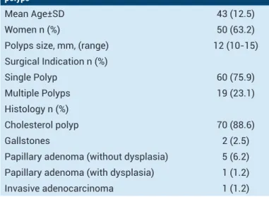
Table 1. Clinicopathologic features of patients operated for gallbladder polyps
Mean Age±SD 43 (12.5)
Women n (%) 50 (63.2)
Polyps size, mm, (range) 12 (10-15)
Surgical Indication n (%) Single Polyp 60 (75.9) Multiple Polyps 19 (23.1) Histology n (%) Cholesterol polyp 70 (88.6) Gallstones 2 (2.5)
Papillary adenoma (without dysplasia) 5 (6.2)
Papillary adenoma (with dysplasia) 1 (1.2)
Invasive adenocarcinoma 1 (1.2)
The postoperative histology examination reported that 70 (88.6%) patients had cholesterol polyps, 2 (2.5%) patients had gallbladder stones, 5 (6.3%) patients had papillary adenomas without dysplasia, 1 (1.2%) patient had papillary adenoma with dysplasia, and 1 (1.2%) patient had gallbladder adenocarcinoma. The patient diagnosed with adenocarcinoma was an 81-year old female and the gallbladder polyp was 15 mm in diameter. During the follow-up period, the patient died after 26 months due to myocardial infarction without recurrence of the original diagnosis.
The patient determined with papillary adenoma with dysplasia was a 76-year old female and the gallbladder polyp diameter was 13mm. The patient was followed up with no problems. In the 6 (7.5%) patients aged >70 years, malignant and premalignant lesions were determined in 2 (33.3%).
DISCUSSION
An increasing number of patients are being diagnosed with gallbladder polyps and undergoing surgical
treatment. Opinions are divided as to whether gallbladder polyps are the precursors to gallbladder adenocarcinoma. In this study, the clinicopathological features of patients who underwent cholecystectomy for GPs were examined and only one patient had adenocarcinoma and one had dysplasia.
Studies have shown that GPs have been detected in 0.5% - 11% of patients applied with cholecystectomy due to various gallbladder diseases (10). In the current series, 3.8% of the patients who underwent cholecystectomy were diagnosed with GPs. In the literature, GPs are more common in females and frequency increases with age (5, 11). Consistent with these findings in literature, 63.2% of the current study patients were female and 60.7% of the patients were aged > 40 years.
Ultrasonography is the least invasive procedure in gallbladder pathologies, and diagnostic sensitivity has been reported to be 45-90% (12). There are studies in literature that have reported that CT and MRI are no more useful than USG in the differentiation of benign and malignant polyps and they are not in the diagnosis algorithm (13, 14). If the diagnosis is suspected, endoultrasonography can be used (1). Since the use of USG became widespread, the number of patients incidentally diagnosed with gallbladder polyps has increased. Consequently, the rates of cholecystectomy performed because of GPs have also increased (15,5). All the patients in the current series were diagnosed with ultrasonography.
Cholesterol polyps are the most common type of GP. Histologically, it consists of histiocytes containing cholesterol, which are covered with columnar single-row epithelium. Although the etiology is not exactly known, it has been suggested that storage of serum cholesterols directly in the gallbladder, as well as the clustering of free sterols from bile or changes in the liver cholesterol synthesis after cholesterolosis, are the mechanisms involved (10). In the current series, 88.6% of the patients who were operated on for gallbladder polyps were found to have cholesterol polyps.
In patients with chronic cholecystitis, precancerous lesions such as metaplasia and dysplasia can be seen in the mucosa. Generally, antral, intestinal and squamous metaplasia are encountered but low-grade dysplasia may also be seen (16,17). These lesions can be seen alone, or as a component of rare adenomas of the gallbladder and biliary tract. These lesions may also sometimes appear with gallbladder adenomas. In the current study, papillary adenoma without dysplasia was detected in 5 (6.3%) patients, while in 1 (1.2%) patient, adenoma with dysplasia was detected.
Gallbladder carcinoma is usually an advanced age disease and it is usually detected in patients over 50 years of age (5,9,18,19). The risk of cancer increases in patients with gallbladder polyps >1 cm and it also increases in those with increasing GP size during follow-ups and with GP that are broad-based (3,20). An initial diagnosis of GP is 1162
Ann Med Res 2019;26(7):1161-3
made in 15% - 25% of patients with gallbladder carcinoma (21-22). There are studies that suggest gallbladder polyps >12 mm may convert to cancer (23). There are also publications that suggest performing cholecystectomy with frozen accompaniment in polyps >20 mm (24). In a study by Bhatt et al., the probability of cancer was stated to be close to zero, when polyp diameter was < 4.15 mm. ıt was also reported that the medical management of polyps 4.15 mm - 10 mm in size was important and 93% of the polyps did not show any growth during follow-up (25). It has also been stated that rapid growth may be associated with malignancy and neoplastic polyps may be observed in 5-10% of patients operated on for polyps (26,27). Consistent with the literature (25-27), surgical treatment is recommended in our clinic for polyps >10mm, patients with polyps <10mm are followed up conservatively with surgery applied if there is an increase in polyp diameter. In 1 (1.2%) patient of the current series who was operated on for a diagnosis of GP, and was then re-diagnosed with cancer, it was noticed that the patient was of an advanced age and the polyp diameter was 15mm. In the current study patients aged ≥ 70 years, a diagnosis of malignant and premalignant lesions was made in 33.3%.
Limitations of this study can be said to be the retrospective design and the lack of data for the etiology. It would be useful to study the surgical indications of gallbladder polyps in a large series showing the relationship between adenocarcinoma and the diameter as well as the characteristics of the gallbladder polyps.
CONCLUSION
In conclusion, as there is a higher risk of malignancy of polyps in the gallbladder which are of large diameter, in females and in older patients, it would be appropriate to operate with frozen if necessary during surgery on these patients.
Competing interests: The authors declare that they have no competing interest.
Financial Disclosure: There are no financial supports
Ethical approval: This work has been approved by the Institutional Review Board.
Refik Bademci ORCID: 0000-0001-9378-4438 Yasar Ozdenkaya ORCID: 0000-0003-0428-2511
REFERENCES
1. Persley KM. Gallbladder polyps. Curr Treat Options Gastroenterol 2005;8:105-8.
2. Yang HL, Sun YG, Wang Z. Polypoid lesions of the gallbladder: diagnosis and indications for surgery. Br J Surg 1992;79:227-9.
3. Jones-Monahan KS, Gruenberg JC, Finger JE, et al. Isolated small gallbladder polyps: an indication for cholecystectomy in symptomatic patients. Am Surg 2000;66:716-9.
4. Park CH, Chung MJ, Oh TG, et al. Differential diagnosis between gallbladder adenomas and cholesterol polyps on contrast-enhanced harmonic endoscopic ultrasonography. Surg Endosc 2013;27:1414-21.
5. Terzi C, Sökmen S, Seçkin S, et al. Polypoid lesions of the gallbladder: report of 100 cases with special reference to
operative indications. Surgery 2000;127:622-7.
6. Azuma T, Yoshikawa T, Araida T, et al. Differential diagnosis of polypoid lesions of gallbladder by endoscopic ultrasonography. Am J Surg 2001;181:65-70.
7. Mainprize KS, Gould SW, Gilbert JM. Surgical management of polypoid lesions of the gallbladder. Br J Surg 2000;87:414-7. 8. Kratzer W, Haenle MM, Voegtle A, et al. Ultrasonographically
detected gallbladder polyps: a reason for concern? A seven-year follow-up study. BMC Gastroenterol 2008;8:41. 9. Cairns V, Neal CP, Dennison AR, Garcea G. Risk
and costeffectiveness of surveillance followed by cholecystectomy for gallbladder polyps. Archives Surg 2012;147:1078-83.
10. Jones-Monahan KS, Gruenberg JC, Finger JE, et al. Isolated small gallbladder polyps: an indication for cholecystectomy in symptomatic patients. Am Surg 2000;66:716-9.
11. Ozdemir A, Ozenc A, Bozoklu S, et al. Ultrosonography in the diagnosis of gallbladder polyps. Br J Surg 1993;80:345. 12. Sadamoto Y, Oda S, Tanaka M, et al. Useful approach to
the differential diagnosis of small polypoid lesions of the gallbladder, utilizing and endoscopic scoring system. Endoscopy 2002;34:959-65.
13. Park JY, Hong SP, Kim YJ, et al. Long-term follow up of gallbladder polyps. J Gastroenterol Hepatol 2009;24:219-2. 14. Kim SW, Kim HC, Yang DM, et al. Gallbladder carcinoma:
causes of misdiagnosis at CT. Clin Radiol 2016;71:e96-109. 15. Csendes A, Burgos AM, Csendes P, et al. Late follow-up of
polypoid Lesions of the gallbadder smaller than 10 mm. Ann Surg 2001;234:657-60.
16. Mazlum M, Dilek FH, Yener AN, et al. Profile of gallbladder diseases diagnosed at Afyon Kocatepe University: a retrospective study. Turk Patoloji Derg 2011;27:23-30. 17. Seretis C, Lagoudianakis E, Gemenetzis G, et al. Metaplastic
changes in chronic cholecystitis: implications for early diagnosis and surgical intervention to prevent the gallbladder metaplasia-dysplasia-carcinoma sequence. J Clin Med Res 2014;6:26-9.
18. Yang HL, Kong L, Hou LL, et al. Analysis of risk factors for polypoid lesions of gallbladder among health examinees. World J Gastroenterol 2012;18:3015-9.
19. Xu Q, Tao LY, Wu Q, et al. Prevalences of and risk factors for biliary stones and gallbladder polyps in a large Chinese population. HPB Oxf 2012;14:373-81.
20. Kwon W, Jang JY, Lee SE, et al. Clinicopathologic features of polypoid lesions of the gallbladder and risk factors of gallbladder cancer. J Korean Med Sci 2009;24:481-7 21. Reid KM, Ramos-De la Medina A, Donohue JH. Diagnosis
and surgical management of gallbladder cancer: a review. J Gastrointest Surg 2007;11:671-81.
22. Rodríguez-Fernádez A, Gómez-Río M, Medina-Benitez A, et al. Application of modern imaging methods in diagnosis of gallbladder cancer. J Surg Oncol 2006;93:650-64.
23. Kozuka S, Tsubone N, Yasui A, et al. Relation of adenoma to carcinoma in the gallbladder. Cancer 1982;50:2226-34. 24. Lee KF, Wong J, Li JC, et al. Polypoid lesions of the
gallbladder. Am J Surg 2004;188:186-90.
25. Bhatt NR, Gillis A, Smoothey CO.. Evidence based management of polyps of the gall bladder: A systematic review of the risk factors of malignancy. Surgeon 2016;14:278-86.
26. Moriguchi H, Tazawa J, Hayashi Y, et al. Natural history of polypoid lesions in the gall bladder. Gut 1996;39:860-2. 27. Koga A, Watanabe K, Fukuyama T, et al. Diagnosis and
operative indications for polypoid lesions of the gallbladder. Archives Surg 1988;123:26-9.