The use of suprapubic cystoscopy in perineal urethroplasty
Perineal üretroplastide suprapubik sistoskopi kullanımı
Rahim Horuz1, Cemal Göktaş2, Ali Cihangir Çetinel2, Oktay Akça2, Ahmet Selimoğlu2, Selami Albayrak1
1Department of Urology, Faculty of Medicine, Medipol University, İstanbul, Turkey 2Department of Urology, Kartal Training and Research Hospital, İstanbul, Turkey Submitted: 17.02.2013 Accepted: 06.05.2013 Correspondence: Rahim Horuz Department of Urology, Faculty of Medicine, Medipol University, Bağcılar 34214 İstanbul, Turkey Phone: +90 212 460 72 86 E-mail: [email protected] ©Copyright 2013 by Turkish Association of Urology Available online at www.turkishjournalofurology.com ABSTRACT
Objective: Posterior urethral strictures are difficult cases to treat for urologists. Anastomotic open
urethro-plasty is the major surgical treatment option, and various modifications/manipulations to the procedure have been recommended in the literature. We aimed to assess the advantages of performing antegrade endoscopy through the suprapubic cystostomy tract during perineal urethroplasty.
Material and methods: Thirty-six perineal urethroplasties combined with suprapubic cystoscopy were
performed in 33 adult male patients between 2005 and 2011. Pre-and peri-operative records of the patients were evaluated, with a particular focus on suprapubic cystoscopy findings during urethroplasty.
Results: The mean patient age was 41 years, and the mean lesion length was 2.6 cm (range 1-10 cm). Lesions
were secondary to pelvic trauma in 21 patients, to a history of prostatic surgery in 8 patients, and to other miscellaneous causes in the 4 remaining patients. In 23 cases with normal bladder necks, the anastomosis was created under the guidance of antegrade cystoscopy with fluoroscopy. In 10 cases, stiff guide wires or paddles were used in addition to endoscopic light to identify the correct anastomosis site. In two procedures, fistula openings were identified during endoscopy, and fistula repair was performed along with anastomosis. In one case involving the longest lesion with involvement of the bladder neck, a combined perineal-transpu-bic urethroplasty was performed. The mean follow up duration was 19 (3-38) months. The overall success rate was 91%, and the re-operation rate was 9%.
Conclusion: Suprapubic cystoscopy is an easy-to-perform adjunctive modality in perineal urethroplasty.
This method provides the benefits of supplementary endoscopic findings and feasibility of certain maneu-vers that facilitate debridement and anastomosis formation concurrent with perineal urethroplasty.
Key words: Anastomosis; cystoscopy; perineum; suprapubic; urethral stricture.
ÖZET
Amaç: Posterior üretra darlıkları ürologlar için tedavisi zor olgular arasında yer almaktadır. Cerrahi
tedavi-de temel seçenek olan anastomotik açık uretroplasti için literatürtedavi-de çeşitli manuplasyonlar ve modifikasyon-lar tanımlanmıştır. Bu çalışmada perineal uretroplasti sırasında suprapubik sistostomi traktından uygulanan antegrad endoskopinin avantajlarını değerlendirmeyi amaçladık.
Gereç ve yöntemler: 2005-2011 yılları arasında toplam 36 seans suprapubik sistoskopi yardımlı perineal
uretroplasti operasyonu uygulanan 33 yetişkin erkek hastanın, özellikle uretroplasti sırasındaki sistoskopik bulgular başta olmak üzere, operatif verileri incelendi.
Bulgular: Hastaların ortalama yaşı 41 yıl ve lezyon uzunluğu 2,6 (1-10) cm idi. Etyolojik faktör olarak 21
hastada travma, 8 hastada prostat cerrahisi, ve 4 hastada diğer nedenler saptandı. Yirmi üç olguda mesane boynu normaldi ve antegrad sistoskopun ışığı kılavuzluğunda anastomoz gerçekleştirildi. On olguda doğru anastomoz hattını saptamak için, ilaveten, sert kılavuz tellerden ve iğnelerden yararlanıldı. İki olguda en-doskopi sırasında fistül saptandı ve anastomoza ilave olarak fistül onarımı uygulandı. Mesane boynunu da kapsayacak şekilde, en uzun darlığa sahip olan 1 olguda combine perineal-transpubik onarım uygulandı. Hastaların ortalama takip süresi 19 (3-38) ay idi. Genel başarı oranı %91 ve re-operasyon oranı %9 idi.
Sonuç: Suprapubik sistoskopi perineal uretroplasti sırasında yararlanılabilecek kolaylıkla uygulanabilen
bir yardımcı yöntemdir. Bu yöntem perineal uretroplasti sırasında debridman ve anastomoz safhalarını ko-laylaştıracak destekleyici endoskopik bulgular sağlaması ve çeşitli manevralara imkan tanıması açısından avantaj sağlar.
Introduction
It is well known in urology that posterior urethral strictures are among the most difficult cases from a treatment standpoint. Additional difficulties in surgical treatment arise because the lesions generally result from a trauma with frequent coexist-ing pubic fracture and because of potential postoperative uro-sexual complications. [1-3] Anastomotic perineal urethroplasty is
the major surgical treatment option, and various modifications and manipulations to the procedure have been proposed in the literature. [1,4-15]
We aimed to assess the advantages of performing antegrade endoscopy through the suprapubic cystostomy tract during peri-neal urethroplasty.
Material and methods
Thirty-three adult male patients diagnosed with posterior ure-thral stricture were included in this study. Thirty-six perineal urethroplasty operations combined with suprapubic cystoscopy sessions were performed on the study patients between May 2005 and January 2011. Informed consent was obtained from all patients. The etiology of the lesions, findings of voiding cys-tourethrography (VUCG) and operative records of the patients were evaluated, with a particular focus on suprapubic cystos-copy findings during urethroplasty. Postoperative outcome data were also assessed.
The procedure
While the patient is in the lithotomy position, after the surgeon operating in the perineal region reaches the distal end of the posterior urethral stricture, a 17 Fr cystoscope is advanced to the bladder through the suprapubic cystostomy tract. The operation table may be repositioned with hyperextension if the anterior abdominal wall limits the movements of the endoscope. An overall endoscopic examination of the bladder is performed. After the bladder neck is visualized and examined, the cysto-scope is advanced to the prostatic urethra. If necessary, a guide wire may be utilized to direct the cystoscope. When the veru-montanum is reached, the obliterated urethral level generally becomes visible. Gentle movements of the endoscope vertically and horizontally may provide guidance to the surgeon perform-ing the urethral/periurethral scar excision on the perineal region, determining the proximal side of the anastomosis. By observing the reflection of the endoscopic light, the surgeon can adequate-ly open the proximal end of the urethra and create an adequate end-to-end anastomosis. When the endoscopic light is not sufficient for guidance of the dissection and anastomosis, two additional manipulations may be utilized. The first manipula-tion involves sending a stiff guide wire from the cystoscope and pulling it out from the perineum after perforating the proximal
end of the stricture. As a second option, a 20 G needle may be used to create a puncture in the proximal stricture site while the tip of the needle is observed via antegrade cystoscopy to define the exact site for anastomosis.
Results
We performed 36 perineal urethroplasty sessions in 33 patients with a mean age of 41 (17-72) years. Of the 36 procedures, tension-free end-to-end anastomosis with urethral mobilization was performed in 33 cases. Patch urethroplasty was performed with the use of buccal mucosa in 2 cases and with skin graft in 1 case.
The mean defect length was 2.6 cm (range 1-10 cm). While the defect was localized to the posterior urethra in 32 of the cases, both the anterior and posterior urethra were involved in the remaining patient, who had a 10 cm defect.
Lesions were secondary to pelvic trauma with pelvic fracture in 21 patients, to a history of prostatic surgery in 8 patients (3 radical, 1 open, and 4 endoscopic prostatectomies), to blunt perineal trauma in 1 patient, to a gunshot wound in 1 patient, to a history of multiple attempts of penoscrotal hypospadias repair in 1 patient, and to iatrogenic injury of the bladder neck and ure-thra surgery performed for rectal carcinoma in one patient with a history of radiotherapy. Three of these patients had previous failed attempts at open urethroplasty performed in other centers.
Findings on suprapubic cystoscopy
In 23 procedures, the bladder neck was found to be normal, and it was possible to advance into the prostatic urethra and to visualize the verumontanum endoscopically. Once dissection of the fibrotic tissues was completed, the proximal end of the anastomosis plane was easily visualized under the guidance of endoscopic fluoroscopy from the perineal side, and the anasto-mosis was created accordingly.
In 5 procedures, it was not possible to advance the endoscope directly into the urethra, and thus, the cystoscope was advanced through the proximal urethra over a guide wire until the veru-montanum was visualized (Figure 1).
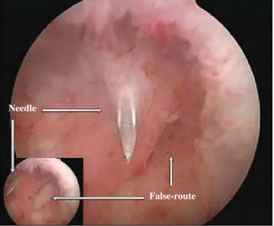
For 5 procedures in which the bladder neck and prostatic ure-thra were irregular and had false routes, anastomosis was made possible using the guidance of a 20 G needle introduced from the perineal side. This needle was observed endoscopically, and anastomosis was performed accordingly (Figure 2).
In two procedures, fistula openings were visualized during endoscopy: one located in the membranous urethra (urethrorec-tal) and the other in the proximal bulbous urethra
(urethrocuta-neous). In these cases, fistula repair was performed along with end-to-end anastomosis.
In one of the cases, the stricture started at the level of the blad-der neck. The defect length was much longer in this patient due to elevation of the bladder neck by a perivesical hematoma, which developed during the trauma and subsequently became fibrosed. Transperineal repair was combined with a transpubic approach in this case, and patch urethroplasty with skin graft was performed.
Clinical outcomes
The mean duration of follow up was 19 (3-38) months. In 24 (73%) of the patients, controlled physiological micturition was achieved without the need for any additional intervention, with a mean Qmax value of 17 mL/s and a mean Qave of 12 mL/s on uroflowmetry. Postoperative mechanical urethral dilatation was required in four cases (12%). In one patient (3%), opti-cal urethrotomy was required on the 7th day after removal of the transurethral catheter. An artificial urinary sphincter was implanted in another patient (3%) due to total urinary inconti-nence after perineal urethroplasty. In 3 patients (9%), urethral stricture recurred, and an additional session of perineal urethro-plasty was required for each patient. Two of these cases had controlled physiological voiding after re-operation. In the third case, the patient had an initial urethral defect 10 cm long and had a history of failed urethroplasty with buccal mucosal graft.
Urethral continuity was achieved in this patient after second and third urethroplasty procedures were performed in our clinic. The incontinent patient was scheduled for artificial urinary sphincter implantation.
Discussion
It is well known that patients with posterior urethral strictures are a heterogeneous group with regards to the etiology and severity of lesions. Posterior urethral strictures may result from traumatic or iatrogenic causes. In the lesions in which the urethral rupture is incomplete and continuous with the lumen, endoscopic surgical treatment may be a first choice. However, reported recurrence rates are as high as 38-75% following both cold-knife and laser endoscopic urethrotomies.[16-18] In addition,
repeated urethrotomies for recurrent strictures may lead to an increase in the length and depth of fibrosis in some cases. Difficult or complex urethral strictures may refer to strictures resulting from traumatic ruptures of the posterior urethra or bladder neck, those resulting from direct invasion by tumors of neighboring organs or from tumor-treating surgery, or those that develop after radiotherapy, prostatectomy, multiple urethroto-mies or gunshot wounds.
For most post-traumatic posterior urethral strictures with con-current pelvic fracture, the exact pathology may be defined as a disruption or gap in the bulbo-prostatic urethra rather than a classical isolated stricture. The term “urethral distraction defect” is suggested in the literature to describe these types of
Figure 1. View of a guide wire passed in an antegrade manner through a cystoscope and pulled out from the perineal side
Figure 2. Antegrade endoscopic view of a false route. Inserting a needle from the perineal side and advancing into the bladder may prevent poor alignment during anastomosis
Needle
lesions.[19] In such cases, the defective region is generally full of
fibrotic tissue resulting from traumatic hematoma.[4] As urethral
continuity is generally lost, endoscopic treatment cannot be performed in most of these cases.[1-3,10]
The most commonly used methods to characterize the lesions preoperatively are antegrade cystourethrography and retro-grade urethrography. Pelvic computerized tomography and magnetic resonance imaging are also helpful in determining the depth of the lesion and bony structures. However, in spite of these modalities, in many cases, the anatomical details of the lesion encountered during the operation may be different than expected. When the bladder neck and proximal urethra cannot be visualized by preoperative methods, a suprapubic cystoscopy performed through the cystostomy tract may provide informa-tion about anatomical details of this region. Hosseini et al.[11]
published a study of 111 cases using suprapubic flexible cys-toscopy to manage posterior urethral stricture. They emphasize that this method provides valuable information regarding the anatomy of the displaced posterior urethra and helps to create a healthy anastomosis by allowing visualization of the proximal ends of the anastomosis.
While the success rate of open urethroplasty in treating pos-terior urethral stricture is reported to be over 90% by some authors, others report a success rate of approximately 70%.[20]
In addition to surgical experience, such heterogeneous results may be attributed to the technique employed. The essential aim of a urethroplasty is to achieve a controlled spontaneous mic-turition with adequate flow rate. The two major requirements for that result are adequate scar debridement and a tension-free end-to-end healthy anastomosis. In the literature, several modifications and/or manipulations have been proposed to sat-isfy these requirements. The most common modification is the advancement of materials such as a sound, bougie, endoscope or endoscopic light through the suprapubic cystoscopy tract to gain the advantage of antegrade guidance for anastomosis.[10-15]
In this study, we combined antegrade suprapubic cystoscopy with perineal urethroplasty to utilize the perioperative endo-scopic findings and additional endoendo-scopic manipulations to create a healthy and safe anastomosis. During perineal urethro-plasty, after determining the distal end of the stenotic urethral segment, it is necessary to accurately define the proximal end. In that stage of the operation, most urologists prefer to use a hard bougie, which is introduced from the cystostomy toward the prostatic urethra, as a guide for the proximal urethra to define the anastomosis site. In most complicated cases like ours (cases with a traumatic etiology and sometimes accompa-nied by pelvic fracture), the lesion may involve an unexpect-edly long segment of the posterior urethra and may be densely fibrotic. Furthermore, VCUG may be limited in determining the
anatomy of bladder neck pre-operatively. In such situations, to ascertain the proximal end of the stricture during the operation, using a hard bougie and moving it transvesically in a blinded manner carries the potential risk of advancing through a false-passage instead of the prostatic urethra.[12,13]
Another complex group of patients undergoing posterior ure-throplasty are recurrent cases with a history of multiple failed endoscopic urethrotomy operations. In these cases, as chronic false-passages may exist adjacent to the stricture, difficulties in defining the appropriate anastomosis site during surgery may result in a disappointing outcome.
It has been reported that utilization of suprapubic cystoscopy in urethroplasty gives the surgeon an opportunity to inspect the proximal end of the stricture, providing the advantage of being able to avoid causing a false-passage or mal-alignment dur-ing the anastomosis.[12,13] By using suprapubic cystoscopy, the
anatomic integrity of the bladder neck can easily be visualized, and when advanced to the membranous urethra after passage through the prostatic urethra, the obliterated end of the proximal urethra can be visualized. When the endoscope has contact with this end, moving the endoscope slightly in all directions can guide the surgeon operating in the perineal side for debridement of the fibrosis and excision of the obliterated proximal urethral tissues. The surgeon can then define the appropriate site for anastomosis by both following the light of the endoscope and by palpating the hard tip of endoscope through the perineum. To our knowledge, the first example of this method was a case report published in 1988. Rich et al reported that a safe and easy debridement of scar tissue and a healthy anastomosis were achievable by utilizing the light guidance of a flexible cysto-scope during posterior urethroplasty in a male child.[21]
Despite these maneuvers, advancing to the proximal end may not be possible, or we may not be confident of the direction of the dissection prior to the anastomosis in patients with heavy fibrosis. In such cases, while the obliterated segment is being antegradely visualized with suprapubic cystoscopy, the direc-tion of the anastomosis can be determined by making a puncture with a 20 G needle from the perineal side through the residual obliterated urethra and introducing it into the bladder until the tip of the needle is visualized with the endoscope. Alternatively, a stiff guide wire may be inserted through the endoscope in an antegrade manner and palpated through the perineum.
The varying success rates of posterior urethroplasty reported in the literature may be due to the heterogeneity in patient characteristics (including age, history of trauma or past opera-tions), duration of follow up, and criteria used to evaluation outcomes. In most studies, a successful procedure is defined as no recurrence of the stricture, and re-operation is considered
a failure. In our study, the re-operation rate was 9%, in accor-dance with the overall success rates of 82-95% reported in the literature.[12,20,22-25] We assume that our success rate of 91% is
associated with the use of suprapubic cystoscopy during peri-neal urethroplasty.
In conclusion; suprapubic cystoscopy is an easy-to-perform adjunctive modality in perineal urethroplasty and can be used without incurring any considerable additional cost. We believe that this method offers significant advantages for a surgeon, including the benefits of supplementary endoscopic findings and the potential use of certain maneuvers while performing both the debridement and the anastomosis during perineal ure-throplasty.
Conflict of Interest
No conflict of interest was declared by the authors.
Peer-review: Externally peer-reviewed.
Informed Consent: Written informed consent was obtained from
patients who participated in this study.
Author Contributions
Concept - C.G.; R.H., C.G.; Design - R.H., C.G.; Supervision - S.A.; Funding - A.C.Ç., O.A., S.A.; Materials O.A., A.C.Ç.; Data Collection and/or Processing - A.S., O.A.; Analysis and/or Interpretation - R.H., C.G.; Literature Review - R.H., C.G., A.C.Ç.; Writer - R.H.; Critical Review - R.H.
Çıkar Çatışması
Yazarlar herhangi bir çıkar çatışması bildirmemişlerdir.
Hakem değerlendirmesi: Dış bağımsız.
Hasta Onamı: Yazılı hasta onamı bu çalışmaya katılan hastalardan
alınmıştır.
Yazar Katkıları
Fikir - C.G.; R.H., C.G.; Tasarım - R.H., C.G.; Denetleme - S.A.; Kaynaklar - A.C.Ç., O.A., S.A.; Malzemeler O.A., A.C.Ç.; Veri toplanması ve/veya işlemesi - A.S., O.A.; Analiz ve/veya yorum - R.H., C.G.; Literatür taraması - R.H., C.G., A.C.Ç.; Yazıyı yazan - R.H.; Eleştirel İnceleme - R.H.
References
1. Koraitim MM. The lessons of 145 post-traumatic posterior urethral strictures treated in 17 years. J Urol 1995;153:63-6. [CrossRef]
2. Mundy AR. Urethroplasty for posterior urethral strictures. Br J Urol 1996;78:243-7. [CrossRef]
3. Turner Warwick R. Principles of urethral reconstruction. In: Webster G, Kirby R, King L, Goldwasser B (ed) Reconstructive urology, Blackwell, Cambridge, Massachusetts; 1993.p.609-42.
4. Turner Warwick R. Prevention of complications resulting from pelvic fracture urethral injuries-and from their surgical manage-ment. Urol Clin North Am 1989;16:335-8.
5. Andrich DE, Dunglison N, Greenwell T J, Mundy AR. The long-term results of urethroplasty. J Urol 2003;170:90-2. [CrossRef]
6. Corriere JN Jr. 1-stage delayed bulboprostatic anastomotic repair of posterior urethral rupture: 60 patients with 1-year followup. J Urol 2001;165:404-7. [CrossRef]
7. Morey AF, McAninch JW. Reconstruction of posterior urethral disruption injuries: outcome analysis in 82 patients. J Urol 1997;157:506-10. [CrossRef]
8. Netto NR Jr, Lemos GC, Claro JF. Internal urethrotomy as a complementary method after urethroplasties for posterior urethral stenosis. J Urol 1989;141:50-1.
9. Webster GD, Ramon J. Repair of pelvic fracture posterior urethral defects using an elaborated perineal approach: experience with 74 cases. J Urol 1991;145:744-8.
10. Webster GD, MacDiarmid SA. Posterior urethral reconstruction. In: Webster G, Kirby R, King L, Goldwasser B, (ed). Reconstructive urology. Blackwell, Cambridge, Massachusetts; 1993.p.687-702. 11. Hosseini SJ, Kaviani A, Jabbari M, Hosseini MM,
Haji-Mohammadmehdi-Arbab A, Simaei NR. Diagnostic application of flexible cystoscope in pelvic fracture urethral distraction defects. J Urol 2006;3:204-7.
12. Kizer WS, Armenakas NA, Brandes SB, Cavalcanti AG, Santucci RA, Morey AF. Simplified reconstruction of posterior urethral disruption defects: limited role of supracrural rerouting. J Urol 2007;177:1378-81. [CrossRef]
13. Hafez AT, El-Assmy A, Sarhan O, El-Hefnawy AS, Ghoneim MA. Perineal anastomotic urethroplasty for managing post-traumatic urethral strictures in children: the long-term outcome. BJU Int 2005;95:403-6. [CrossRef]
14. Jordan GH, Schlossberg SM. Surgery of the penis and urethra. In: Walsh PC, Retik AB, Vaughan ED Jr, et al (ed). Campbell’s Urology. 8th ed. WB Saunders, Philadelphia; 2002.p.3886-954. 15. Lewis RM, McCullough DL. Use of the flexible fiberoptic
neph-roscope in the preoperative evaluation and delayed repair of trau-matic urethral strictures. J Urol 1985;133:1036-7.
16. Gibod BL, Le Portz B. Endoscopic urethrotomy: Does it live up to its promises? J Urol 1982;127:433-5.
17. Albers P, Fichtner J, Brühl P, Müller SC. Long-term results of internal urethrotomy. J Urol 1996;156:1611-4. [CrossRef]
18. Holm-Nielsen A, Schultz A, Moller-Pedersen V. Direct vision urethrotomy: A critical review of 365 operations. Br J Urol 1984;56:308-11. [CrossRef]
19. Mundy AR. Transperineal bulbo-prostatic anastomotic urethro-plasty. World J Urol 1998;16:164-70. [CrossRef]
20. Koraitim MM. On the art of anastomotic posterior urethroplasty: a 27-year experience. J Urol 2005;173:135-9. [CrossRef]
21. Rich M, Brock WA. Flexible cystoscopy in surgical repair of traumatic urethral strictures in children. Urology 1988;31:129-31. [CrossRef]
22. Pratap A, Agrawal CS, Tiwari A, Bhattarai BK, Pandit RK, Anchal N. Complex posterior urethral disruptions: management by combined abdominal transpubic perineal urethroplasty. J Urol 2006;175:1751-4. [CrossRef]
23. Al-Rifaei MA, Zaghloul S, Al-Rifaei AM. Bulboprostatic anas-tomotic urethroplasty with preservation of potency: anatomical study, operative approach and clinical results. Scand J Urol Nephrol 2005;39:163-8. [CrossRef]
24. Koraitim MM. Failed posterior urethroplasty: lessons learned. Urology 2003;62:719-22. [CrossRef]
25. Aghaji AE, Odoemene CA. One-stage urethroplasty for strictures: Nigerian experience. Int J Urol 2001;8:380-5. [CrossRef]