Current Topics in Medicinal Chemistry, 2018, 18, 1-33 1
REVIEW ARTICLE
1568-0266/18 $58.00+.00 © 2018 Bentham Science Publishers
Prediction and Targeting of Interaction Interfaces in G-Protein Coupled
Receptor Oligomers
Anke C. Schiedel
1,#, Meryem Köse
1#, Carlos Barreto
2, Beatriz Bueschbell
1, Giulia Morra
3,4,
Ozge Sensoy
5,*and Irina S. Moreira
2,6,*1
Pharmaceutical Chemistry I, PharmaCenter Bonn, University of Bonn, 53121 Bonn, Germany;
2Data-driven Molecular
Design, CNC - Center for Neuroscience and Cell Biology, University of Coimbra;
3Weill-Cornell Medical College,
De-partment of Physiology and Biophysics, 1300 York Ave, New York, NY 10065, USA;
4ICRM-CNR Istituto di Chimica del
Riconoscimento Molecolare, Consiglio Nazionale delle Ricerche, Via Mario Bianco 9, 20131 Milano, Italia;
5Istanbul
Medipol University, The School of Engineering and Natural Sciences, 34810, Istanbul, Turkey;
6Bijvoet Center for
Bio-molecular Research, Faculty of Science - Chemistry, Utrecht University, Utrecht, 3584CH, The Netherlands
A R T I C L E H I S T O R Y Received: February 16, 2018 Revised: May 14, 2018 Accepted: May 15, 2018 DOI: 10.2174/1568026618666180604082610
Abstract: Background: Communication within a protein complex is mediated by physical interactions
made among the protomers. Evidence for both the allosteric regulation present among the protomers of
the protein oligomer and of the direct effect of membrane composition on this regulation has made it
essential to investigate the underlying molecular mechanism that drives oligomerization, the type of
in-teractions present within the complex, and to determine the identity of the interaction interface. This
knowledge allows a holistic understanding of dynamics and also modulation of the function of the
re-sulting oligomers/signalling complexes. G-protein-coupled receptors (GPCRs), which are targeted by
40% of currently prescribed drugs in the market, are widely involved in the formation of such
physio-logical oligomers/signalling complexes.
Scope of the Review: This review highlights the importance of studying protein-protein interactions
(PPI) by using a combination of data obtained from cutting-edge experimental and computational
meth-ods that were developed for this purpose. In particular, we focused on interaction interfaces found at
GPCR oligomers as well as signalling complexes, since any problem associated with these interactions
causes the onset of various crucial diseases.
Major Conclusions: In order to have a holistic mechanistic understanding of allosteric PPIs that drive
the formation of GPCR oligomers and also to determine the composition of interaction interfaces with
respect to different membrane compositions, it is essential to combine both relevant experimental and
computational data. In this way, efficient and specific targeting of these interaction interfaces in
oli-gomers/complexes can be achieved. Thus, effective therapeutic molecules with fewer side effects can be
designed to modulate the function of these physiologically important receptor family.
Keywords: GPCRs, dimerization, PPI, oligomers, ghrelin, molecular dynamics, umbrella sampling, hot spot.
1. INTRODUCTION
Determining key players that govern proteprotein
in-teractions and also understanding the underlying molecular
mechanism of oligomerization are essential for modulating
various physiological functions in the cell such as signal
transduction pathways, in which various proteins do function
in coordination to respond to the stimulus reliably and
timely. Evidences have shown that a protein, when is part of
an oligomer, can modulate the function of the other members
*Address correspondence to these authors at the Data-driven Molecular Design, CNC - Center for Neuroscience and Cell Biology, University of Coimbra, Coimbra, Portugal and Bijvoet Center for Biomolecular Research, Faculty of Science - Chemistry, Utrecht University, Utrecht, 3584CH, The Netherlands; E-mail: [email protected] (S. Moreira); Istanbul Medi-pol University, The School of Engineering and Natural Sciences, 34810, Istanbul, Turkey; E-mail: [email protected] (O. Sensoy)
#Authors contributed equally to this work
present in the complex. In this respect, G-Protein Coupled
Receptors (GPCRs) constitute ideal systems for this
phe-nomenon. According to the current knowledge, they are
functional in monomeric and dimeric/oligomeric forms
(ei-ther homo or hetero) [1] and also they form complexes with
a wide array of signalling partners such as G-proteins [2],
arrestins, GPCR-kinases, PDZ-domain [3] containing
pro-teins to function properly. As to the GPCR oligomerization,
it has been shown that protomers within the oligomer can
allosterically cross-talk to each other either to alter the ligand
binding affinity or efficacy of the other members present in
the complex [4]. Considering the fact that GPCRs are
tar-geted by approximately 40% of currently prescribed drugs in
the market and also oligomers modulate the function of
indi-vidual GPCRs it is crucial to understand the molecular
mechanism of oligomer formation and also to determine
in-teraction interfaces that emerge under different
environ-mental conditions, e.g. membrane composition.
The first step before determining the interaction interface
and studying PPIs is the identification of the constituents of
the complex/oligomer. There are a variety of experimental
methods which are developed for this purpose. Among many
others, proteomics approaches have been widely used despite
the inherent problems in studying membrane proteins due to
complex biochemical properties associated with these
sys-tems. Nevertheless, the cell-based and genetic assays have
been found successful for identifying numerous interaction
partners of GPCRs [5–13]
.Once the partners and interaction interfaces are
deter-mined, computational methods can be used to complement
experimental data as they provide atomistic information
re-garding both the structure dynamics of these physiological
complexes/oligomers [14]. In particular, one can determine
the set of residues involved in interaction interfaces and also
have an insight on the molecular mechanism of allosteric
interactions present among the protomers [15]. Moreover,
one can also achieve a molecular level understanding of the
effect that the membrane composition elicits on the
dynam-ics and the identity of the resulting interfaces. Here, it is
im-portant to emphasize that since the relaxation times of such
systems are large it is crucial to test if the results obtained
from in silico calculations are statistically reliable and
com-parable to experimental data.
In spite of existing experimentally determined structures
of GPCR oligomers (in particular, dimers) and signalling
complexes (with either G-protein or arrestin) they are scarce.
These structures reveal that some GPCR interfaces are
favoured over the others, in particular, those that are formed
by either transmembrane (TM) TM4-TM5 or TM1, TM2 and
TM8 suggesting that similar mechanisms might mediate the
oligomer formation in this receptor family [16,17].
Consider-ing the fact that GPCR oligomers are involved in various
pathophysiological pathways, in particular, neurological
dis-orders, cancer, an atomistic level knowledge regarding these
interfaces can lead to breakthroughs in the field of neurology
and also oncology.
In this review, we aim to make an extensive review on
recent experimental and computational methods that have
been widely used to determine interaction partners in GPCR
oligomers/signalling complexes and also those that are
de-veloped to investigate the identity and dynamics of the
inter-action interfaces. In addition, we present several examples of
software that are widely used for hot-spot prediction,
inhibi-tor design that target interaction interfaces in GPCRs. Lastly,
we finish by giving an example of one of the GPCRs that has
been known to form oligomers, namely Ghrelin receptor. We
also discussed the methods that have been used to target
di-mers formed by this receptor.
2. IN SILICO APPROACHES APPLIED TO THE
STUDY OF GPCR DIMERIZATION
2.1. Structural Determination and Characterization of
the Dimerization Interface
If any experimental data regarding the interaction
inter-face is available then it can be used to guide molecular
dock-ing calculations, instead of performdock-ing blind dockdock-ing whose
success has been shown to be far below than that of the
guided one. Alternatively, coarse-grained molecular
dynam-ics (CGMD) simulations can also be used to determine the
most probable interface. However, such calculations may
end up with more than one interface each of which having a
similar frequency. Under such circumstances, the stability of
each of these interfaces can be determined by using umbrella
sampling [18] or steered molecular dynamic (MD)
simula-tions [19–21]. These methods can also be used to
discrimi-nate between the native oligomer and other oligomers that
might be present in crystal structures of GPCR complexes as
a result of crystallization artefacts. Below, we discuss
above-mentioned computational techniques in the context of
identi-fication and assessment of the stability of protein-protein
interface(s) in GPCR oligomers.
2.1.1. Coarse-grained Molecular Dynamics Simulations: A
Computational Tool for Estimating Interaction Interface(s)
in GPCR Oligomers
Coarse-grain modelling can be used to represent a given
atomistic system by a reduced number of degrees of
free-dom. As a result of the reduction in the degrees of freedom
and elimination of fine details, one can simulate systems
with larger length scales and can access longer time scales at
the expense of losing atomistic details. Martini force field
[22] has been widely used for performing CGMD
simula-tions of GPCRs in an explicit membrane environment.
Ac-cording to the force field, each residue is represented by one
backbone bead and zero or more side-chains beads
depend-ing on the type of the amino acid. The protein in question is
allowed to change its tertiary arrangement; however, the
local secondary structure, which has an effect on the bead
type and also on the bonded parameters, is pre-defined and
so it is fixed throughout the simulation. Therefore, for
in-stance, one cannot study ligand-induced conformational
changes in the GPCR using CGMD simulations. Instead, the
exact conformational state of the receptor (active or inactive)
must be defined and assigned a-priori to each residue of the
receptor. The Martini force field allows [22] usage of a time
step in the range of 20-40 fs depending on the system
prop-erties. In particular, a four-to-one mapping is used where
four heavy atoms and associated hydrogens are on average
represented by a single interaction center. As a result, a
stan-dard conversion factor of 4, which corresponds to the
effec-tive speed-up factor in Martini water diffusion dynamics, is
used. For modelling non-bonded interactions, standard
cut-off schemes are used where Lennard Jones interactions are
shifted to zero in the range of 0.9-1.2 nm whereas
electro-static interactions in the range of 0.0-1.2 nm. The studies on
test systems have shown that while the translational and
rota-tional diffusion of a Class A GPCR, namely Rhodopsin,
have been shown to be in good agreement with experimental
data [23] the sampling of the local configurational space of a
lipid molecule [24] and the aggregation rates of lipids into
bilayers however have [25] been accelerated. Before
per-forming CGMD simulation of any GPCR-membrane system,
corresponding Martini time-scales of the system
compo-nents, protein, water, lipid, should be compared to available
experimental data to have an insight on the speed-up factor.
The self-assembly of GPCRs involves the slow diffusion
of lipid and receptor molecules, which may lead to problems
in achieving convergence due to lack of binding/unbinding
events [26]. This can be partially overcome by simulating
different replicas of the same system in parallel, in each of
which individual GPCRs are placed differently with respect
to each other. A recently developed high-throughput
simula-tion method, namely, docking assay for transmembrane
components (DAFT) [27,28], provides an automated
exten-sive sampling of different GPCR dimerization interfaces,
which is shown to be in excellent agreement with
experi-ments [28,29]. According to the method, multiple CG
simu-lations of the GPCR dimer, which is embedded in an explicit
membrane environment, are performed simultaneously. The
two GPCRs are initially placed at a fixed distance but at
dif-ferent starting orientations. By means of this ensemble
simu-lation setup, one can achieve statistically meaningful results
on the dimerization interface. Once the convergence issue
has been fixed in order to discriminate between random
con-tacts and recurrent interfaces
root-mean-square-difference-based clustering can be used [30]. According to the method,
first, the dimer pairs are fitted and then matrix of positional
root-mean-square-difference of the backbone beads of the
dimers is calculated. Subsequently, the number of
neighbouring dimers in the set is counted for each dimer
conformation. The dimer with the highest number of
neighbours is removed from the system together with its
neighbours. The process is repeated until the pool is empty.
2.1.2. In Silico Determination of Potential of Mean Force
(PMF) to Measure the Strength of Interaction interface(s)
in GPCR Oligomers
CGMD simulations of self-assembly of GPCRs may end
up with more than one oligomerization interface as
men-tioned above. In order to determine the relative stability of
these interaction interfaces, one can calculate the potential of
mean force (PMF) between corresponding GPCR monomer
or oligomer pairs. In addition, PMF can also be used to
dis-criminate between the native oligomer and the others present
in the crystal, which might be formed artificially because of
the crystallization conditions. In principle, PMF can be
com-puted from probability distribution functions of
conforma-tions that are sampled in unbiased simulaconforma-tions; however, the
lack of binding/unbinding events, even in CGMD
simula-tions, prevents one to compute statistically meaningful PMF.
In such circumstances, umbrella sampling [18] or steered
MD simulations [19,20] can be used together with Martini
force field [22], which has been shown to reproduce
reason-able protein-protein interaction energies upon a reduction in
Lennard Jones interaction term in the force field [31].
To perform an umbrella sampling, first, a series of initial
configurations of the GPCR dimer is generated along an
ap-propriate reaction coordinate, which is usually taken as the
distance between the pair of the receptor. In a study by
Johnston et al. [32], the authors carried out metadynamics
simulations to generate starting configurations for using in
umbrella sampling. In each of these configurations, one of
the protomers in the GPCR dimer is harmonically restrained
with respect to the other at increasing center-of-mass
dis-tance from a reference starting point. In this way, the GPCR
dimer is allowed to sample a defined region of the
configura-tional space along the selected reaction coordinate. After
preparation of initial configurations in each window,
simula-tions are started in parallel. Until achieving a good overlap
between neighbouring windows, which is important for the
proper reconstruction of the PMF, the simulations are
per-formed. In a recent study, it has been shown that
replica-exchange between windows can be used for a better
conver-gence [33]. Finally, the change in free energy in each
win-dow can be calculated by means of sampled distributions
along the reaction coordinate. The windows can be combined
by using weighted histogram analysis method (WHAM)
[34]. However, in order to estimate errors bootstrap method
can be preferably used [35].
Steered MD simulations, in contrast to umbrella
sam-pling, are performed under non-equilibrium conditions,
where the motion is guided continuously along the reaction
coordinate by an external potential function. This is done to
drive the system from state A to B (in the case of GPCR
di-mer, bound to unbound state). In this technique, the pulling
of molecules is usually done by applying a force on one
sin-gle atom. Alternatively, it can also be done by applying a
force between the center of mass (CM) of the protomers in
the GPCR dimer. The latter approach, which corresponds to
applying a force uniformly to each atom in the given
mole-cule in proportion with its mass, is not appropriate for big
protein complexes such as GPCR, in which the protomers
are bound to each other by a strong interaction. The method
for such systems can induce distortion of the tertiary
struc-ture or partial unfolding before unbinding occurs. Moreover,
if the interaction between the protomers is spread over a
large surface, which is perpendicular to the pulling direction,
the applied force may cause rotation of the two protomers
with respect to each other. In order to overcome either
possi-ble distortions or rotation artefacts an alternative scheme can
be used [36]. According to the method, the reference
posi-tion of an atom is determined with respect to CM of the unit
to which it belongs. A harmonic potential is applied only to
the Z coordinate of the atom, while the movements in either
X or Y direction remain free. Finally, the positions of the
restrained atoms in the two protomers are uniformly shifted
in opposite directions only along the Z coordinate, which
leads an increment in CM distance.
The free energy differences from steered MD simulations
can be recovered using the Jarzynski identity [37].
Accord-ing to the method, multiple simulations, each of which starts
with different initial velocity, are performed and the work
done in each of these trajectories are calculated, thus having
independent canonical distributions. Subsequently, the free
energy change can be estimated by taking the ensemble
av-erage of the exponential of the work, which can be calculated
using the exponential average method, as shown in Eq.1:
Eq.1
The initial conformations used in each steered MD run
can be obtained either from a long reference run at
equilib-rium or from different replicas each of which started with
different initial velocity. The latter approach can provide a
better convergence over the other because the conformations
coming from individual runs do not deviate much from the
reference structure and also more structural diversity can be
achieved at the end of independent runs. Finally, the bias and
errors can be calculated using the scheme developed in Gore
small number of pulling experiments as long as the
collec-tion of individual runs displays Gaussian-like distribucollec-tions.
2.1.3. The Effect of Membrane Nano-domains and Lipid
Composition on GPCR Oligomerization
GPCR-mediated signal transduction is mainly performed
by specific interactions between the receptors, G-proteins,
adenylyl cyclases, channel proteins, phospholipases or GTP
exchange factors [40]. On the other hand, these components
have been reported to be expressed at low concentrations in
the cell which suggests the compartmentalization of the
components of GPCR signalling for producing effective
signalling and also for increasing the probability of
oli-gomerization [41]. GPCRs, as well as above-mentioned
sig-nalling components, have been shown to co-localize in
dy-namic membrane nano-domains, namely, lipid rafts which
are densely packed, and are rich in glycosphingolipids and
cholesterol [42,43]. Caveolae are composed of similar lipid
composition, but they also contain the protein caveoline on
the inner leaflet of the bilayer [44]. As being one of the
dominant components in nano-domains cholesterol can
modulate GPCR oligomerization by: 1) introducing higher
order, preferentially, to saturated lipid tails, thus increasing
the membrane thickness, 2) directly binding to specific parts
of the receptor surface, eg. CRAC motif [45], thus
preclud-ing some areas from bepreclud-ing involved at the interface or 3)
intercalating between GPCR protomers to stabilize specific
quaternary structures [46]. In addition to cholesterol,
polyun-saturated fatty acid chains and also palmitoyl groups also
affect the oligomerization of GPCRs. In particular,
polyun-saturated omega-3 fatty acid docosahexaenoic (DHA) causes
low lipid order due to the high conformational flexibility of
the molecule, which allows the membrane to adopt various
conformational organizations without remarkable energetic
penalty [47,48]. The palmitoyl group(s), which is added
post-translationally to carboxyl-terminal cysteine residue(s)
of GPCRs, triggers compartmentalization of receptors in
membrane nano-domains. They also preferably interact with
cholesterol molecules [3,49], thus adjusting the membrane
insertion depth of Helix-8, which is one of the domains
in-volved in interaction interfaces of GPCR oligomers [3,49].
In particular, the assembly of GPCRs in membrane
nano-domains is mediated by hydrophobic mismatch, which is
defined as the difference between the thickness of the lipid
bilayer and the hydrophobic part of the transmembrane
do-main [50]. Using CGMD simulations on systems containing
multiple copies of Rhodopsin it has been shown that shorter
lipid tails cause more hydrophobic mismatch induced
defor-mation of the lipid bilayer [23]. To alleviate hydrophobic
mismatch, the GPCR can: 1) associate with another receptor,
2) translate into a membrane region with increased thickness
or 3) do both simultaneously.
2.1.4. Molecular Docking Approaches
The number of experimentally determined structures of
GPCR dimers is still low and homology modelling can be
used as a reliable computational approach to feel this gap
and build accurate models of GPCRs [51]
.Template
selec-tion, the first step of homology modelling, is extremely
im-portant for the production of robust GPCR models [51]
.The
similarity between the template and the target protein
se-quence must be at least 30-40% in order to obtain accurate
models [52]. Low sequence identity leads to inaccuracies in
the alignment of sequences that result in dislocation of
resi-dues and impairment of important contacts [51]
.Addition-ally, the activation state of the receptor must also be
consid-ered [51]. However, there are few active or pre-active crystal
structures [53]. Inactive structures instead could be used as
templates for active models if the ECL2 is modelled in the
presence of a ligand [15,51]. Also, constraints such as
disulphide bonds and transmembrane domains should be
assigned for the geometric optimization [51]. Ligand
similar-ity can also be used for template selection. Lin et al.
orga-nized family A of GPCRs into dendrograms considering the
similarity of ligands and of the ligand binding site of
recep-tors. This organization demonstrated that GPCRs which
seem to be distantly related with respect to sequence can
become closely related if they are grouped with respect to
ligand similarity [54].
Kaczor et al. reviewed several docking tools applied to
modelling of GPCR complexes, most of which originally
used rigid-body docking approach; however, most currently
used tools incorporate also protein side-chain flexibility,
which has been showed to increase the quality of the results
[55].
2.1.5. Other Approaches
Sequence-based bioinformatics methods such as
statisti-cal coevolution analysis (SCA)
can also be used to infer
functional coupling between distant sites manifested by
co-evolution, and to define networks, which are indirectly
asso-ciated with allostery in all its aspects, including dynamic
modulation [56,57]. Beyond the prediction of allosteric and
dynamic coupling that define “sectors” within a single chain,
the latter method has been also applied for identifying
inter-action interfaces through the co-evolution analysis of distinct
interacting partners [58]. An example to the latter is the
ap-plication done by McCammon’s group on the human CXC
chemokine receptor type 4 (CXCR4) [59]. The authors
con-sidered a number of crystallographic dimers emerging from
experiments and analysed the co-evolution properties of their
residues, in order to identify the so-called sectors. Here, the
predominant coevolution sector which lies along the
ob-served dimer interface, suggesting that the dimers are
evolu-tionarily conserved because of their functional relevance.
Furthermore, coevolution scoring also provided a basis for
determining significant nodes in the network which are
formed by residues found along the interface of the
ho-modimer, namely hot-spots (HS).
Alternatively, methods which are based on machine
learning (ML) techniques that benefit from the Big Data Era
can also be used to predict interaction interfaces. The method
can be applied to study membrane-proteins, in particular
GPCRs. Indeed, several ML algorithms that are based on
various system properties such as transmembrane helices,
helix-helix contacts and burial propensity, have been
devel-oped to predict interaction interfaces [60]. For example,
TMHindex is a method that predicts interacting helices by
considering only the amino acid sequence [61] of
transmem-brane regions. A much more complex method, named
WRF-TMH, uses singular value decomposition to combine amino
acid composition as well as their relevant physicochemical
properties to efficiently predict the TM segments [62]. Other
servers like TransMembrane eXposure (TMX) [63] and
Pro-tein Solvent Accessible Surface Area Predictor (ASAP) [64]
focus on the accessibility of the amino acids found on the
helices. The former is based on uses evolutionary
conserva-tion while the latter predicts accessible surface area (SASA)
values using PSI-BLAST profile. Predicting accessibility is
important to understand which transmembrane residues are
most likely to establish contacts with the other receptors. A
neural network, which is developed by Fuchs et al. [65], is
shown to successfully predict helix-helix contacts. The
dataset used not only included commonly used features like
residue distance in the sequence but also membrane protein
specific features like residue orientation towards the
mem-brane. By combining all of these methods, Ahmad et al. [66]
trained multiple structural features in an integrated model.
This algorithm seems to be able to predict one-dimensional
structural features like SASA, dihedral angles and
amino-acids helical topology.
Once the interaction interface has been determined
nor-mal mode analysis [55] can be used to investigate the effect
of oligomerization on the dynamics of GPCRs. The principle
is that vibrational nodes exhibiting low frequencies describe
the largest movements in the protein and are the ones
rele-vant to function [67]. Niv et al. used elastic network model
to compare dynamics of monomer, dimer and tetramer of
Rhodopsin and they showed that oligomerization alters
GPCR dynamics. They also identified which residues are
important for dynamics and the stability of the dimer [68].
2.2. Conformational Modification Upon Dimerization
2.2.1. Dynamic Perspective
Protein function and activation are determined by the
in-terplay between structure and dynamic modulation, which, in
the case of GPCRs, can lead to a change in affinity favouring
or impairing the binding of the effector. Such modulation is
fundamentally allosteric in nature, as it is generated at the
binding site of the ligand and propagated through the TM
domains towards the intracellular side [69]. Allostery can
have both a structural and a dynamical component. Besides
ligand induced conformational changes, which can be
identi-fied by high-resolution structural information and predicted
by computational methods, the rearrangements that underlie
allosteric functional regulation often include dynamic
modu-lation [69]. This includes increased or decreased fluctuations
at the allosteric site, which can increase affinity for the
bind-ing partner.
The dynamic component of allostery can be addressed
computationally through structural approaches based on
elas-tic network models (ENM) [70] that predict the intrinsic,
structure-driven fluctuations. A network model is a
represen-tation of a biological macromolecule as an elastic
mass-and-spring network used to characterize its long-time and
large-scale dynamics, which is encoded in the lowest frequency
normal modes of the model. The springs are usually defined
for residue pairs closer than a given cut-off [71] and full
atom description is neglected, in favour of a coarse grained
representation as function of C
α or Cα-Cβ atoms [72]. For
instance, Kolan et al. [73] built an elastic network
represen-tation in a number of GPCR monomer molecules, including
M
2and M
3muscarinic receptors, A
2Aadenosine receptor,
beta2 adrenergic and CXCR4 chemokine receptors, and
rhodopsin. The normal modes of the elastic network were
used to highlight the determinants of the intrinsic dynamics
of the receptors, which in this study were related to
activa-tion. The collective motions described by the lowest
fre-quency modes highlight a modulation of the GPCR vestibule
in terms of dilation and contraction which is associated with
ligand passage, and activation, respectively. Contraction of
the vestibule on the extracellular side is correlated with
cav-ity formation of the G-protein binding pocket on the
intracel-lular side, which is connected to the initiation of intracelintracel-lular
signalling.
More generally, albeit with a higher computational
ex-pense, Molecular Dynamics can virtually address any
con-formational evolution in the protein and specific dynamic
response. Instead of focusing on the intrinsic dynamic
prop-erties that are encoded in the protein topology, Molecular
Dynamics-based approaches can account for the effect of a
chemical perturbation such as a mutation, or the binding of a
small molecule or of an interacting partner, and predict both
conformational and dynamic modulation. Molecular
Dynam-ics was applied, for instance, in an attempt to describe the
intra- and intermolecular communication between a GPCR,
thromboxane A2 receptor (TXA2R), as induced by an
acti-vating ligand, and structure and dynamics properties of a
GDP-bound heterotrimeric G protein in response to receptor
binding. Here, the dynamic modulation of the complex is
analysed by extracting the global motions through PCA of
the MD trajectory to highlight the most significant collective
motions [74]. Several studies have focused on GPCR
monomers to help elucidate the mechanism of propagation
from the binding site to the intracellular side upon activation,
as shown in studies of Shan et al. [75] and Perez-Aguilar et
al. [76]. This approach could, in principle, be transferred to
oligomers, provided that the computational power is high
enough to allow one to simulate a multi-molecular complex.
Thereby, collective motions can help elucidate the long
range dynamic modulation and cross-talk between the units.
Moreover, local fluctuation analysis that focuses on the
RMSF spectra or distance fluctuations can also be applied to
identify local modulation of hotspots and predict mutation
sites to alter the dimerization interface.
2.2.2. Allostery and Networks
One popular computational approach aimed at describing
the propagation of allosteric signals from the orthosteric
binding site to a distal region involves the construction of a
network, describing the communication propensity among
residue pairs. This can either be based on proximity criteria
(i.e. interatomic distances) or on dynamical features, such as
the mutual information content or generalized correlation
emerging from the spatial fluctuations of each residue. The
fluctuation pattern, in turn, can be obtained by Molecular
Dynamics or by Gaussian Network Models [77–79]. Besides
illuminating the global motions, the information derived
from the elastic network approach can be used to map the
allosteric communication pathways and identify the critical
residues –hotspots- that are coordinated and involved in the
signal propagation underlying activation. This approach
combines dynamics and topological properties, hence
inves-tigating the intrinsic dynamics (structure-induced) of the
system. A higher resolution methodological approach has
been proposed by Levine et al. [80], the N-body Information
Theory (NbIT) analysis, which is based on information
the-ory and uses measures of configurational entropy derived
from MD simulations, to identify residues involved in the
signal propagation. Originally applied to the Leucine
trans-porter LeuT, the method relies on all atom MD simulations
and can be generally used to highlight sets of amino acids
collectively involved in the coordination process, and can be
in principle used to analyze dynamic coordination
underly-ing the stability of dimer interfaces as well.
2.2.3. Networks and Dimerization
The occurrence of multimeric GPCR complexes,
includ-ing intracellular and extracellular proteins, might imply that
the propagation of conformational and dynamic changes
induced by the ligand is also affected by the other partners
and specifically in the case of homodimers, by the cognate
receptor [81]. Therefore, when applying the network
ap-proach to GPCR dimers, the aims of the network-based
al-losteric analysis are twofold: on one hand, one wants to
vali-date the dimerization interface, by comparing the allosteric
activation pathway in the monomer to the one in the dimer,
in order to assess whether are both compatible with a
func-tional network. On the other hand, the interface itself can
affect the network, hence the function of the GPCR; the
analysis can therefore provide insight into the biological role
of the dimerization process in sustaining receptor activation.
Fanelli et al [82]. applied the strategy of defining the
net-work structure for different assemblies of A
2Adimers to
pre-dict their biological relevance. In this study, MD simulations
on three selected dimers combined with protein structure
network (PSN) analysis was aimed at predicting the effects
of homodimerization on the structural network of the
mono-mer that is underlying activation. The PSN method,
intro-duced by Vishveshwara and co- workers [83] is based on a
graph theory approach applied to protein structures. A graph
is defined by a set of points (nodes) and connections (edges)
between them [84]. In a protein structure graph (PSG), each
amino acid is represented as a node and these nodes are
con-nected by edges based on the strength of non- covalent
inter-actions between residues [40], defined with a contact
crite-rion among their atoms. Hubs are defined as highly
con-nected residues, and connectivity clusters can be defined, as
well as the shortest communication pathways. Such
path-ways are then interpreted in terms of allosterically connected
units. Putative dimers, obtained by means of rigid docking
[85] were subjected to 10 ns MD simulation in implicit
sol-vent in order to relax the structure at equilibrium. Then, on
the equilibrated snapshots of the trajectory, the PSN analysis
was performed to identify allosteric pathways involved in the
GPCR activation.
As a reference, in the A
2Amonomer, both in the presence
and in the absence of the antagonist ZMA all possible
short-est communication paths connecting extracellular and
intra-cellular halves of the targeted monomer were searched by
combining PSN data with cross-correlation of atomic
fluc-tuations calculated by using the Linear Mutual Information
(LMI) method [86]. The latter approach estimates allosteric
connection between two sites by evaluating the quantity of
coupled information, which is associated with allostery. The
outcome of this mapping highlights a residue set involving
mainly TM1, TM2, TM6-TM7, which is substantially
con-served in the three dimer forms considered. Nevertheless, the
path composition within each considered monomer in the
context of the TM6–TM6/TM6–TM7 dimer differs from that
of the same monomer simulated in isolation or in the TM1–
TM1/TM2–TM2 and TM1–TM4/TM2–TM2 dimer
architec-tures. In particular, the TM6–TM6/TM6–TM7 architecture
relatively reduces the ZMA-mediated communications
be-tween ligand binding site and cytosolic region. TM1 turns
out to play a significant role in mediating A2AR
dimeriza-tion as two out of the three predicted dimers share TM1 at
the inter-monomer interface. Moreover, these dimers retain
the typology of the most frequent communication paths seen
in the complexed form of the monomer, but increasing the
overall coordination compared to the MONO form. In this
respect, the TM1–TM4/TM2–TM2 architecture shows the
most diffuse communication among all the ZMA-
com-plexed forms. In contrast, the TM6–TM6/TM6–TM7 dimer
is characterized by a dramatic reduction in the total number
of paths compared to the MONO form, suggesting an
im-paired functionality. This analysis can therefore be used to
validate the plausibility of the dimerization interface.
Another approach aimed at the validation of the
dimeri-zation surface in GPCRs and relying on a network approach
was proposed by Nichols et al. [59] in the case of human
CXC chemokine receptor type 4 (CXCR4). Here the network
is built upon a sequence-based statistical method, the SCA
analysis [87] coupled to MD simulations to detect the
sig-nificant contacts. The network is used to highlight
co-evolutionarily related residues acting as hubs, which are
identified as hotspots stabilizing the interface, thereby
vali-dating the functional relevance of the experimentally
ob-served dimer.
2.3. PPI Inhibition Through Hot-spot Targeting
Interfaces of protein-protein complexes consist of buried
surface areas, which are mostly hydrophobic in nature [88].
These complexes are stable if the complex formation results
in an increase in entropy, and a decrease in de-solvation
en-ergy [89,90]. The energetic contribution of individual
resi-dues at the interaction interface is not uniform and only a
tiny fraction of these residues contributes to binding
free-energy of complexes [91]. These key residues are known as
hot-spots (HS) and are defined as sites where alanine
muta-tions result with an increase of at least 2.0 kcal/mol in
bind-ing free energy [92]. The amino acid composition of
hot-spots is very unique. The most representative residues that
frequently act as hot-spots are tryptophan, arginine and
tyro-sine [93]. Bogan and Thorn hypothesized that they are
shel-tered from the solvent by surrounding residues, together
which form an O-ring type packing structure [93].
Disease-causing non-synonymous single nucleotide
polymorphism (nnSNPs) often occurs at proteprotein
in-terfaces and is highly linked to hot-spots [94]. As such,
iden-tification of these residues is of utmost importance for
inves-tigating the molecular mechanism of various crucial diseases
[95]. Various hot-spot databases have been constructed over
the years. Among them are the alanine energetics database
(ASEdb) [92], the binding interface database (BID) [96], the
protein-protein interactions thermodynamic database (PINT)
[97] and structural, kinetic and energetic database of mutant
proteins interactions (SKEMPI) [98] which have been
widely used. Nevertheless, targeting hot-spots remains
chal-lenging as they are mostly “undruggable” due to their large
surface areas and non-classical chemical/physical properties
[95].
Computational methods can be used as alternatives for
high-throughput hot-spot identification compared to more
expensive experimental methods [99]. Molecular-dynamics
(MD) simulations can be used to predict free energy changes
occur upon complex formation by calculating the differences
between the monomers and the complex [100,101].
How-ever, these methods are computationally expensive due to
large size of the systems studied [101]. Instead, rigid-body
molecular docking, which uses physics-based models to
search for binding poses having favourable energies and
complementarity, can be used as alternative computational
methods. However, the accuracy of the method is limited by
the accuracy of the force field itself and the complexity of
the search space [102].
Machine learning methods developed for prediction of
hot-spots have been known for their computational
effi-ciency [101,103–105]. These methods, which can be
se-quence- or structure-based, are very sensitive to the type of
the features which are used to characterize the hot-spot
resi-dues [99,106]. Sequence-based methods explore the identity,
physicochemical properties, and conservation and interface
propensities of the amino acid residues. On the other hand,
structure-based methods gather information about chemical
composition, interface size and geometry, SASA and atomic
interactions [99]. The latter has typically a better
perform-ance but is dependent on the knowledge of the
three-dimensional structure of protein complexes, which are scarce
for GPCRs. In addition, the structure of GPCR changes upon
ligand binding but most of the crystal structures available are
in the apo state raising the question that structural features of
the unbound state may not represent the active structure
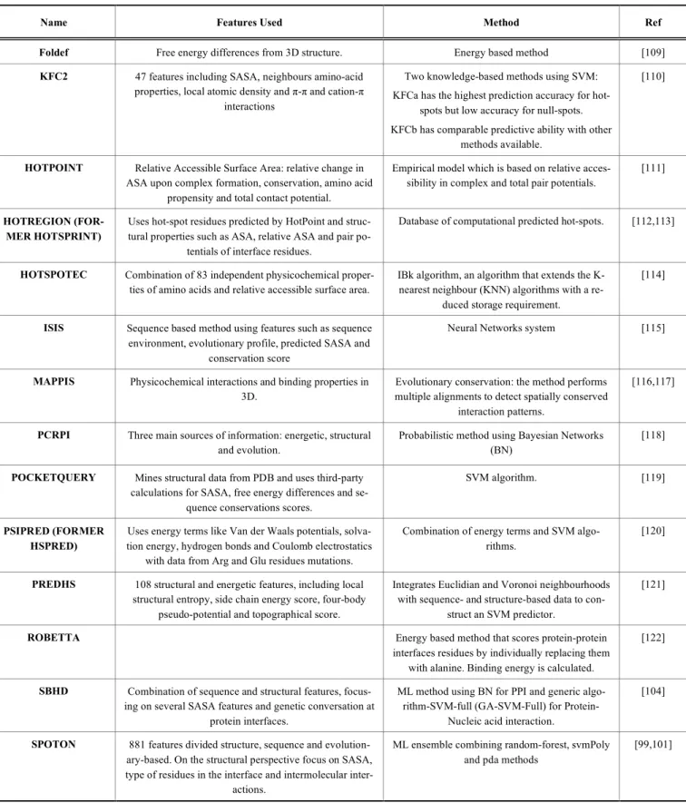
[107]. Table 1 summarizes recently developed
soft-ware/servers which are used for hot-spot prediction.
The occurrence of hot-spots at protein-protein interfaces
provides the opportunity to inhibit complex/oligomer
forma-tion by targeting these residues by means of therapeutic
agents. In this respect, computational methods are extremely
valuable for drug-design since it helps filter most of the
non-relevant compounds without a therapeutic value [123].
The workflow that can be used to develop therapeutic
molecules is depicted in Fig. (1). Docking protocols are one
the most widely used computational tools in the early stages
of drug development. This technique provides a faster and
cheaper way of screening a library of compounds [124].
Docking most recently has been used not only as a screening
tool but also as a method for target identification. Hot-spot
identification is a crucial step when designing inhibitors. The
methods used for this purposed were discussed previously.
Once the hot-spots are determined, structure- or ligand-based
virtual screening can be done, along with protein-protein
docking [125]. However, ligand-based screenings are rarely
used for such purposes due to lack of significant numbers of
known inhibitors [125].
Structure-based pharmacophore design can be done by
using softwares, such as LigandScout [126] or Phase [127].
In addition, it can also be calculated by means of potential
interaction sites which are derived by DSX [128] or
Super-Star [129]. Alternatively, determination of pharmacophores
can be based on hot-spots. Zerbe et al. compared hot-spots
which are predicted by either alanine scanning mutagenesis
or small molecule fragment screening. The authors showed
that high correlation exists between the two groups while
only a small subset of hot-spots, which are predicted by
alanine mutagenesis, could be used for potential binding of
inhibitors [130]. After achieving a pharmacophore model,
various ligand databases can be searched for finding
poten-tial hits. The top poses can be identified by clustering the
docking results according to their spatial arrangement and
energy values. The inhibitors obtained in this way can be
classified into three groups: antibodies, peptides and small
molecules. Often the process starts with a peptide and then it
is converted to a small molecule by incorporating important
functional groups. Secondary structures like α-helices,
β-sheets,β-turns, extended structures and proline-rich segments
function as scaffolds for the design of inhibitors [123]. An
example to such successful inhibitors is the one that can
dis-rupt the interaction between the anti-apoptotic BCL-XL and
its pro-apoptotic partners. Identification of such an inhibitor
was done by using virtual screening which is based on
struc-ture-based pharmacophore modelling and sequential docking
[131]. Mysinger et al. [132] were also able to identify 4
in-hibitors which were developed against chemokine receptor
CXCR4 using structure-based methods. Ligands retrieved
showed high specificity towards the receptor. The same
method was also used to develop ligands that can provide
preferential coupling of the receptor to its cognate signalling
partner such as G-protein or Arrestin by using biased ligands
[21]. In (Fig. 2) we illustrated the use of ours SpotOn
soft-ware, which classifies interfacial residues as hot-spots and is
able to highlight key binding determinants for the coupling
of the 2 binding partners of a typical GPCR [99,101]. These
type of information can also be used to develop new and
more specfic ligands.
Consequently, preclinical and clinical studies have been
initiated for development of effective biased agonists that
target GPCRs, in particular, opioid receptors. Development
of such specific ligands towards these receptors is necessary
to overcome drug resistance and treat substance abuse [133].
3. EXPERIMENTAL APPROACHES APPLIED TO
THE STUDY OF GPCR DIMERIZATION
Investigation of PPIs, in particular in GPCR
com-plexes/oligomers, is a challenging task. In order to find the
most appropriate method for the system the following points
should be considered [10]:
• If the study is discovery-driven, then, a
high-throughput-screening-suitable (HTS-suitable) method should be
pre-ferred to allow for exploring of interactomes or
alterna-tively, screening of whole libraries;
• For targeted approaches with defined interaction
part-ners’ assays which use tagged proteins are desirable;
Table 1. A list of software/servers that are currently used for prediction of hot-spots which is given along with the relevant features
and algorithm/methods used. Adapted from Moreira et al. [108].
Name Features Used Method Ref
Foldef Free energy differences from 3D structure. Energy based method [109] KFC2 47 features including SASA, neighbours amino-acid
properties, local atomic density and π-π and cation-π interactions
Two knowledge-based methods using SVM: KFCa has the highest prediction accuracy for
hot-spots but low accuracy for null-hot-spots. KFCb has comparable predictive ability with other
methods available.
[110]
HOTPOINT Relative Accessible Surface Area: relative change in ASA upon complex formation, conservation, amino acid
propensity and total contact potential.
Empirical model which is based on relative acces-sibility in complex and total pair potentials.
[111]
HOTREGION (FOR-MER HOTSPRINT)
Uses hot-spot residues predicted by HotPoint and struc-tural properties such as ASA, relative ASA and pair
po-tentials of interface residues.
Database of computational predicted hot-spots. [112,113]
HOTSPOTEC Combination of 83 independent physicochemical proper-ties of amino acids and relative accessible surface area.
IBk algorithm, an algorithm that extends the K-nearest neighbour (KNN) algorithms with a
re-duced storage requirement.
[114]
ISIS Sequence based method using features such as sequence environment, evolutionary profile, predicted SASA and
conservation score
Neural Networks system [115]
MAPPIS Physicochemical interactions and binding properties in 3D.
Evolutionary conservation: the method performs multiple alignments to detect spatially conserved
interaction patterns.
[116,117]
PCRPI Three main sources of information: energetic, structural and evolution.
Probabilistic method using Bayesian Networks (BN)
[118]
POCKETQUERY Mines structural data from PDB and uses third-party calculations for SASA, free energy differences and
se-quence conservations scores.
SVM algorithm. [119]
PSIPRED (FORMER HSPRED)
Uses energy terms like Van der Waals potentials, solva-tion energy, hydrogen bonds and Coulomb electrostatics
with data from Arg and Glu residues mutations.
Combination of energy terms and SVM algo-rithms.
[120]
PREDHS 108 structural and energetic features, including local structural entropy, side chain energy score, four-body
pseudo-potential and topographical score.
Integrates Euclidian and Voronoi neighbourhoods with sequence- and structure-based data to
con-struct an SVM predictor.
[121]
ROBETTA Energy based method that scores protein-protein
interfaces residues by individually replacing them with alanine. Binding energy is calculated.
[122]
SBHD Combination of sequence and structural features, focus-ing on several SASA features and genetic conversation at
protein interfaces.
ML method using BN for PPI and generic algo-rithm-SVM-full (GA-SVM-Full) for
Protein-Nucleic acid interaction.
[104]
SPOTON 881 features divided structure, sequence and evolution-ary-based. On the structural perspective focus on SASA, type of residues in the interface and intermolecular
inter-actions.
ML ensemble combining random-forest, svmPoly and pda methods
[99,101]
• The sensitivity of the assay is important: for weak,
tran-sient interactions only very few assays are suitable, if
stable/strong interaction will be studied, most assays can
be used;
• Determination of the stoichiometry of the complex- that
is to say- if consideration of binary PPIs is enough or the
whole protein complex is of interest should be
consid-ered as well;
• The dependence of the results on the type of the medium
in which the sample is preserved should be checked. For
instance, experiments will be done in cells or native
tis-sues, or can the cells be lysed and proteins solubilized?
• The necessity of certain (co-)factors, auxiliary proteins
or micro-environments for interactions to occur should
also be determined;
• Does the whole protein need to be analysed or is a part of
it (either short peptides or domains that represent the
whole protein’s properties) sufficient?
Fig. (1). Workflow used for computational design of PPI inhibitors.
Adapted from Sable et al. [123].
First indications of PPIs can be achieved by using
bio-chemical (co-) immunoprecipitation or pull-down
experi-ments. When working with recombinant proteins mostly tags
are used, such as glutathione-S-transferase (GST), human
influenza hemagglutinin (HA) or myc tags [10,134,135]. To
further characterize true interactions mostly
fluorescence-based methods are applied, such as FRET (fluorescence
resonance energy transfer), BRET (bioluminescense
reso-nance energy transfer), BiFC (biomolecular fluorescence
complementation assays) or more recently developed
meth-ods which emerged from the standard methmeth-ods, like
time-resolved FRET (Tr-FRET). However, all these methods have
in common that they do not address the questions about the
interfaces involved in the oligomers/complexes, but rather
they only confirm the interaction itself. In addition, these
methods are not suitable for analysing interactions in native
tissues or those that are being transient. For dynamic
moni-toring of transient interactions a novel technique, namely
total internal reflection fluorescence microscopy (TIRFM),
which can be used with the SNAP-tag technology can be
used to label GPCRs at the cell surface of living cells [136].
Alternatively, BioID [137] can also be used for detecting
transient interactions; however, it has not been used for the
study of GPCRs yet. For deciphering the interaction sites
experimentally, cleverly designed mutagenesis studies are
essential. In some cases, especially for interactions between
receptors and specific domains, microarrays can be well
suited to decipher such interaction sites.
Especially for receptors activated by peptides the
devel-opment of PPI inhibitors interfering or preventing ligand
binding can be of high interest for the treatment of several
diseases or to reduce side effects by tailoring the drug
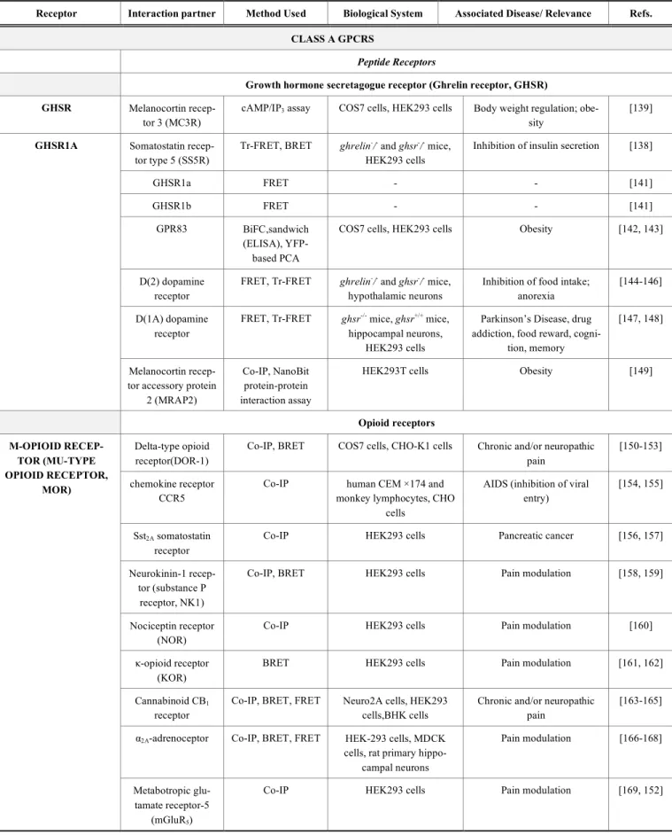
sponses to selective pathways. For example, for ghrelin
re-ceptors different heterodimers have been described, such as
GHS-R1a-SST5, which are involved in controlling the
glu-cose homeostasis [138] or GHSR-MC3R heterodimers,
which are important for hypothalamic weight regulation
[139] (check Table 2 and section C).
For the design of inhibitors, the nature of the interaction
as well as the type of modulation of PPIs must be considered
and the type of assay should be chosen accordingly. The
Table 2. A list of protein-protein interactions taken from GPCR oligomers and GPCR signalling complexes.
Receptor Interaction partner Method Used Biological System Associated Disease/ Relevance Refs. CLASS A GPCRS
Peptide Receptors
Growth hormone secretagogue receptor (Ghrelin receptor, GHSR) GHSR Melanocortin
recep-tor 3 (MC3R)
cAMP/IP3 assay COS7 cells, HEK293 cells Body weight regulation;
obe-sity
[139]
Somatostatin recep-tor type 5 (SS5R)
Tr-FRET, BRET ghrelin-/- and ghsr-/- mice, HEK293 cells
Inhibition of insulin secretion [138]
GHSR1a FRET - - [141]
GHSR1b FRET - - [141]
GPR83 BiFC,sandwich
(ELISA), YFP-based PCA
COS7 cells, HEK293 cells Obesity [142, 143]
D(2) dopamine receptor
FRET, Tr-FRET ghrelin-/- and ghsr-/- mice, hypothalamic neurons
Inhibition of food intake; anorexia
[144-146]
D(1A) dopamine receptor
FRET, Tr-FRET ghsr-/- mice, ghsr+/+ mice,
hippocampal neurons, HEK293 cells
Parkinson’s Disease, drug addiction, food reward,
cogni-tion, memory
[147, 148] GHSR1A
Melanocortin recep-tor accessory protein
2 (MRAP2)
Co-IP, NanoBit protein-protein interaction assay
HEK293T cells Obesity [149]
Opioid receptors Delta-type opioid
receptor(DOR-1)
Co-IP, BRET COS7 cells, CHO-K1 cells Chronic and/or neuropathic pain
[150-153]
chemokine receptor CCR5
Co-IP human CEM ×174 and
monkey lymphocytes, CHO cells
AIDS (inhibition of viral entry)
[154, 155]
Sst2A somatostatin
receptor
Co-IP HEK293 cells Pancreatic cancer [156, 157]
Neurokinin-1 recep-tor (substance P
receptor, NK1)
Co-IP, BRET HEK293 cells Pain modulation [158, 159]
Nociceptin receptor (NOR)
Co-IP HEK293 cells Pain modulation [160]
κ-opioid receptor (KOR)
BRET HEK293 cells Pain modulation [161, 162]
Cannabinoid CB1
receptor
Co-IP, BRET, FRET Neuro2A cells, HEK293 cells,BHK cells
Chronic and/or neuropathic pain
[163-165]
α2A-adrenoceptor Co-IP, BRET, FRET HEK-293 cells, MDCK
cells, rat primary hippo-campal neurons Pain modulation [166-168] Μ-OPIOID RECEP-TOR (MU-TYPE OPIOID RECEPTOR, MOR) Metabotropic glu-tamate receptor-5 (mGluR5)
Co-IP HEK293 cells Pain modulation [169, 152]
Receptor Interaction partner Method Used Biological System Associated Disease/ Relevance Refs. CLASS A GPCRS
Gastrin-releasing peptidereceptor
(GRPR)
Co-IP HEK 293 cells,Mice spinal cord
Morphine-induced scratching (MIS)
[170]
5HT1A Co-IP, BRET HEK 293 cells, COS7 cells Pain modulation [171]
Galanin receptor subtype Gal1
(Gal1R)
BiFC, BRET HEK293T cells, rat ventral tegmental area
Opioid use disorders [172]
Negative elongation factor A
MYTH screen, Co-IP, BRET
yeast, HEK293 cells - [11]
α2A-adrenoceptor Co-IP, BRET HEK293 cells,rat spinal
cord
Pain modulation [173, 174]
β2-adrenoceptor Co-IP, BRET HEK293 cells, CHO cells Alteration of β2-adrenoceptor internalization
[175, 176]
chemokine receptor CCR5
Co-IP human CEM ×174 and
monkey lymphocytes
AIDS [154]
Sensory Neuron-Specific Receptor-4
(SNSR-4)
BRET HEK293 cells Pain modulation [177]
Cannabinoid CB1
receptor
Co-IP, BRET Neuro2A cells, HEK293 cells
Altered subcellular localiza-tion of CB1 receptor, enhanced
CB1 receptor desensitization
[163, 178]
CXCR4 chemokine receptor
Co-IP, FRET MM-1 cells, HEK293 cells Inflammation, Pain, sensing HIV-infection [179] Δ-OPIOID RECEP-TOR(DELTA-TYPE OPIOID RECEPTOR, DOR) κ-opioid receptor (KOR)
Co-IP, BRET peripheral sensory neurons, HEK293 cells
Pain modulation, allodynia [180, 176, 151]
β2-adrenoceptor Co-IP, BRET HEK293 cells, CHO cells - [175, 176]
chemokine receptor CCR5
Co-IP human CEM ×174 and
monkey lymphocytes
AIDS [154]
Apelin receptor (APJ)
Co-IP, BRET HEK293 cells Increase in cell proliferation [181] Κ-OPIOID RECEPTOR (KAPPA-TYPE OPIOID RECEPTOR, KOR) Bradykinin B2 re-ceptor
BRET, PLA HEK293 cells Increase in cell proliferation [182]
NOCICEPTIN RE-CEPTOR (NOR, KAPPA-TYPE 3 OPIOID RECEP-TOR,(KOR-3), OPIOID RECEPTOR-LIKE 1 RECEPTOR (ORL1)) Ceramide synthase 6 (CerS6)
MYTH screen, Co-IP, BRET
yeast, HEK293 cells - [11]
Protease activated receptor 2 (PAR-2, also known as thrombin receptor-like 1) Regulator of
G-protein signalling 8 (RGS8)
GST pull-down, BRET
HEK293 cells, Neuro2a cells
- [183]
PAR-2
Major prion protein (PrP)
MYTH screen, Co-IP, BRET
yeast, HEK293 cells - [11]
Receptor Interaction partner Method Used Biological System Associated Disease/ Relevance Refs. CLASS A GPCRS Sarcoplasmic/ endo-plasmic reticulum calcium ATPase 2 (SERCA2)
MYTH screen, Co-IP, BRET
yeast, HEK293 cells - [11]
Heat shock 70 kDa protein 1B
(HSP70-2)
MYTH screen, Co-IP, BRET
yeast, HEK293 cells - [11]
Type-1 angiotensin II receptor (AT1R) Bradykinin B2
re-ceptor
Co-IP HEK293 cells, mesangial cells (rat)
Hypertension [184, 185]
Cannabinoid CB1
receptor
Co-IP, BRET HEK293 cells,Neuro2A cells,HSCs
Fibrosis [186, 151]
α2C- adrenoceptor BRET, FRET HEK293 cells Hypertension, heart failure [187]
Sodium/potassium-transporting ATPase
subunit beta-1
MYTH screen, Co-IP, BRET
yeast, HEK293 cells - [11]
DnaJ homolog subfamily C
member 8
MYTH screen, Co-IP, BRET
yeast, HEK293 cells - [11]
Ceramide synthase 6 (CerS6)
MYTH screen, Co-IP, BRET
yeast, HEK293 cells - [11]
AT1R
Ornithine decar-boxylase antizyme 1
(ODC-Az)
MYTH screen, Co-IP, BRET
yeast, HEK293 cells - [11]
5a anaphylatoxin chemotactic receptor 2 (C5a-R, GPR77) Calmodulin-1, 2, 3 MYTH screen,
Co-IP, BRET
yeast, HEK293 cells - [11]
uncharacterized protein C4orf3 (Hepatitis C virus F protein-transactivated pro-tein 1)
MYTH screen, Co-IP, BRET
yeast, HEK293 cells - [11]
Mitochondrial 2-oxoglutarate/malate
carrier protein (OGCP)
MYTH screen, Co-IP, BRET
yeast, HEK293 cells - [11]
C5A-R
Synaptogyrin-2 MYTH screen, Co-IP, BRET
yeast, HEK293 cells - [11]
Oxytocin receptor (OTR) Oxytocin receptor Co-IP, BRET,
Tr-FRET
COS7 cells , rat mammary glands - [188, 189] Vasopressin V1 receptor (V1R) Co-IP, BRET, tr-FRET
HEK293T cells, CHO cells, COS7 cells, rat mammary
glands - [190, 189] OXYTOCIN RECEP-TORS Vasopressin V2 receptor (V2R) Co-IP, BRET, tr-FRET
HEK293T cells, COS7 cells, rat mammary glands
- [190, 189]
Receptor Interaction partner Method Used Biological System Associated Disease/ Relevance Refs. CLASS A GPCRS
Thyrotropin-releasing hormone receptor (TRHR)
TRHR TRHR BRET HEK293 cells, COS1 cells - [191]
Gonadotrophin-releasing hormone receptors (GnRHR)
GNRHR GnRHR BRET HEK293 cells, COS1 cells - [191]
Protein receptors
Thyrotropin receptor (Thyroid-stimulating hormone receptor) (TSH-R)
TSH-R BRET HEK293T cells - [192]
Mid1-interacting protein 1
MYTH screen, Co-IP, BRET
yeast, HEK293 cells - [11]
TSH-R
Synaptotagmin-1 MYTH screen, Co-IP, BRET
yeast, HEK293 cells - [11]
C-C chemokine receptors Myelin basic protein
(MBP)
MYTH screen, Co-IP, BRET
yeast, HEK293 cells - [11]
CCR1
Major prion protein (PrP)
MYTH screen, Co-IP, BRET
yeast, HEK293 cells - [11]
Lipid receptors
Platelet-activating factor receptor (PAF-R) Lipid Myelin basic protein
(MBP)
MYTH screen, Co-IP, BRET
yeast, HEK293 cells - [11]
Major prion protein (PrP)
MYTH screen, Co-IP, BRET
yeast, HEK293 cells - [11]
Plasmolipin MYTH screen, Co-IP, BRET
yeast, HEK293 cells - [11]
Rhomboid domain-containing protein 2
MYTH screen, Co-IP, BRET
yeast, HEK293 cells - [11]
PAF-R
Transmembrane protein 120A
MYTH screen, Co-IP, BRET
yeast, HEK293 cells - [11]
Thromboxane A2 receptor (TXA2-R), also known as Prostanoid TP receptor TXA2-R G-protein coupled
receptor-associated sorting protein 1-3, 7 (GASP-1-3, 7) GST-pull down experiments, Co-IP HEK293 cells - [9] Aminergic receptors Dopamine receptors D(2) dopamine receptor
Co-IP, FRET, BRET Rat striatal neurons, HEK293 cells,striatal
post-mortem brain samples
Depression, schizophrenia, addiction [193, 194, 151, 195] D(1) DOPAMINE RE-CEPTOR D(3) dopamine receptor BRET, FRET, Tr-FRET
HEK293T cells, rat brain striatum
Basal-ganglia disorders [196, 197]