Light and Electron Microscope Studies of Species of Plant
Pathogenic Basidiomycota Isolated from Plants in Kıbrıs
Village Valley (Ankara, Turkey)
Tuğba EKİCİ , Makbule ERDOGDU , Zeki AYTAÇ and Zekiye SULUDERE1 2* 1 1
1 2
Gazi University Faculty of Science, Department of Biology, Teknikokullar, Ankara-TURKEY Ahi Evran University, Faculty of Science and Literature Department of Biology, Kırsehir-TURKEY
,
,
Abstract: A search for present in Kıbrıs Village Valley (Ankara, Turkey) was carried out during the period 2009-2010.
Kıbrıs Village Valley Morphological data obtained by light and scanning electron microscopy of identified fungi are presented.
SEM.
Microstromatales, Uredinales,
basidiomycetous plant parasites
Twenty-two basidiomycetous plant parasites were identified from .
Key Words: Basidiomycota, Ustilaginales,
Kıbrıs'ın Köyü Vadisi'nde (Ankara, Türkiye) Bitkilerden İzole Edilmiş
Bitki Patojeni Basidiomycota Türlerinin Işık ve Elektron Mikroskobu
Çalışmaları
Özet:
Anahtar Kelimeler:
Kıbrıs Köyü Vadisi' nde (Ankara, Türkiye) bulunan bazidiyumlu bitki paraziti mantarların araştırılması 2009-2010 yıllarında yapılmıştır. bazidiyumlu bitki paraziti Teşhis edilmiş mantarların ışık ve taramalı elektron mikroskobuna dayalı morfolojik verileri sunulmuştur.
SEM
Microstromatales, Uredinales,
Kıbrıs'ın Köyü Vadisi' nde yirmi iki tespit edilmiştir.
Basidiomycota, Ustilaginales,
Introduction
The order with the
single family was erected for
species having simple-septate hyphae and local interaction zones without the formation of interaction apparatus. Haustoria or other intracellular fungal organs are lacking (Begerow et al. 2006).
The Niessl the single genus
currently placed in the , is
represented by two species ( (Desm.) Sacc. and
(Bérenger) Sacc.) in Turkey (Göbelez 1967). Rust fungi ( ) are one of the largest natural taxa within the kingdom Eumycota. More than 7000 species belonging to 100-125 genera and 14 families are accepted currently. Microstromatales Microstromataceae Microstroma , Microstromataceae Microstroma album Microstroma juglandis Uredinales
The largest genus, Pers., contains ca. 4000 spp., 650 of which occur on (Abbasi 1996). Recently, Bahcecioglu & Kabaktepe (2012) listed species of rust fungi and their hosts in Turkey. 351 species of rust fungi were registered on 778 species of high plants from 325 genera of 63 families.
The order comprises the
majority of smut fungi including the large genera (Pers.) Roussel and
Ehrenb. ex Link Most species of this group sporulate in the reproductive parts of their hosts (Begerow et al. 2006). About 30 species, belonging to the genera Bref.
Ehrenb. ex Link
Woronin ex J. Schröt. Lavrov
and (Pers.) Roussel have been
reported from Turkey (Şahin and Tamer 1998; Kırbağ 2003; Bahçecioğlu et al. 2006; Kabaktepe and Bahçecioğlu 2006).
This research was carried out in valley of Kıbrıs Village belonging to Mamak district which about 20 km southeast of Ankara province. Kıbrıs Village Valley is situated in the Irano–Turanian phytogeographic region and according to the grid square system adopted by Davis (1965–1985), it is located in the squares B4. The climate of the province is Mediterranean. Kıbrıs Village Valley is 1st degree field of natural sites and its three area are 1st archaeological site.
As an aid in identification and classification of fungi, the scanning electron microscope (SEM) allowing the observation of surface structures of various organs is becoming increasingly available (Udagawa & Hoire 1973). The discovery of additional features with the SEM has provided useful support for identification when crucial characters are not clear with the light microscope (LM). The aim of this study is to investigate micromorphology of spores of basidiomycetous plant parasites present in Kıbrıs Village Valley using SEM and LM.
Infected plant specimens were collected from Kıbrıs Village Valley in Ankara province of Turkey. The host specimens were prepared according to established herbarium techniques. Host plants were identified using the Flora of Turkey and East Aegean Islands (Davis 1965–1985). The fungal specimens were isolated from the host plants by obtaining thin sections or scraping. For microscopic examination and microphotographs a Leica DM E light microscope was used. Spores were measured using a Leica DM E light microscope. Lenght and width of 20 spores were measured for each sample. Leica EZ4D stereo microscope was used for close-up photo of the uredinia and/or telia on leaf surface. The microfungi were identified using relevant literature (Azbukina 2005; Kuprevich and Ulijanishchev 1975; Ulijanishchev 1978; Ulijanishchev et al. 1985; Wilson and Henderson 1966). All specimens examined were deposited in the mycological collection of the Department of Biology, Faculty of Science, Gazi University, in Ankara province of Turkey.
For scanning electron microscopy (SEM), 8–10- mm-square pieces of infected leaves were mounted on the SEM stubs with double-sided adhesive tape. They were coated with gold using a Polaron SC 502 Sputter Coater and were examined with a Jeol JSM 6060 scanning electron microscope operated at 5-10 kV in the Electron Microscopy Unit, Faculty of Science, Gazi University (Turkey).
Twenty-two microfungi were identified in the research area. Morphological data which was obtained by light and scanning electron microscopy of these fungi was provided. The author abbreviations of fungi are according to Kirk and Ansell (1992). The systematics of taxa were listed according to Index Fungorum (www.speciesfungorum.org, accessed 2013). Family and species names are listed in alphabetical order in the text.
Puccinia Poaceae Ustilaginales Ustilago Sporisorium . Anthracoidea , Sporisorium , Tolyposporium , Tranzscheliella Ustilago ,
Materials and Methods
Results
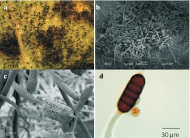
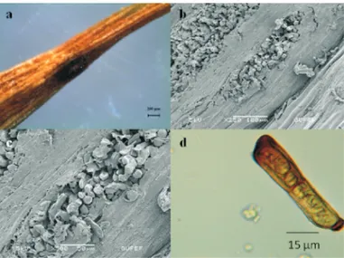
List of Taxa 1-Basidiomycota Exobasidiomycetes Microstromatales Microstromataceae Microstroma Microstroma album Niessl (1861) (Desm.) Sacc. Leaf spots circular to angular, small, speckled with yellow. Basidium hypophyllous,
clavate, hyaline, 20-25 x 10 µm in size, bursting out through the ruptured cuticle. Basidiospores one celled, fusiform to ellipsoid, guttulate, hyaline, 7.5-12.5 x 2.5-3.5 µm in size, wall smooth (Fig. 1).
B4 Ankara: Kıbrıs Village, 39°52'7,7''N, 33°00'14,7''E, 1105-1115 m, roadside, stepe, on
living leaves of Willd.
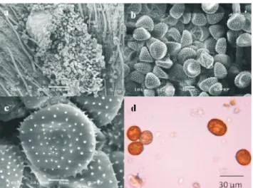
( ), 24.09.2009, TE 1102. Quercus pubescens Fagaceae Pucciniomycetes Pucciniales Melampsoraceae Melampsora 2-Melampsora salicis-albae Melampsora euphorbiae Kleb. 3-Castagne (1843)
Spermogonia: generally hypophyllous, rarely on the stems, lenticular, scattered. Uredinia: generally amphigenous, sometimes on the stems, scattered or confluent, causing yellow or orange spots on the leaves, paraphyses c a p i t a t e , h y a l i n e , 1 2 - 1 5 µ m w i d t h . Urediniospores: yellow, globoid, oblong or pyriform, 20-27.5 x (15-) 17.5-20 (25) µm in size, wall densely echinulate. Telia: amphigenous, subepidermal, dark brown. Teliospores: prismatic, rounded at both ends, yellowish, (30) 35-40 x 10-12.5 µm in size.
B4 Ankara: Kıbrıs Village, Akçadere location, 1100-1200 m, on living leaves
of L. 24.9.2009, TE
1112.
(Ficinus & C. Schub.) Castagne
Uredinia: amphigenous, generally hypophyllous, rarely on the stems, yellow, scattered, minute, surrounded by many capitate paraphyses. Urediniospores: yellow, globoid, oblong, ellipsoid, 20-25 (-30) x (15-) 17.5-20 (22.5) µm in size, wall densely echinulate, hyaline. Telia: amphigenous, subepidermal, brown. Teliospores: cylindrical-prismatic, rounded at both ends, brown, 25-55 x 10-13.5 µm in size (Fig. 2).
B4 Ankara: Kıbrıs Village, 39°52'7,7''N, 32°00'14,7''E, 1050 m, steppe, on living leaves of L. (Euphorbiaceae), 09.08.2009, TE 1088; A B4 Ankara: Kıbrıs Village, Akçadere location, 1062 m, riverside, on
living leaves of L.
( ), 17.4.2009, TE 1049. riverside,
Salix alba (Salicaceae),
Euphorbia macroclada
Euphorbia stricta Euphorbiaceae
Fig.1. : a-Leaf spot;b-Basidium and basidiospores(SEM); c-Basidiospores (SEM);d- Basidiospores
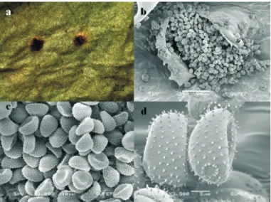
Phragmidiaceae Phragmidium
4-Phragmidium bulbosum
Link (1816)
(Fr.) Schltdl. Spermogonia: epiphyllous, in minute clusters, orange. Uredinia: hypophyllous, yellowish-brown, scattered or confluent, pulverulent. Urediniospores: yellow, globoid, oblong or ovoid, 17.5-22.5 (25) x 12.5-15 (17.5) µm in size, wall echinulate. Telia: hypophyllous, black, scattered or in groups, rounded, pulvinate, pulverulent. Teliospores: black, broadly ellipsoid
to cylindrical, 4- to 7 (mostly 5-6)-celled, rounded above with a hyaline apiculus 11-15 µm long, rounded at the base, not constricted or slightly constricted, 60-70 x 22.5-27.5 µm in size, wall verrucose, 2.5-5 µm thick at the side, pedicel hyaline, 100-110 µm long, clavate in lower half, persistent (Fig. 3).
B4 Ankara: Kıbrıs Village, 39°52'4,9''N, 33°00'18,7''E, 1050-1090 m, riverside, shady places, on living leaves of
Schreb. ( ), 24.09.2009, TE 1096. Rubus sanctus Rosaceae
Nisan(2014)5(1)7-21
Fig. 2. : a-General appearance;b-Uredinia(SEM); c,d-Urediniospores and paraphyses(SEM)
Melampsora euphorbiae
Fig. 3. : a-General appearance; b,c-Teliospores(SEM);d-Teliospore and urediniospore
5- (Pers.) Schltdl.
Phragmidium mucronatum
Uredinia: amphigenous, pale orange, scattered or in groups. Urediniospores: pale yellow, globoid, ellipsoid, ovoid or angular, (20) 22.5-27.5 (-30) x 17.5-22.5 µm in size, wall echinulate, or verruculose. Telia: hypophyllous, scattered or in groups, rounded, black, 500 µm diam., pulverulent. Teliospores: blackish-brown,
ellipsoid to cylindrical, 6- to 7-celled, not constricted, 85-100 x 32.5-37.5 , wall smooth or verrucose. The hyaline pedicel was swollen, clavate in lower half, 150-175 µm (Fig. 4).
B4 Ankara: Kıbrıs Village, 39°52'22''N, 33°00'01''E, 1027-1050 m, riverside, shady places, on living leaves of L. ( ), 24.09.2009, TE 1093. μm in size Rosa canina Rosaceae Pucciniaceae Gymnosporangium Gymnosporangium confusum Puccinia 7-Puccinia acarnae R. Hedw. ex DC. (1805) - Dietel
Spermogonia: epiphyllous, orange, in small groups, subgloboid, subepidermal in origin. Aecidia: hyphophyllous, yellowish brown, 4-5 mm diam. Peridial cells in surface view lanceolate, in lateral view rhombic, elongate, obliquely arranged warts and ridges. Aecidiospores: cinnamon-brown, globoid, angular, (20) 22.5-27.5 x (20) 22.5-27.5
5).
B4 Ankara: Kıbrıs Village, 39°52'07,66''N, 33°00'12,43''E, 1128 m, roadside, steppe, on fruit and living leaves of
Jacq. var. ( ), 17.IV.2009,
TE 1036.
Pers. (1801)
P. Syd. & Syd. Uredinia: hypophyllous, chestnut-brown, roundish. Urediniospores: cinnamon-brown, globoid, ellipsoid, 25-30 x (20) 22.5-27.5
echinulate. 6
μm in size, wall verruculose (Fig.
μm in size, wall
Crataegus monogyna monogyna Rosaceae
Telia: amphigenous, blackish brown, rounded, pulverulent. Teliospores: chestnut-brown, ellipsoid, broadly ellipsoid, ovoid, oblong, 33.5-45 x 25-30 µm in size, constricted in septate, wall verruculose, pedicel hyaline, short, fragile (Fig. 6).
B4 Ankara: Kıbrıs Village, Cellinin kayası location, 1100m, on living leaves of
(L.) Cass. ( 17.04.2009, TE 1031.
steppe.
Asteraceae), Picnomon acarna
Fig. 4. : a-General appearance;
8-Puccinia behenis J. Schröt.
Uredinia: amphigenous, scattered, yellow, chestnut-brown, roundish, pulverulent. Urediniospores: subgloboid to ellipsoid, 25-30 x (20) 22.5-27.5 size, wall echinulate, with 3 or 4 pores. Teliospores not seen (Fig. 7).
B4 Ankara: Kıbrıs Village, Akçadere location, riverside, on living leaves
of (Rafn) Godr. subsp.
(Boiss.) McNeill & H.C.Prent.
( ), μm in 1100-1150 m, Silene pratensis eriocalycina Caryophyllaceae 01.08.2010, TE 1187. Nisan(2014)5(1)7-21
Fig. 5., : a-General appearance; b-Aecidiospores(SEM);c- aecidiopsores; d-Peridial cells
Gymnosporangium confusum
Fig. 6., : a-General appearance; b,c-Urediniospores(SEM);d-Teliospores
9-Puccinia calcitrapae DC.
Telia: amphigenous, generally epiphyllous, scattered or confluent, rounded, 400-1200 µm diam., black, pulverulent. Teliospores: ellipsoid, oblong, 32.5-40 x (20) 22.5-27.5 (30)
rounded or attenuate both ends, slightly constricted in septate, wall 2.5-5
pedicel, filiform, hyaline, fragile (Fig. 8).
B4 Ankara: Kıbrıs Village, Kavakderesi location, 1000-1080 m, riverside, on living leaves
of L. subsp
(M.Bieb.) Kazmi (Asteraceae), 17.04.2009, TE 1025.
μm in size, brown,
μm thick,
verruculose,
Carduus pynocephalus . albidus
10-Puccinia cynodontis Lacroix ex Desm. Spermogonia: amphigenous, pale yellow, 70-90 µm diam. Aecia: hypophyllous, globoid, 1-3 µm diam., orange. Aeciospores: oblong, 23-29 x 19-24 µm, wall 1,5-2 µm thick. Urediniospores and
teliaspores on (L.)Pers.
B4 Ankara: Kıbrıs Village, Dipsiz gölü location, 1200 m, riverside, shady places, on living leaves of L. subsp
(Gilib.) Lange ( ),
24.09.2009, TE 1124. Cynodon dactylon
Plantago major .
intermedia Plantaginaceae
Fig. 7., : a-Uredinia and urediniospores(SEM); b,c-Urediniospores(SEM);d-Urediniospores
Puccinia behenis
Fig. 8., : a-General appearance; b,c-Teliospores(SEM);d-Teliospores
11-Puccinia eryngii G. Winter
Uredinia: hypophyllous, scattered, yellow, chestnut-brown, roundish, pulverulent. Urediniospores: globoid, ellipsoid, 25-32.5 x (20) 22.5-25
higenous, dark brown, scattered. Teliospores: chestnut-brown, broadly ellipsoid, ovoid, oblong, (32.5) 37.5-42.5 (45) x 22.5-27.5
, wall smooth, pedicel short, hyaline, fragile (Fig. 9).
B4 Ankara: Kıbrıs Village, Cellinin kayası location, 1105-1120 m, slopes, stepe, on living
leaves of L. var. Link
( ), 17.04.2009, TE 1014. μm in size, wall echinulate. Telia:
amp
μm in size, rounded at apex, attenuate at the base, slightly constricted
Eryngium campestre virens Apiaceae 12- (Röhl.) H. Mart. f. 13-Puccinia hieracii hieracii Puccinia jasmini
Uredinia: amphigenous, yellowish, small, scattered or in groups, ovate or irregular, 0.2-1 mm long, pulverulent. Urediniospores: cinnamon-brown, ellipsoid, globoid, ovate, 22.5-30 (32.5) x (20) 22.5-25 all echinulate,
1- 0.3-0.8 mm diam., pulverulent. Teliospores: chestnut-brown, ovoid, ellipsoid, oblong, 27.5-32.5 x (17.5) 20-22.5 µm in size, rounded at both ends, sometimes attenuate at the base, wall verruculose, pedicel short, hyaline (Fig. 10).
B4 Ankara: Kıbrıs Village, Kaynar gölü location, 1140 m, riverside, on living leaves of
Weber ( ),
24.09.2009, TE 1098.
DC.
Te l i a : a m p h i g e n o u s , g e n e r a l l y hypophyllous, in dense groups on the stems and petioles, rounded or ovoid, gray or black. Teliospores: oblong, ellipsoid or ovoid, (40) 42.5-50 x 20-25
onstricted in septate, wall smooth, up to 3.5
10 11).
B4 Ankara: Kıbrıs Village, 39°52'09,73''N, 33°00'18,81''E, 1111 m, on living leaves of L. ( ), 17.04.2009, TE 1038.
μm in size, w 2 μm
thick. Telia: amphigenous, blackish brown to black, scattered or rarely in groups, roundish,
μm in size, brown, apex often tapering, conic or obtuse, narrowed below, c
μm thick at the side, 7.5 μm thick at the apex, pedicel up to 0 μm long, filiform, hyaline, strong (Fig.
Taraxacum officinale Asteraceae
Jasminum fruticans Oleaceae
Nisan(2014)5(1)7-21
Fig. 9., : a-General appearance; b-Telia and teliospores(SEM);c-Teliospores(SEM); d-TeliosporesPuccinia eryngii
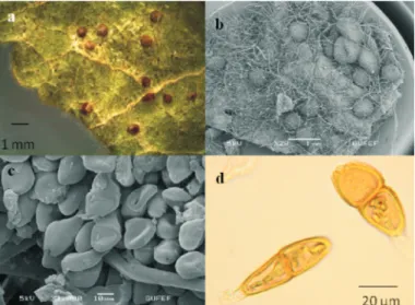
14- 15-Puccinia malvacearum Puccinia nevodovskii Bertero ex Mont.
Telia: hypophyllous and on the stems and petioles, scattered, rounded, pulvinate, compact, hard, 0.2-1 mm diam., often 2.5 mm diam. in groups, at first gray then black. Teliospores: subfusoid to ellipsoid,
in size, brown, attenuate at both ends, slightly constricted in septate, wall smooth, up to 2-3
. 12). B4 Ankara: Kıbrıs Village, Akçadere location, 1000-1200 m, riverside, on living leaves
of L. ( ), 24.09.2009,
TE 1106; B4 Ankara: Kıbrıs Village, Akçadere location, 1079 m, riverside, on living leaves of Winterl. ( ), 17.04.2009, TE 1058.
Gamalizk. Uredinia: amphigenous, yellowish, small, scattered or often confluent, ovate or irregular. Urediniospores: chestnut-brown, ellipsoid, globoid, ovate, 17.5-25 x 17-22.5 size, wall echinulate, pale chestnut-brown. Telia: amphigenous, blackish brown to black, scattered or rarely in groups, globoid.
(22.5) 35.5-55 x 17.5-22.5 μm
μm thick at the side, 8 μm thick at the apex, pedicel hyaline, strong (Fig
μm in
Malva sylvestris Malvaceae
Alcea biennis Malvaceae Fig. 10., f. : a-General appearance;
b-Telia and teliospores(SEM);c-Teliospores and urediniospores(SEM); d-Teliospores
Puccinia hieraciif hieracii
Fig. 11., : a,b-General appearance; c-Telia and teliospores(SEM); d-Teliospores(SEM)Puccinia jasmini
Teliospores: chestnut-brown, ovoid, broadly ellipsoid, oblong, 40-67.5 x 20-25 (30) µm in size, rounded or sometimes attenuate at the apex, attenuate at the base, constricted in septate, guttulate, wall smooth, 5-12.5 µm thick
at the apex, pedicel persistent, hyaline (Fig. 13). B4 Ankara: Kıbrıs Village, 39°52'01,71''N, 33°00'19,58''E, 1111 m, steppe, on stem and
living leaves of Sm. subsp
( ), 17.04.2009, TE 1040.
Galium floribundum .
floribundum Rubiceae
16-Puccinia poarum E. Nielsen
Spermogonia: epiphyllous, brownish-black, in the middle of thickened irregular spots on the leaf. Aecidia: hyphophyllous, globoid, 2-3 µm diam., orange. Aecidiospores: chestnut-brown, globoid, ellipsoid, ovate, 25-30 x 15-27.5
µm in size, wall verruculose. Urediniospores and teliaspores on L. species (Fig. 14).
B4 Ankara: Kıbrıs Village, 39°52'7,7''N, 33°00'14,7''E, 1000-1100 m, slope, stony place,
on living leaves of L. ( ), 24.09.2009, TE 1085. Poa Tussilago farfara Asteraceae Nisan(2014)5(1)7-21
Fig. 12., : a-General appearance; b-Telia and teliospores(SEM);c-Teliospores(SEM); d-TeliosporesPuccinia malvacearum
Fig. 13., a-General appearance;
b-Telia and teliospores(SEM);c-Teliospores and urediniospores(SEM); d-Teliospores
17-Puccinia rubiae-tataricae Syd. & P.
Syd.
Uredinia: amphigenous, chestnut-brown, globoid. Urediniospores: cinnamon-brown, ellipsoid, globoid, ovate, 22.5-25 x 17.5-25
pale chestnut-brown. Telia: amphigenous, black, roundish, pulvinate.
Teliospores: brown, ovoid, oblong, 37.5-50 x 15-22.5 µm in size, wall verruculose, pedicel short, hyaline, fragile (Fig. 15).
B4 Ankara: Kıbrıs Village, 39°52'11,40''N, 33°00'13,59''E, 1084 m, on living leaves of
24.09.2009, TE 1111. μm in
size, wall echinulate,
riverside,
Rubia tinctorum L. (Rubiaceae), Fig. 14., a-General appearance;
b- c-Aecidiospores;d-Aecidiospores(SEM)
Puccinia poarum
Aecidiospores(SEM);
Fig. 15., a-General appearance; b-Uredinia and urediniospores(SEM);c,d- Urediniospores(SEM)
18-Puccinia striiformis Westend. Uredinia: amphigenous, chestnut-brown, small, scattered or often confluent, ovate or irregular. Urediniospores: chestnut-brown, ellipsoid, globoid, oval, (17.5) 25-27.5 x 17.5-25
h brown to black, oblong. Teliospores: pale brown, cylindrical, broadly ellipsoid, oblong, 42.5-62.5 x 12.5-25 µm
in size, wall smooth, rounded or sometimes attenuate at the apex, attenuate at the base, constricted in septate, pedicel hyaline, fragile (Fig. 16).
B4 Ankara: Kıbrıs Village, Akçadere location, shady places, on living
leaves of (Opiz) Melderis
subsp ( ),
μm in size, wall echinulate, pale chestnut-brown. Telia: amphigenous, blackis
1000-1050 m, Elymus hispidus . hispidus Poaceae 24.09.2009, TE 1105. Uromyces Uromyces anthyllidis Uromyces polygoni-avicularis (Link) Unger (1833) 19- 20-(Grev.) J. Schröt.
Uredinia: amphigenous, chestnut-brown, small, scattered, in the concentric rings, roundish, pulverulent. Urediniospores: chestnut-brown, globoid, 20-22.5 (28) x 20-22.5
Teliospores not seen. Spermogonia, aecidia on L. species (Fig. 17).
B4 Ankara: Kıbrıs Village, Dipsiz gölü location, 1100 m, steppe, on living leaves of
Boiss. ( ),
01.08.2010, TE 1183.
(Pers.) P. Karst.
Uredinia: amphigenous, cinnamon-brown, scattered, rounded, pulverulent. Urediniospores: pale-brown, globoid, ellipsoid, (17.5) 22.5- 27.5 x 17.5-20 µm in size, wall echinulate. Telia: hypophyllous, dark-brown, scattered or in the concentric rings, pulverulent. Teliospores: chestnut-brown, globoid, obovoid, oblong, 22.5-25 x 17.5-22.5
remotely echinulate, pedicels hyaline, short, persistent.
B4 Ankara: Kıbrıs Village, Akçadere location, riverside, on living
l e a v e s o f L .
( ),
μm in size, wall echinulate, 1.5-4 μm thick.
μm in size, wall Euphorbia
Onobrychis hypargyrea Fabaceae
1000-1200 m,
P o l y g o n u m a v i c u l a r e Polygonaceae 24.09.2009, TE 1084.
Nisan(2014)5(1)7-21
Fig. 16., a-General appearance; b,c-Telia and teliospores(SEM);d- TeliosporePuccinia striiformis:
21-Uromyces rumicis (Schumach.) G. Winter
Uredinia: amphigenous, sometimes in the concentric rings, cinnamon-brown, scattered, rounded, 200-1000 µm diam., pulverulent. Urediniospores: pale-brown, globoid, ellipsoid, sometimes angular, 22.5-32.5 (35) x 20-25 µm in size, wall sparsely echinulate, 1.5-2 µm thick. Telia: hypophyllous, dark-brown, scattered or in
the concentric rings, pulverulent. Teliospores: brown, ovate, ellipsoid, globoid, obovoid, oblong, 30-37.5 x (17.5) 20-25 µm in size, wall sparsely echinulate, 2.5-3 µm thick, pedicels hyaline, short, fragile (Fig. 18).
B4 Ankara: Kıbrıs Village, Cellinin kayası location, 1128 m, rocky slope, on living leaves of
L . ( ) ,
24.09.2009, TE 1034.
R u m e x s c u t a t u s P o l y g o n a c e a e Fig. 17., a-Uredinia and urediniospres(SEM);
b,c-Urediniospores(SEM);d- Urediniospores
Uromyces anthyllidis:
Fig. 18., a-Uredinia;b-Uredinia(SEM); c-Urediniospores(SEM);d- Teliospores and urediniosporesUromyces rumicis:
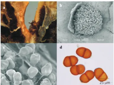
Ustilaginomycetes Ustilaginales Ustilaginaceae (Pers.) Roussel (1806) 22- Berk. Ustilago Ustilago bullata
Sori in the spikelets, usually in all spikelets on the inflorescence, replacing the floral parts and including the bases of the glumes, covered by a green to grey membrane of host tissue
which at maturity ruptures to expose the spore mass. Spore mass at first firm then dusty, dark brown to purplish-black, consisting of spores only. Spores globose to subglobose or angular,
dark chestnut-brown, ,
wall densely verrucose (Fig. 19).
B4 Ankara: Kıbrıs Village, Cellinin kayası location, 1150 m, steppe, on spikes of
L. (Poaceae), 17.04.2009, TE 1048. 7.5-10 x 5-7.5 μm in size Bromus sterilis Discussion Kıbrıs Village Valley
But the plants are completely covered by the dense dust mass caused from the activities of the stone quarries in the research area. This dust mass is a mechanical barrier for the penetration and distribution to the host plant of the fungi. This was detected as a decreasing factor on the fungi diversity and the rate of contamination.
As a result of field work carried out between 2009-2010 in Kıbrıs Village Valley (Ankara), 3 classes, 3 orders, 5 families, 7 genera and 22 species of the Basidiomycota divisio have been identified. As a classification of species within genera in class Pucciniomycetes:
12 species assigned to genus , 3
species to , two species to
, two species to and
one species to .
Exobasidiomycetes is represented by
and by a single
species of genus . As a result of present
study, , a microfungus
attributed to this divisio, is carpotroph in terms of trophic structure while the others are biophyllotroph. All of them consider as parasite.
Puccinia Uromyces Melampsora Phragmidium Gymnosporangium Microstroma Ustilaginomycetes Ustilago Ustilago bromivora
In terms of ecological relationships of microfungi, their lifes on a substrate is also interesting in addition to the host. In this case, living together of species belonging to different systematic groups and genera, as well as different species classified in the same genus is the subject.
The was chosen as a
research area, because its climatic conditions and plant distributions are suitable for the growth of microfungi.
Nisan(2014)5(1)7-21
During the present investigation some fungi were recorded growing together on the
same substratum. U. Braun was
found developing together with J. Schröt. on living leaves of
(Rafn) Godr. subsp. (Boiss.) McNeill & H.C.Prent., DC. together with
(Pers.) P. Karst. on living leaves of
L. (Fuckel)
Petr. together with
Syd. & P. Syd. on living leaves of
L. In addition, some species of microfungi are known on different hosts. Bertero ex Mont. was found on living leaves of L. and
Winterl.
Gamalizk. on the
leaves of Sm. subsp.
have only been given recently as new record (Ekici et al. 2010). We believe this study will contribute to mycoflora of Turkey which will be prepared in the future.
Erysiphe buhrii Puccinia behenis Silene pratensis eriocalycina Erysiphe polygoni Uromyces polygoni-avicularis Polygonum aviculare , Sporonema punctiforme
Puccinia rubiae-tataricae Rubia tinctorum Puccinia malvacearum Malva sylvestris Alcea biennis Puccinia nevodovskii Galium floribundum floribundum References
Abbasi M., , Iranian Journal of Plant Pathology, 32:244
267(1996).
Azbukina Z.M., , Dalnauka,
Vladivostok(2005).
Bahçecioğlu Z., Kabaktepe Ş., Yıldız B., , Turkish
Journal of Botany, 30:419-434(2006).
Bahcecioğlu Z., Kabaktepe Ş., Mycotaxon, 119:494(2012). (Link page). Begerow D., Stoll M., Bauer R.,
, Mycologia, 98(6)906-916(2006).
Davis P H Edinburgh Univ Press, Edinburgh(1965–1985).
Ekici T., Erdoğdu M., Aytaç Z., XX. Ulusal Biyoloji Kongresi, 21-25 Haziran 2010, s. 963-964,Denizli(2010).
Göbelez M Mycopathologia et Mycologia Applicata 23(1)47-67(1967)
Kabaktepe Ş., Bahçecioğlu Z., Turkish Journal of
Botany, 30:251 265(2006).
Kırbağ S., , Turkish Journal of Botany, 27:153-154(2003). Kirk P.M., Ansell A.E., , CAB International, Wallingford(1992).
Kuprevich V.F., Ulıjanishchev V., , Nauka i Tekhnika, Minsk, Belarus(1975).
Şahin N Tamer A Ü The Journal of Turkish Phytopathology 27:151-156(1998) Tamer A.Ü., Şahin N., Uğurlu E., , XIV Ulusal Biyoloji Kongresi, Bitki ekolojisi-Bitki
sistematiği seksiyonu, 07-10 Eylül 1998, s. 395 408, Samsun(1998).
, 39:313-319(1973).
Ulijanishchev V.I., Nauka, Leningrad(1978).
Ulijanishchev V.I., Babajan D N Melia M S Elm, Bakü(1985) Wilson L M Henderson D M University Press, Cambridge(1966)
www.speciesfungorum.org
Contribution to the knowledge of Puccinia species in Iran
-Nizshije Rastenija, Griby i Mokhoobraznye Dal'nego Vostoka Rossii. Griby, Vol: 5
Microfungi isolated from plants in Kahramanmaraş province, Turkey
Checlist of rust fungi in Turkey
A phylogenetic hypothesis of Ustilaginomycotina based on multiple gene analyses and morphological data
Flora of Turkey and East Aegean Islands, Vol: 1-9
Kıbrıs Köyü Vadisi (Ankara) Pas Mantarları,
La Mycoflore de Turquie II
Microfungi identified from the flora of Ordu Province in Turkey,
Two New Records for the Mycoflora of Turkey Authors of Fungal Names
Key to the rust fungi in SSSR Smut species determined in Turkey
Türkiye'de belirlenen pas mantarları
Udagawa S., Horie Y., Surface ornamentation of ascospores in Eupenicillium species, Antonie van Leeuwenhoek
Opredelitel' Rzhavchinnykh Gribov SSSR
Opredelitel' Rzhavchinnykh Gribov Zakavkazja British Rust Fungi
, . ., , ., , , . , . ., . ., , . . ., . ., , . -., . ., , , .