Gene Therapy and Molecular Biology Vol 15, page 85
85
Gene Ther Mol Biol Vol , 85-91 2013
The
analyses
of
C677T
and
A1298C
Polymorphisms on the MTHFR gene and Factor
V Leiden mutation in Pseudotumor Cerebri
Patients
Research Article
Murat Cosar
1, Betul Eser
2, Mujgan Ozdemir Erdogan
3, Olcay Eser
4, Adem
Aslan
5, Handan Yildiz
3, Serhat Korkmaz
6, Guliz Fatma Yavas
7, Mustafa
Solak
31Canakkale 18 March University, Faculty of Medicine, Department of Neurosurgery, Canakkale / Turkey 2Balıkesir University, Faculty of Medicine, Department of Medical Genetic, Balıkesir / Turkey
3Kocatepe University, Faculty of Medicine, Department of Medical Genetic, Afyonkarahisar / Turkey 4Balıkesir University, Faculty of Medicine, Department of Neurosurgery, Balıkesir / Turkey 5Kocatepe University, Faculty of Medicine, Department of Neurosurgery, Afyonkarahisar / Turkey 6Ministry of Health, Sandıklı Hospital, Department of Neurosurgery, Afyonkarahisar / Turkey 7Kocatepe University, Faculty of Medicine, Department of Ophtalmology, Afyonkarahisar / Turkey
*Correspondence: Canakkale 18 March University, Faculty of Medicine, Department of Neurosurgery
Canakkale/TURKEY Phone: +90 545 975 2828 Fax: +90 286 2180393 email: [email protected]
Keywords: C677T Polymorphism, A1298C Polymorphism, Factor V Leiden, MTHFR, Pseudotumor Cerebri Abbrevations: Pseudotumor cerebri: PC, idiopatic intracranial hypertension: IHH, Cerebrospinal Fluid:CSF,
methylen-thetrahydropholate reductase:MTHFR
Received: 15 November 2013; Revised: 16 December 2013
Accepted: 17 December 2013; electronically published: 19 December 2013 Summary
Background: In this study, we aimed to investigate the genetic factors which may cause to thrombosis such as C677T and A1298C methylen-thetrahydropholate reductase (MTHFR) gene polymorphisms and Factor V Leiden mutation.
Methods: A case-control study was performed to detect C677T and A1298C MTHFR and Factor V Leiden gene polymorphisms in 30 patients which have headache and prediagnosed as pseudotumor cerebri (PC) after neurological examination and cranial magnetic resonance imaging. The intracranial pressures of the patients were measured via lumbar punction and 16 of 30 patients were diagnosed as PC and the rest 14 patients diagnosed as normal patients (control group). The C677T and A1298C polymorphisms of the MTHFR gene and Factor V Leiden polymorphisms in PC and normal patients were analyzed.
Results: The 677 CC/CT and 677 CC/TT MTHFR genotypes between the PC patients and controls were significant. However, 677 CT/TT MTHFR genotypes between the PC patients were not significant. We were not found 677 TT genotype in control groups. We compared 1298 AA/AC/CC genotype frequencies between the PC patients and controls groups, and the difference was not significant. There were no participants with AA genotype of Factor V Leiden. A significant association was not found between GA/GG of Factor V Leiden genotypes between PC patients and controls groups. (!2=0.01, P=0.72).
Conclusion: We think that C677T polymorphisms of the MTHFR can be regarded as a risk factor for PC patients. However, A1298C polymorphisms of the MTHFR and factor V Leiden polymorphisms cannot be regarded as major risk factors for PC in the Turkish population.
!"#!$%&'()*%+'$,
Idiopatic intracranial hypertension (IIH), also known as pseudotumor cerebri (PC), is a syndrome of raised intracranial pressure with headache and papilloedema, usually without localising signs, obstruction, or deformation of the ventricular system, in an alert and oriented patient, for which no cause can be found (Dandy, 1937; Hoffman, 1982; Corbett and Mehta 1983) Increased CSF pressure can be caused by intracranial venous sinus thrombosis; unrecognized nonocclusive venous thrombosis might impede CSF drainage (Daif et al., 1995; Sussman et al., 1997; De Bruijn et al., 1998; Munts et al., 1998). The risk of thrombosis is increased by factors that cause hypercoagulability or venous stasis, such as oral contraceptives, pregnancy or post-partum period, trauma, prolonged immobilization. However, a predisposition to venous thrombosis is a feature of the familial thrombophilic states (deficiency of antithrombin, protein C, protein S), and the recently described resistance to activated protein C associated with inheritance of factor V Leiden, the prothrombin and the antiphospholipid syndrome (Greaves and Davies-Jones, 1993). Martinelli et al. recently described the association of thrombophilia, particularly hyperhomocysteinemia, and overt cerebral vein thrombosis (Martinelli et al., 2003). The C!T 677 transition in the methylen-thetrahydropholate reductase (MTHFR) gene (Margaglione et al., 1998) have widened the spectrum of inherited thrombophilia through hyper-
homocysteinemia.Yıldız et al. (Yıldız et al., 2012) reported a case with recurrent, transient attacks due to thrombosis of the dural venous sinuses and the cortical veins. Homozygous mutations were found for methylenetetrahydrofolate reductase (MTHFR) A1298C and MTHFR CG677T.The aim of this study is to investigate the association between of the thrombophilic factors (Factor V leiden mutation, C677T and A1298C polymorphisms of MTHFR gene) and IIH disease.
II. Material and Method:
The subjects of this study included 30 participants which have headache and prediagnosed as pseudotumor cerebri from Departments of Neurosurgery, Afyon Kocatepe University of Turkey. The intracranial pressures of the patients were measured via lumbar punction and sixteen of 30 patients were diagnosed as PC and the rest 14 patients diagnosed as normal patients (control group). This study was approved by the Human Ethics Committee of Afyon Kocatepe University (Afyonkarahisar / Turkey).
Genetic analysis
Peripheral blood was collected and genomic DNA extracted from the samples using High Pure
Template Preparation (Roche Diagnostics,
Indianapolis, IN, USA) kit. DNA amount and DNA purity were quantified for each DNA sample by spectrophotometry (Nanodrop ND-100). DNA samples stored at -20 ºC until use.
Gene Therapy and Molecular Biology Vol 15, page 87
87
LightCycler® FastStartPLUS DNA Master
Hybridization Probes (Roche Diagnostics), LightMix® (TIB MOLBIOL, Berlin, Germany) and LightCycler® Instrument 1.2 were used for analyzing the C677T and A1298C polymorphisms in the MTHFR gene and G1691A mutation of Factor V gene.
The genotyping of the MTHFR gene (C677T and A1298C), and the Factor V Leiden (G1691A) polymorphisms was performed using designed MTHFR C677T and MTHFR A1298C Mutation Detection Kit (TIB MOLBIOL GmbH, Berlin, Germany), and Factor V Leiden Kit (Roche Molecular Systems, Branchburg, New Jersey,
USA). Single-nucleotide polymorphism
amplification assays were performed according to the manufacturer’s instructions. During PCR, the amplicon was detected using two specific hybridization probes, one labeled with fluorescein
and one with Light-Cycler® Red 640
(LCRed640). Based on a detailed melting curve analysis of PCR products, gene polymorphism was detected. The 677C>T polymorphism was determined by running a melting curve with a specific melting point (Tm) of 55 °C (±1.5 °C) for the mutant and 62.5 °C (±1.5 °C) for the wild type in channel 640. The A1298C polymorphism was determined by running a melting curve with a specific melting point (Tm) of 59.0 °C (±1.5 °C) for the mutant and 65.0 °C (±1.5 °C) for the wild type in channel 640. The Factor V Leiden (G1691A) was determined by running a melting curve with a specific melting point (Tm) of 57.0 °C (±1.5 °C) for the mutant and 65.0 °C (±1.5 °C) for the wild type in channel 640.
Statistical analysis
The chi-square test was performed to compare genotype frequencies of the C677T and A1298C polymorphisms in the MTHFR gene and the Factor V Leiden mutation between patient and control groups. A P value of less than 0.05 was considered statistically significant.
III. Results:
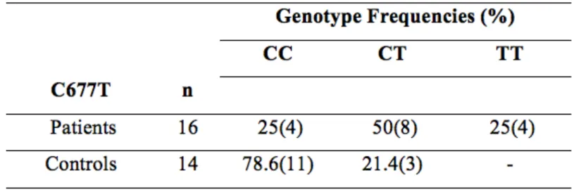
C677T genotype frequencies of the MTHFR gene are shown in Table 1. 677 CC/CT and 677 CC/TT MTHFR genotypes between the PC patients and controls were significant (!2
=5.41, P=0.023; !2
= 6.96, P=0.018; respectively). However, 677 CT/TT MTHFR genotypes between the PC patients were not significant (!2=1.36, P=0.36). We were not
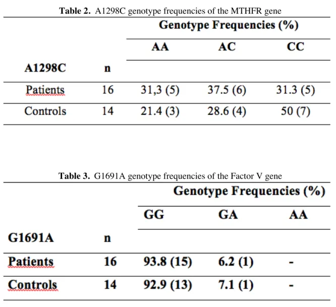
found 677 TT genotype in control groups. We compared 1298 AA/AC/CC genotype frequencies (Table 2) between the PC patients and controls groups, and the difference was not significant (!2= 0.83, P=0.32; !2=0.01,
P=0.64; !2
=0.73, P=0.33; respectively). There were no participants with AA genotype of Factor V Leiden (Table 3). A significant association was not found between GA/GG of Factor V Leiden genotypes between PC patients and controls groups. (!2
=0.01, P=0.72).
IV. Discussion:
Idiopathic intracranial hypertension (IIH) (Jacome, 2001), also known as pseudotumor cerebri, primarily affects young obese women; no single mechanism explains the disorder. (Duncan et al., 1988; Brazis and Lee, 1998; Bandyopadhyay, 2001; Jacome, 2001; Kesler and Gadoth, 2001; Kesler et al., 2001) The diagnosis is based on findings of papill edema; increased intracranial pressure with normal CSF composition; normal results of central nervous system imaging studies, including normal or small ventricles; and the absence of any intracranial mass (Duncan et al., 1988; Brazis and Lee, 1998; Bandyopadhyay, 2001; Jacome, 2001; Kesler and Gadoth, 2001; Kesler et al., 2001). One of the possible causes of IIH may be due to intracranial venous sinus thrombosis (Sussman et al., 1997), although cerebral angiograms could be normal in patients affected by IIH associated with
conditions highly predisposing to venous thrombosis. This raises the possibility that unrecognised non-occlusive venous thrombus might impede cerebral spinal fluid drainage (Grun et al., 1995). Thrombophilia-promoting oral contraceptives and heritable thrombophilic disorders synergistically increase the risk of cerebral sinus thrombosis (De Bruijn et al., 1998).
The present study is reported in 30 patients with IIH for the prevalence of coagulation polymorphisms that are thrombophylic risk factors (C677T MTHFR, A1298C MTHFR and FVL) in Turkish population.
MTHFR C677T and A1298C point mutations result in reduced enzyme activity that leads to elevated homocysteine concentrations and decreased folate concentration in plasma, which may represent important risk factors in thromboembolic diseases (Kang et al., 1993). The C!T 677 transition in the methylen-thetrahydropholate reductase (MTHFR) gene have caused inherited thrombophilia through hyperhomocysteinemia.
We were not found 677 TT genotype in control groups. However, 8% and 4% were found to be 677CT and 677TT genotype in PC patients group, respectively. Kesler et al. (Kesler et al., 2010) reported 30 (37.5%) carried a heterozygous mutant allele and 8 (10%) were found to be homozygous. Glueck et al. detected that C677T homozygous was present in 38% in patients with polycystic ovarian syndrome (Glueck et al., 2003). De Lucia et al. found that 18% were homozygous for the T allele of the MTHFR gene in IIH patients group (De Lucia et al., 2006)
In our study, 677 CC/TT MTHFR genotype frequencies between the PC patients were significant. Similarly, Kesler et al. obtained that the high prevalence of the MTHFR C677T mutation significantly increased the risk (Kesler et al., 2010). By contrast, no association were found between IIH and the C677T MTHFR polymorphism by Glueck et
al. (Glueck et al., 2003).
The 1298A>C polymorphism affects the capacity of the MTHFR enzyme less than the C677T polymorphism. We determined three genotypes of MTHFR A1298 C polymorphism. We compared 1298 AA/AC/CC genotype frequencies between the PC patients and controls groups, and the difference was not significant.
Among the familial thrombophilic states, altered activity of mutated Factor V (FV) is the most common hereditary blood coagulation disorder (Bertina, 1999; Nicolaes and Dahlbäck, 2002). FV Leiden (G1691A substitution) mutation is the most frequently reported abnormality of FV gene associated with increased risk of thrombosis.
Recently, the first study of a genetic polymorphism related to an increased risk of IIH was presented (Dogulu et al., 2003). The prevalence of three different factor V polymorphisms (Factor V Leiden, G1628A substitution and R2 allele) was found to be significantly higher in IIH compared with healthy controls. De Lucia et al. reported heterozygous FV mutation R506Q in one patient (6%) (De Lucia et al., 2006). Our findings were consist with these results. Our results revealed a prevalance of 6% heterozygosis for FV Leiden mutation in PC patients. On the other hand, no association between the FV Leiden mutation and increased prevalance of trombophilia in IIH was found by Kesler et al. (Kesler et al., 2010) and Glueck et al. (Glueck et al., 2003). Similarly, a significant association was not found between GA/GG of Factor V Leiden genotypes between PC patients and controls groups in present study. Additionally we did not determine AA genotype of Factor V Leiden. Glueck et al. (Glueck et al., 2005), De Lucia et al. (De Lucia et al., 2006) and Kesler et al. (Kesler et al., 2010) also did not show homozygous FV Leiden mutation.
Gene Therapy and Molecular Biology Vol 15, page 89
89
polymorphisms of the MTHFR can be regarded as a risk factor for PC patients in Turkish population. However, A1298C polymorphisms of the MTHFR and factor V
Leiden polymorphisms cannot be regarded as major risk factors for PC patients. A limitation of our study was the relatively small sample size.
The present study need to be replicated in larger population samples and investigated
changes in biochemical markers of hypercoagulability.
Table 2. A1298C genotype frequencies of the MTHFR gene
Gene Therapy and Molecular Biology Vol 15, page
91 References:
Bandyopadhyay S (2001). Pseudotumor cerebri. Arch. Neurol. 58: 1699-1701.
Brazis PW, Lee AG (1998). Elevated intracranial pressure and pseudotumor cerebri. Curr. Opin. Ophthalmol. 9: 27-32.
Bertina RM (1999). Molecular risk factors for thrombosis. Thromb. Haemost. [Review] 82: 601–609.
Corbett JJ, Mehta MP (1983). Cerebrospinal fluid pressure in normal obese subjects and patients with pseudotumor cerebri. Neurology. 33:1386-1388.
Daif A, Awada A, Al-Rajeh S, Abduljabbar M, et al. (1995). Cerebral venous thrombosis in adults. A study of 40 cases from Saudi Arabia. Stroke. 26: 1193-1195.
Dandy WE (1937). Intracranial pressure without brain tumor. Ann. Surg. 106: 492-513.
De Bruijn SF, Stam J, Koopman MM, Vandenbroucke JP (1998). Case control study of risk of cerebral sinus thrombosis in oral contraceptive users and in carriers of hereditary prothrombotic conditions. The Cerebral Venous Sinus Thrombosis Study Group. BMJ. 316:589-92.
De Lucia D, Napolitano M, Di Micco P, Niglio A, et al. (2006).
Benign intracranial hypertension associated to b lood coagulation derangements. Thromb. J. 4: 21.
Dogulu CF, Kansu T, Leung MY, Baxendale V, et al. (2003). Evidence for genetic susceptibility to thrombosis in idiopathic intracranial hypertension. Thromb. Res. 111: 389–395. Duncan FJ, Corbett JJ, Wall M (1988). The incidence
of pseudotumor cerebri. Population studies in Iowa and Louisiana. Arch. Neurol. 45: 875-877. Greaves M, Davies-Jones GAB (1993). Neurological
abnormalities associated with coagulopathies. Elsevier.
Glueck CJ, Iyengar S, Goldenberg N, Smith LS, et al. (2003). Idiopathic intracranial hypertension: associations with coagulation disorders and polycystic–ovary syndrome. J. Lab. Clin. Med.142: 35–45.
Glueck CJ, Goldenberg N, Golnik K, Sieve L, et al. (2005).
Idiopathic intracranial hypertension: association s with thrombophilia and hypofibrinolysis inme n. Clin. Appl. Thromb. Hemost. 11(4): 441-448. Grun L, Vinker S, Amitel M, Amir T, et al. (1995). Pseudotumor cerebri in Systemic Lupus Erythematosus Gemin. Arthritis. Rheum. 25(2):
103-108.
Hoffman HJ (1982). How is pseudotumor cerebri diagnosed? Arch. Neurol. 43:167-168.
Jacome DE (2001). Idiopathic intracranial hypertension and hemophilia A. Headache. 41: 595-598.
Kang SS, Passen EL, Ruggie N, Wong PW, et al. (1993). Thermolabile defect of methylenetetrahydrofolate reductase in coronary artery disease. Circulation. 88: 1463-1469. Kesler A, Goldhammer Y, Gadoth N (2001). Do men
with pseudomotor cerebri share the same characteristics as women? A retrospective review of 141 cases. J. Neuroophthalmol. 21: 15-7.
Kesler A, Gadoth N (2001). Epidemiology of idiopathic intracranial hypertension in Israel. J. Neuroophthalmol. 21: 12-4.
Kesler A, Kliper E, Assayag EB, Zwang E, et al. (2010).
Thrombophilic factors in idiopathic intracranial hypertension:a report of 51 patients and a meta-analysis. Blood. Coagul. Fibrinolysis. 21(4): 328-333.
Margaglione M, D'Andrea G, D'addedda M, Giuliani N, et al. (1998). The methylenetetrahydrofolate reductase TT677 genotype is associated with venous thrombosis independently of the coexistence of che FV Leiden and the prothrombin A2010 mutations. Thromb. Haemost. 79: 907-911.
Martinelli I, Battaglioli T, Pedotti P, Cattaneo M, et al. (2003). Hyperhomocysteinemia in cerebral venous thrombosis. Blood. 102: 1363-1366. Munts AG, Vangenderen PJ, Dippel DW, Van
Kooten F, et al. (1998). Coagulation disorders in young adults with acute cerebral ischaemia. J. Neurol. 245: 21-25.
Nicolaes GA, Dahlbäck B (2002). Factor V and thrombotic disease: description of a janus-faced protein. Arterioscler. Thromb. Vasc. Biol. [Review]. 22: 530– 538.
Sussman J, Leach M, Greaves M, Malia R, et al. (1997). Potentially prothrombotic abnormalities of coagulation in benign intracranial hypertension. J. Neurol. Neurosurg. Psych. 62: 229-233.
Yıldız OK, Cevik S, Cil G, Oztoprak I, et al. (2012). Cerebral Venous Sinus Thrombosis Presenting as Transient Ischemic Attacks in a Case With Homozygous Mutations of MTHFR A1298C and CG677T. J. Stroke. Cerebrovasc. Dis. 21(1):75-77.