Prostate-specific markers are required to identify rare prostate
cancer cells in liquid biopsies
EE van der Toom
3, H Axelrod
1,2, JJMCH de la Rosette
3, ThM de Reijke
3, KJ Pienta
1, and KC
Valkenburg
11
The James Buchanan Brady Urological Institute, Johns Hopkins University School of Medicine,
Baltimore, MD 21287
2Graduate Program in Cellular and Molecular Medicine, Johns Hopkins
University School of Medicine, Baltimore, MD 21287
3Department of Urology, Academic Medical
Center, Amsterdam, The Netherlands
Abstract
Despite early detection and treatment advancements, prostate cancer patients continue to succumb
to their disease. Minimal residual disease may lead to relapse and distant metastases, and
increasing evidence suggests that circulating and bone marrow disseminated tumor cells (CTCs
and BM-DTCs) can offer clinically relevant biological insights into prostate cancer. In this review,
we emphasize the pitfalls of using epithelial markers to accurately detect CTCs and BM-DTCs and
discuss the pressing need for prostate-specific markers in the detection of these cells using rare cell
assays. We have assembled a comprehensive list of published putative prostate-specific markers
and posit an ideal strategy for staining rare cancer cells from liquid biopsies. The ideal
prostate-specific marker is expressed on every CTC/BM-DTC throughout disease progression (high
sensitivity), and is not expressed on non-prostate cancer cells in the sample (high specificity). We
conclude that some markers are likely not specific enough to the prostate to be used as individual
markers of prostate cancer cells, whereas other genes may be truly prostate-specific and would
make ideal markers for rare cell assays. The goal of future studies is to utilize sensitive and
specific prostate markers to consistently and reliably identify rare cancer cells.
Keywords
prostate-specific markers; prostate cancer; circulating tumor cells; disseminated tumor cells; rare
cells; bone marrow
Corresponding author. KCV. Author Contributions
EEvdT: Wrote approximately half of the manuscript; helped assemble figures and tables; edited manuscript, figures, and tables HA: Edited manuscript, figures, and tables; added written sections
JJMCHdlR: Edited manuscript; provided input and funding TMdR: Edited manuscript; provided input and funding KJP: Edited manuscript; provided input and funding
KCV: Wrote approximately half of the manuscript; helped assemble figures and tables; edited manuscript, figures, and tables
HHS Public Access
Author manuscript
Nat Rev Urol
. Author manuscript; available in PMC 2019 July 01.
Published in final edited form as:
Nat Rev Urol. 2019 January ; 16(1): 7–22. doi:10.1038/s41585-018-0119-5.
A
uthor Man
uscr
ipt
A
uthor Man
uscr
ipt
A
uthor Man
uscr
ipt
A
uthor Man
uscr
ipt
Introduction
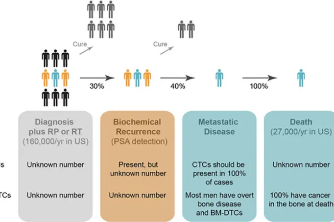
Prostate cancer (PCa) is the most common cancer and the second most common cause of
cancer-related deaths in men in the US
1. Despite advances in PCa screening, surgery,
hormone-related therapies, and chemotherapies, approximately 27,000 men still die of
metastatic PCa each year in the U.S. Of the patients diagnosed with early-stage PCa, nearly
half of them will not die of their disease without treatment. The other half of patients will
undergo treatment, by either radical prostatectomy or radiation therapy, with the goal to cure
their disease. Unfortunately, approximately 30% of these patients recur biochemically, based
on rising prostate specific antigen (PSA) levels in blood (FIG. 1)
2. Approximately 40% of
men with biochemical recurrence will develop metastatic disease, and 100% of those
patients will succumb to their disease
2. Notably, 100% of men who died of PCa and who
were autopsied had PCa present in their bones
3. Metastases often appear years after primary
treatment, indicating that tumor cells must have escaped the primary tumor prior to therapy
and disseminated to distant sites
4–6. Tumor cell dissemination and metastasis is a
complicated multi-step process
7that requires primary tumor cells to enter the vasculature,
where they are referred to as circulating tumor cells (CTCs). Most CTCs are unable to
withstand the shear stress, immune surveillance, and lack of cell-cell adhesion in the
circulation and will die prior to reaching distant sites. CTCs that are able to exit the
circulation and establish residence at a distant site, such as the bone marrow (BM), are
called disseminated tumor cells (DTCs; we will refer to DTCs in the BM as BM-DTCs). The
specific timing of this cellular dissemination process in the natural history of PCa
progression prior to metastatic development is largely unknown but highly intriguing (FIG.
1). Metastatic PCa remains incurable, and current imaging modalities are not sensitive
enough to detect individual cancer cells or small colonies of disseminated cells. If CTCs and
BM-DTCs can be identified prior to the formation of overt metastatic lesions, treatments can
be aimed at preventing metastasis altogether
8–10.
Fine needle biopsies are the standard for PCa diagnosis and prognosis, but they are invasive
and can cause significant morbidity. Therefore, there is much appeal for investigating the
clinical utility of minimally invasive liquid biopsies to use CTCs and BM-DTCs as
biomarkers of disease
11–13. Accurate detection of these cells will also allow for their
biological characterization, in which therapies can be more precisely targeted to the
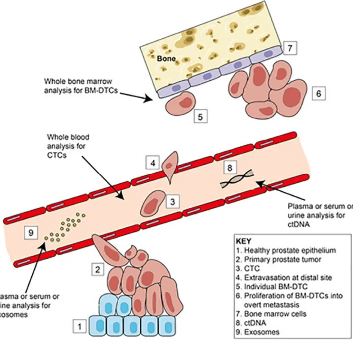
mechanisms leading to recurrence. Although we will focus mostly on CTCs and BM-DTCs,
liquid biopsies can also provide clinically relevant information in the form of cell-free
circulating tumor DNA (ctDNA) and exosomes, both of which can also be present in urine
(FIG. 2)
14–16. Liquid biopsies can provide a real-time non-invasive snapshot of the total
tumor burden of a patient and can furthermore provide important complementary
information on therapeutic targets and mechanisms of drug resistance. De Bono et al.
previously reported that the number of CTCs found in patients with castration-resistant
prostate cancer (CRPC) can predict overall survival. Patients with ≤5 CTCs (per 7.5 mL of
blood) survived 10.2 months longer than patients with >5 CTCs (using EpCAM-based
purification methods)
17. Other studies have correlated the number of CTCs in metastatic
PCa to therapeutic response and survival, while limited, but emerging, studies have been
paralleled in pre-metastatic PCa patients
18–23. As such, CTC data from blood draws are
A
uthor Man
uscr
ipt
A
uthor Man
uscr
ipt
A
uthor Man
uscr
ipt
A
uthor Man
uscr
ipt
extremely clinically relevant, and will continue to be so. Clinical correlations have not been
as rigorously assessed for BM-DTCs, as bone marrow is more difficult to obtain, and it is
more difficult to identify BM-DTCs than CTCs due to decreased marker specificity. While
CTCs will likely play a more important role in providing clinically relevant data real-time,
BM-DTCs may represent a more important cell population, as they have successfully
migrated from the primary tumor to a distal site. We propose that BM-DTC data will provide
much-needed information about timing of dissemination, as well as the genetic and
epigenetic qualities of a successfully disseminated and proliferating cancer cell. As such, our
ultimate goal is to determine prostate-specific markers that sensitively and specifically
identify BM-DTCs for downstream analysis.
It is important to understand the lethal characteristics and clinical application of CTCs and
BM-DTCs after they are reliably detected. The two most commonly used methods for CTC
detection are reverse transcription PCR (RT-PCR) and fluorescence-based immunostaining
(referred to as immunofluorescence, or IF). FISH (fluorescence in-situ hybridization) can be
used as a tool similar to IF and PCR to identify CTCs via RNA expression, thereby helping
to define the different gene expression patterns within these cells
24. Each of these methods
has its own set of advantages and limitations (TABLE 1), but IF has certain advantages that
allow for further biological characterization of functional activity at the time of detection.
Many different assays exist for the detection of CTCs (very few exist for BM-DTCs), and
most rely on positive selection of cancer cells or negative selection of leukocytes, though
selection-free methods also exist
25–27. Most also involve the separation of red blood cells
from white blood cells and cancer cells, which is commonly done via microfluidics chips,
red blood cell lysis buffers, and/or centrifugation-based separation
26–29. The type of
detection methodology will change the resulting cell population and molecular composition
that is analyzed, as certain cell types may be enriched or lost based on the experimental
conditions. For instance, analyzing whole blood RNA for a specific marker without
including a selection step will not yield meaningful results about the specificity of that
marker to cancer cells. Many studies have used selection methods (usually via epithelial
selection based on EpCam expression or size-based selection using a microchip) to detect
CTCs from blood using RT-PCR, multiplex PCR, or digital droplet PCR
30–37. These studies
show that RT-PCR is extremely sensitive for CTCs, but no such success has been found in
BM-DTCs.
Current standard markers used for CTC detection via IF include a nuclear marker (usually
DAPI), a marker for white blood cells (WBCs; usually CD45) and one or more epithelial
markers (usually EpCAM and/or pan-cytokeratin)
17,38. A major limitation of relying on
epithelial markers for CTC identification is that several studies have shown previously that
these markers are not always highly expressed on cancer cells, and have also been shown to
be expressed on cells of hematopoietic lineage
39–44. Furthermore, it is thought that CTCs
lose their epithelial phenotype after undergoing epithelial to mesenchymal transition (EMT)
to escape the primary tumor, and thus they may lose EpCAM and/or cytokeratin (CK)
expression
45–50. While EpCAM-based detection methods have been the most common
method to identify CTCs, it is unknown how frequently this loss of epithelial characteristics
occurs. In addition, the blood, and particularly the BM, contains a vast heterogeneity of
cells, many of which are stem or other cells that can epigenetically alter their phenotype.
A
uthor Man
uscr
ipt
A
uthor Man
uscr
ipt
A
uthor Man
uscr
ipt
A
uthor Man
uscr
ipt
This can lead to false positive immunostaining, in which the detection marker is no longer
specific for prostate cells. Once CTCs are isolated, further characterization can be performed
by using different functional assays, such as EPISPOT, which detects specific proteins
during the in vitro culturing of CTCs
51. Another example is the cancer cell line-derived
xenografts (CDXs), by which cancer cells from cell lines or patient-derived CTCs are
injected into immune-compromised mice, after which metastases will develop
52,53, although
this has not been successful in PCa. This can give important in vivo information for more
individualized treatment of cancer patients.
We posit that the use of prostate-specific markers to identify prostate CTCs and BM-DTCs
will allow for more sensitive and specific detection of these rare cancer cells. So far, the
identification of these markers for rare tumor cells has been challenging, as some reported
prostate-specific markers are not very sensitive (not expressed in all PCa cells) or specific
(also expressed by other cells in the blood or BM) (SUPPL. TABLE 1). Many studies on
these markers have only assessed expression of protein at the tissue level (e.g. IHC on
formalin-fixed paraffin-embedded tissue) or RNA in whole blood (e.g. RT-PCR), neither of
which represents true sensitivity or specificity at the rare cell level. Therefore, this
manuscript makes clear that each of these markers should be assessed in rare cell assays in
blood and BM samples before any conclusion can be made as to their utility in liquid
biopsies. Also, dedifferentiation and loss of prostate-specific markers can occur in a
significant proportion of poorly differentiated prostatic adenocarcinomas
47,54,55. It is thus
imperative that we find highly sensitive and specific prostate markers that are expressed
during all the stages of a patients’ disease, expressed on every tumor cell, and not expressed
on any blood or BM cells. In this review, we will discuss what is known about the putative
prostatic lineage markers and highlight their pros and cons in the detection of CTCs and
BM-DTCs (TABLE 2).
Prostate Specific Markers
Prostate specific antigen and other kallikreins
Prostate specific antigen (PSA, also known as kallikrein related peptidase 3, or KLK3, and
human glandular kallikrein 3, or hK3) is currently the most important and clinically useful
marker in PCa screening. It is produced by secretory epithelial cells in the prostate
56and is
an androgen-regulated serine protease expressed in both benign and malignant prostatic
tissue. PSA is one of the oldest prostatic markers used in immunohistochemistry (IHC) to
confirm that a metastatic carcinoma is prostatic in origin
57. It has been widely shown that
PSA has a high specificity for PCa, but that its expression also tends to decrease with cancer
progression. PSA expression may be absent in around 5% of patients with high-grade PCa
and distant metastases, as well as in around 10% of lymph node metastases
47,58–61. The
staining pattern for PSA is cytoplasmic, which can present an issue for IHC because diffuse
cytoplasmic staining can generate false positives during analysis and is known to occur in
IHC
62. While PSA expression has been reported in a variety of non-prostatic tissues and
tumors, including breast and lung carcinomas
58,63–67, others have reported high sensitivity
and specificity of PSA in PCa using monoclonal and polyclonal anti-PSA antibodies
68. PSA
expression from PCa patient blood has been correlated with cancer at the RNA level via
RT-A
uthor Man
uscr
ipt
A
uthor Man
uscr
ipt
A
uthor Man
uscr
ipt
A
uthor Man
uscr
ipt
PCR, but neither study used a selection protocol to ascertain which cells expressed PSA
32,69.
Overall, PSA is a promising marker for rare cell assays, as it seems to be sensitive for most
PCa cells while its expression has not been reported in blood or BM cells (unlike AR
expression), although this must be tested in rare cell assays. Coupled with evidence that PSA
can be controlled in an AR-independent manner
70, addressing sensitivity issues, PSA could
potentially be a more promising rare cell marker than AR. PSA is also a widely-used
biomarker of primary prostate tumor growth as well as for biochemical recurrence following
radical prostatectomy or radiation therapy. As with all of the proteins we will discuss in this
article, its full utility as a rare cell marker in blood and BM has yet to be ascertained in PCa
and non-cancer patients.
PSA belongs to the kallikrein serine protease family, which contains 15 family members.
Besides PSA, two other kallikrein family members, KLK2 and KLK4 (also known as
prostase and KLK-L1), also seem to be prostate specific
71–78. There is less known about the
clinical utility of these markers, but both have been found in PCa patient tissue and serum.
Both KLK2 and KLK4 seem to have a proteolytic function in activating PSA from its
precursor pro-PSA form to its active PSA form. These kallikreins should be assessed in rare
cell assays in addition to PSA.
Androgen receptor
The androgen receptor (AR) is the most widely studied protein related to prostate
development and PCa. AR is a powerful transcription co-factor that affects the development
and growth of male sex organs, including the prostate
79. Androgen-mediated nuclear
localization and activation of AR is required for the development and growth of the prostate
gland
80–83, and deprivation of androgens inhibits proper ductal development of the gland
84.
These phenotypes can be seen during embryonic development, where fetal testicular
secretion of androgens promotes prostate development
82. The adult prostate’s structural
maintenance and reproductive function also requires androgens and AR activity
85. Binding
of dihydrotestosterone to AR causes it to translocate to the nucleus and bind androgen
response elements in genomic DNA to initiate
86,87or down-regulate transcription of target
genes
88,89. AR also has non-transcription-related functions, but these are less well
understood and have only been reported in cancer tissue
90. Expression of many other
prostate-specific genes that we will discuss in this article is transcriptionally regulated by
AR. Due to its crucial roles in the development, growth, and maintenance of the prostate, it
is not surprising that AR plays critical roles in PCa. Some groups have reported tumorigenic
properties of AR in mouse models
91,92. However, mice lacking AR specifically in the
murine prostate had increased cellular proliferation, indicating that the role of AR in cancer
initiation is still not fully understood
93. Interestingly, while PCa is one of the most prevalent
cancers in men, there are almost no cancers of the seminal vesicle or bulbourethral gland,
both of which express AR
94. AR is strongly expressed in most PCa tumors, and PCa
maintenance seems to depend on AR signaling
95–98. Androgen deprivation therapy (ADT)
and AR targeting therapies have significant survival benefits in advanced PCa patients and
are widely used in the clinical setting
99–101. Importantly, AR expression can be lost in some
PCa tumors, particularly those with neuroendocrine or small cell PCa pathology
102–105. Of
great interest and potential utility in rare cell assays are the AR splice variants. It has been
A
uthor Man
uscr
ipt
A
uthor Man
uscr
ipt
A
uthor Man
uscr
ipt
A
uthor Man
uscr
ipt
shown that expression of the AR-V7 variant increases in castration resistant PCa
106.
Moreover, expression of the full-length version of AR versus the AR-V7 variant in PCa
CTCs can predict ADT response
21,47,107,108, and this has led to its use in guiding therapeutic
strategy
19. However, the use of AR solely as a CTC/BM-DTC marker for rare cell assays
poses specificity issues because it is expressed on BM cells and platelets, as well as in other
tissues
109–112(SUPPL. TABLE 1). We believe that AR is not specific enough for prostate
tumor cells to be used as an individual marker for rare PCa cells, but has potential as an
adjuvant marker for clinical management. Furthermore, because AR is expressed in certain
blood and BM cells, and AR regulates the expression of many other putative
prostate-specific markers, each of these markers must be rigorously assessed for its expression in
blood and BM to determine specificity. A non-androgen-regulated prostate-specific gene
would be an ideal marker in prostate CTC and BM-DTC detection assays, but such markers
are seemingly rare.
Prostate specific membrane antigen
Prostate specific membrane antigen (PSMA, also known as folate hydrolase 1, or FOLH1) is
a membrane-bound glycoprotein with high specificity for both benign and malignant
prostatic tissues. In contrast to other androgen-regulated prostate genes, PSMA is suppressed
by androgens in an AR-dependent manner
113. The initial cloning of the gene of PSMA was
accomplished by Israeli et al. in 1993 using the LNCaP PCa cell line
114. PSMA is currently
being explored extensively as a promising target for molecular imaging as well as a
therapeutic target in prostate and renal cancers. For PCa, it may be useful in the setting of
biochemically recurrent disease, where PSMA-targeted radiotracers seem to be superior to
conventional imaging for detection of metastatic PCa
115–117. PSMA is expressed at low
levels in benign prostatic epithelium and is strongly expressed in most prostate
carcinomas
118. PSMA is, in contrast with PSA, highly up-regulated in high-grade tumors
and corresponding metastases
119. Normal prostate epithelium often has a low level of diffuse
cytoplasmic staining, while high-grade and metastatic tissues mostly have a very intense
cytoplasmic and focal membrane staining
61,119. Unfortunately, as it was originally thought
to be strictly expressed in prostatic tissue, it is now known that PSMA is widely expressed in
a variety of non-prostatic solid tumors and vasculature, including urothelial, renal,
gastrointestinal, and breast carcinomas, in addition to bone diseases such as Paget’s disease
and healing bone fractures
120–130(SUPPL. TABLE 1). PSMA expression in non-prostatic
cancer cells is mostly restricted to the cytoplasm
61. Furthermore, a study by Kinoshita et al.
reported the detection of the PSMA protein in an exceptional variety of healthy tissues,
including the urinary bladder and proximal tubules of the kidney
122. Uhlén et al.
demonstrated mRNA expression of PSMA in normal male and female BM, but no protein
expression
112. PSMA expression in PCa and non-PCa patient blood was ascertained in a
selection-free way via RT-PCR of whole blood RNA, and its sensitivity and specificity were
reported as 59% and 47%, respectively; however, due to lack of selection, there was no way
to ascertain which cells expressed the marker
131. While PSMA is a promising marker for
overt prostate tumor detection, the application of PSMA as a marker for rare PCa cells needs
further assessment, as its true specificity is still in question.
A
uthor Man
uscr
ipt
A
uthor Man
uscr
ipt
A
uthor Man
uscr
ipt
A
uthor Man
uscr
ipt
Prostate stem cell antigen
Prostate stem cell antigen (PSCA) is an androgen-regulated
glycosylphosphatidylinositol-anchored membrane-bound glycoprotein, originally identified as a prostate-specific
tumor-promoting antigen in 1998
132,133. Its expression is restricted to the basal layer of the
prostate, and it is the only protein in this article that is expressed by basal cells
132. It is
expressed in approximately 88–94% of primary PCa specimens
132,134, one study observed
100% (9/9) of bone metastatic lesions to be PSCA-positive
134. Another study by Lam et al.
found a PSCA protein expression in 87.2% (41/47) of cases of bone metastases
135. PSCA
may be a useful marker for PCa prognosis
135–137, as one study reported PSCA mRNA
expression in the peripheral blood of 71% of PCa samples, 13% of benign prostatic
hyperplasia samples, and 0% of non-prostate disease controls
138. A similar study reported a
sensitivity of 40% in patients with gastrointestinal tumors
139. However, because there was
no selection process in these studies, whole blood RNA was assessed, so it is unclear
whether the PSCA-positive cells were actually prostate cells or another type of cell. As we
have discussed, this is one significant drawback to RT-PCR compared to IF assays. Though
there are several reports showing absence of PSCA expression in non-prostatic
tissues
132,134, others have found expression in the normal epithelium of various tissues,
including the urinary bladder, kidney, and intestine
112,134,140–142(SUPPL. TABLE 1).
PSCA is also overexpressed in various cancers, including urothelial, kidney, and lung
143–147.
In some cancers, it is down-regulated, indicating it may also play a tumor suppressive role,
depending on the tissue
140,142,148–150. Overall, data suggest that PSCA expression is not
actually specific to the prostate, which makes it a less desirable marker for rare cells assays
on its own. However, its expression in the basal cell compartment of the prostate indicates
that it could potentially be used for certain subsets of PCa that are of basal cell origin.
Alpha-methylacyl-CoA racemase
Alpha-methylacyl-CoA racemase (AMACR, also known as P504S) is a peroxisomal and
mitochondrial enzyme involved in bile acid biosynthesis and beta-oxidation of
branched-chain fatty acids, and it is not androgen-regulated
151,152. Its expression is granular and
cytoplasmic. Apart from the prostate, AMACR is expressed in other normal tissues,
including BM cells
112(SUPPL. TABLE 1). AMACR is also overexpressed in almost every
type of carcinoma assessed, including over 95% of PCa cases
112,153,154. It is thus not useful
in distinguishing PCa from other malignancies. However, it is still commonly used as a
diagnostic biomarker for PCa due to its stronger expression in malignant relative to normal
tissue, and it is often used in combination with a negative marker for PCa such as the basal
cell marker p63
155–157. In an RNA-based study from patient blood, AMACR expression was
found in only 16/22 PCa patients, as well as 11/20 non-PCa patients, indicating poor
sensitivity and specificity, although there was no selection process, so there is no way to
assess which cells were expressing the marker
158. AMACR can be detected (in tissue
studies) in approximately 80% of atypical, non-hormonally-regulated PCa, such as small
foci prostate adenocarcinomas and pseudohyperplastic carcinomas
157,159. AMACR is also
overexpressed in non-cancerous prostate diseases, such as adenosis, post-atrophic
hyperplasia, partial atrophy, and prostatic intraepithelial neoplasia
160. AMACR RNA is
expressed in the BM
112; therefore, it cannot be used for BM-DTC detection in PCR assays.
A
uthor Man
uscr
ipt
A
uthor Man
uscr
ipt
A
uthor Man
uscr
ipt
A
uthor Man
uscr
ipt
However, more work needs to be done to determine its sensitivity and specificity in rare cell
assays.
Prostate specific acid phosphatase
Prostate specific acid phosphatase (PSAP, also known as prostatic acid phosphatase (PAP)
and prosaposin) is a glycoprotein that hydrolyzes esters under acidic conditions to yield
inorganic phosphates, and it is one of the major proteins that is secreted by the
prostate
161,162. It is an androgen-regulated protein that was first discovered in 1938 by
Gutman et al. who showed that the level of PSAP was increased in the blood of patients with
localized PCa, and was even more highly expressed in metastatic disease, relative to healthy
individuals
163. It thus became the first serum tumor marker for biochemical testing to
diagnose and monitor progression of PCa. Later, PSA was found to be a more sensitive and
specific biomarker and replaced PSAP in these assays. A study by Walsh et al. evaluated 460
localized PCa cases, and only 0.9% of cases were PSAP-positive and PSA-negative,
indicating that PSAP detection would not capture additional cancer cells that would not
already be detected by PSA
164. PSAP is still occasionally used for the evaluation of PCa
tissue by IHC, where it shows granular cytoplasmic staining. PSAP is expressed at moderate
to high levels in normal prostate tissue and is strongly expressed in >95% of malignant
prostatic tissue
165–167. While these studies are tissue-based, and not cell-based, they suggest
that PSAP may be a sensitive marker for PCa in general. However, a study by Perner et al.
showed that PSAP was expressed in only 84% and 77% of lymph node and distal
metastases, respectively, suggesting that express may be lost in a clonal fashion during
metastasis
61. It is also expressed in a variety of other cancers, including melanoma,
lymphoma, cancer of the testis, and urothelial cancer
112(SUPPL. TABLE 1). Several studies
have reported expression of PSAP protein in normal non-prostatic tissues, including
granulocytes
112,165,167–171. Importantly, Uhlén et al. detected protein and mRNA in normal
female and male BM tissues, indicating decreased specificity for BM-DTC detection
112.
Despite its high expression in most prostate carcinomas, the distribution of PSAP expression
in other healthy tissues, particularly immune cells and other BM cells, indicates that PSAP is
not as prostate-specific as was initially suggested, and may not be specific enough to be used
alone as a detection marker for CTCs or BM-DTCs.
TMPRSS2-ERG
The transmembrane protease, serine 2 (TMPRSS2) gene is androgen-regulated and is
located close to the erythroblastosis virus E26 transformation specific related gene (ERG) on
chromosome 21. In about 50% of PCa patients a gene rearrangement occurs between
TMPRSS2 and ERG, which produces the androgen-regulated over-expressed fusion protein
TMPRSS2-ERG, where ERG is the driving oncogene
172. The TMPRSS2-ERG fusion is
typically assessed via FISH, and is nearly 100% specific for prostate tissue (SUPPL. TABLE
1). ERG expression by IHC can also be used as a surrogate for expression of the fusion
gene
173, and ERG staining has been associated with worse prognosis for PCa patients
174.
Even before the discovery of the TMPRSS2-ERG gene fusion, the presence of ERG in PCa
was reported
175. Similar to PCA3, TMPRSS2-ERG has utility as a biomarker in urine tests
with 37% sensitivity and 93% specificity
176. When TMPRSS2-ERG and PCA3 detection in
urine samples was combined, sensitivity increased to 73%, which still falls short of the ideal
A
uthor Man
uscr
ipt
A
uthor Man
uscr
ipt
A
uthor Man
uscr
ipt
A
uthor Man
uscr
ipt
sensitivity for a rare cell assay. However, due to their high specificity for PCa cells, both of
these markers have value moving forward, likely in combination with other markers. The
biggest advantage of using TMPRSS2-ERG to detect PCa cells is that it is specific to cancer
cells, and has not been found in normal prostate tissue. Most of the other candidate
prostate-specific markers discussed in this article have been detected in benign tissue, making it
difficult to differentiate cancer from benign. In rare cell assays, it is likely that only cancer
cells will be present in blood or BM, but that has not been definitively proven. It is possible
that non-cancer cells could slough into the blood and be identified as cancer cells based on
expression of prostate-specific markers. In patients known to have TMPRSS2-ERG
expression in their primary tumor, including TMPRSS2-ERG as an additional marker for
CTC/BM-DTC detection would eliminate doubt about the origin of the rare cells in
question. It is important to note that other gene fusions exist in PCa, including a
prostein-ERG fusion
177, TMPRSS2 fusion with other ETS family genes such as
TMPRSS2-ETV4
178, as well as many other fusions that have not been assessed for their sensitivity but
could be useful in identifying cancer cells in a multiplex FISH staining strategy
179.
Prostate cancer antigen 3
Prostate cancer antigen 3 (PCA3, initially known as differential display clone 3, or DD3), is
an androgen-regulated long non-coding RNA (lncRNA) that was discovered in 1999
180,181.
PCA3 down-regulates expression of the tumor suppressor PRUNE2, thereby promoting
tumor progression
182,183. PCA3 is overexpressed in around 95% of PCa cases and is thought
to be prostate-specific, as it was not detected in 18 other normal tissues in a major study
(although blood and BM were not assessed)
180(SUPPL. TABLE 1). As a lncRNA, PCA3
cannot be detected by IHC or IF, and its detection is limited to RT-PCR or fluorescent in situ
hybridization (FISH) assays
29,184. PCA3 is currently being tested as a urinary biomarker for
PCa, although its sensitivity is limited, even when combined with urinary
biomarkers
176,185,186. Overall, PCA3 holds some promise as a marker of rare PCa cells, but
because the combination of IF with FISH is technically challenging, we are less enthusiastic
about this marker for rare cell assays.
Homeobox protein NKX3.1
NKX3.1 is a homeobox-containing transcription factor. It is androgen-regulated and is
therefore largely prostate-specific, although – like PSA – its expression can be regulated
independent of AR. It is often used as an IHC marker of prostatic origin in metastatic
tumors
187. NKX3.1 is primarily detected in secretory prostatic epithelia, and its staining
pattern is primarily nuclear, though it can also be seen in the cytoplasm
188. It is one of the
earliest known markers of prostate development
189. It is a putative tumor suppressor in PCa,
as it functions to inhibit prostate cell growth and proliferation in a context dependent
manner, and one allele is frequently deleted in patients with PCa
189. It has been reported that
NKX3.1 expression is high in primary PCa tumors, but low in high-grade tumors and absent
in metastatic PCa
190,191. However, Gurel et al. assessed the performance of NKX3.1 as a
marker of hormone naïve metastatic PCa and found that the sensitivity for NKX3.1
expression was 98.6%
187, as 68/69 of cases were positive. The same study showed the
specificity of NKX3.1 was 99.7% as only 1/349 non-prostatic tumors was positive. This
discrepancy with previous studies is most likely explained by the use of different antibodies,
A
uthor Man
uscr
ipt
A
uthor Man
uscr
ipt
A
uthor Man
uscr
ipt
A
uthor Man
uscr
ipt
where the latter study used an ostensibly better antibody
190,191. NKX3.1 has been found in
rare invasive lobular breast carcinomas and in benign testis
189,192,193(SUPPL. TABLE 1).
Uhlén et al. detected mRNA expression in a plethora of healthy tissues, including the
salivary glands, kidney, testis, and importantly, the bone marrow, but did not assess protein
expression
112. Altogether, these data suggest that NKX3.1 is relatively sensitive for PCa
cells, but potentially not specific enough to differentiate PCa cells from BM cells, although
this has yet to be tested at the protein level.
Homeobox B13
Homeobox B13 (HOXB13) is a transcription factor that is involved in prostate development
and is one of the few markers discussed here whose expression is
androgen-independent
194,195. HOXB13 may physically interact with AR in the nucleus of prostate
cells, potentially in an inhibitory fashion
196,197. It is expressed in normal prostatic tissue
198,
and overexpressed in PCa
197,199. It is used to identify metastatic prostate tissue
200. The
HOXB13 G84E variant mutation is associated with significantly increased risk of hereditary
PCa
201. The fact that there is a reported lack of any truncating mutations in HOXB13 and
the recurrent nature of the G84E change, suggest a carcinogenic mechanism that is most
likely of oncogenic nature (gain of function) than of tumor-suppressor nature (loss of
function). The staining pattern of HOXB13 is primarily nuclear, but can also be seen in the
cytoplasm. Weak to moderate cytoplasmic staining has been observed in some non-prostatic
cancers, such as in liver and lung cancers
112(SUPPL. TABLE 1). Furthermore, Uhlén et al.
reported low expression of HOXB13 in patients with lymphoma
112. A recent study by
Barressi et al. compared the diagnostic value of HOXB13 and PSA protein expression to
determine if metastatic tissue was of prostatic origin
202. HOXB13 immunostaining was
strong in >75% of the neoplastic cells in 100% (15/15) of the prostatic metastases, and weak
staining was found in <25% of the neoplastic cells in 17% (2/12) of urothelial carcinoma
metastases. The sensitivity and specificity of HOXB13 for metastatic PCa were 100% and
94%, respectively. Furthermore, the sensitivity and specificity of PSA for these metastatic
PCa tissues were 53% and 100%, respectively
202. A study by Varinot et al. also assessed
HOXB13 sensitivity, and reported that while all 400 PCa tumors they assessed expressed
some level of HOXB13, bone metastases had less frequent HOXB13 expression, although
this could have been due to decalcification of the bone tissue
200. Another group showed that
HOXB13 expression was found in 52% of 10,216 PCa patient samples, and that stronger
staining was associated with PCa cells relative to normal prostate cells, giving it prognostic
relevance
197. Interestingly, it appeared that HOXB13/AR interaction resulted in a reduction
of PSA expression, indicating that HOXB13 and PSA could be used together in rare cell IF
assays. Overall, these data suggest that HOXB13 is a promising candidate marker for the
detection of prostate CTCs and BM-DTCs due to its specificity and androgen-independence
in tissue-based assays, but work needs to be done in rare cell assays to fully ascertain its
utility.
Prostatic secretory protein of 94 amino acids
Prostate secretory protein of 94 amino acids (PSP94, gene name MSMB) is one of the first
three secretory proteins in the prostate to be identified, in addition to PSA and PSAP
203.
PSP94 was originally identified as beta-microseminoprotein (MSMB)
204, or beta-inhibin
205,
A
uthor Man
uscr
ipt
A
uthor Man
uscr
ipt
A
uthor Man
uscr
ipt
A
uthor Man
uscr
ipt
and is an androgen-regulated immunoglobulin-binding factor that is secreted into seminal
plasma
206–209. Its specific function is still uncertain, but it has been suggested that it
increases sperm quality
210and acts as a fungicidal agent in sperm
211. PSP94 protein has
been found in numerous additional secretions, including mucous gland secretions
212. Its
expression has also been detected in tonsil, skin, bronchus, stomach, testis, and seminal
vesicle tissue
112(SUPPL. TABLE 1). PSP94 expression in cancer is somewhat unclear.
Overexpression of PSP94 has been observed in ovarian cancer
213, while several studies have
shown that it acts as a tumor suppressor in PCa
214–218. One study in PCa showed that while
PSP94 expression was inversely correlated with Gleason score, its expression persisted after
hormone therapy while PSA expression decreased, indicating that PSP94 expression can be
up-regulated in the absence of androgens
219. Support for its putative role as a tumor
suppressor comes from the observed association of the loss of function of variant MSMB
alleles with increasing PCa risk
220,221, as well as its antifungal, and therefore
anti-inflammatory properties
221. It has also been shown that a driver of PCa, EZH2, targets and
silences PSP94
222. Finally, a synthetic peptide corresponding to certain PSP94 amino acids
has been shown to decrease vascular endothelial growth factor (VEGF) expression in
endothelial cells, indicating PSP94 may have anti-angiogenic effects
223. All in all, PSP94 is
not likely a suitable candidate for CTC and BM-DTC detection due to its varied expression
throughout PCa progression.
Prostein
Prostein (also known as p501s, and solute carrier family 45 member 3, or SLC45A3) is one
of the latest prostate-specific markers to be discovered, having been found via a
genome-based approach in 2001
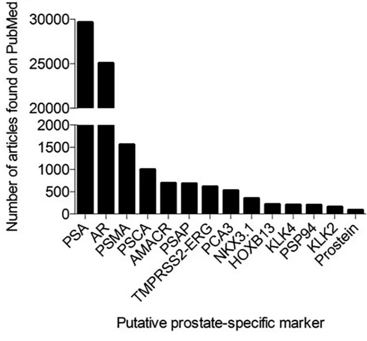
224. It is also the least published marker in this article, with only 86
results in PubMed, compared to 29,628 results for PSA (FIG. 3). Prostein is an
androgen-regulated type IIIa transmembrane protein located in the Golgi apparatus with functions
related to macromolecule transport
225. Prostein is expressed in normal prostate tissue as well
as PCa tissue
61,226, even when PSA is negative
225,227. It has a unique granular staining
pattern, which helps to distinguish it from other markers and increases confidence of true
staining. Prostein has been used to differentiate PCa (prostein-positive, p63-negative) from
urothelial cancers (prostein-negative, p63-positive) in tissue IHC
55. Along with HOXB13,
prostein is one of the most prostate-restricted proteins in tissue-based assays, though its
expression has also been found in lung and bladder cancer
228(SUPPL. TABLE 1). To date,
prostein expression has been analyzed on different normal non-prostatic tissue, but none of
these tissues expressed this marker, though it has not been extensively characterized. One
study compared tissue expression of prostein to expression of PSA, PSAP, PSMA, AR, and
ERG in primary PCa and metastatic tumors, and found that prostein sensitivity was
decreased in metastatic tumors, although it was still expressed in 89% of tumors
61. They
also found that when PSA was absent in tumors, prostein and AR were present, indicating
that more than one prostate-specific marker should be used to increase sensitivity in IHC and
certainly in rare cell assays. Taken together, we believe that prostein is a promising marker
for use in IF-based rare PCa cell assays, although this has not been directly tested.
A
uthor Man
uscr
ipt
A
uthor Man
uscr
ipt
A
uthor Man
uscr
ipt
A
uthor Man
uscr
ipt
Murine Prostate Markers
Mice are used extensively as in vivo models of prostate cancer metastasis, and rare cell
assays have recently been developed for xenograft, syngeneic, and transgenic mouse
models
29. Xenograft models utilize human cancer cells, for which the markers we have thus
far discussed are applicable. However, when using mouse models that develop murine
prostate cancer (syngeneic models or genetically engineered mouse models (GEMMs)), one
must consider the similarities and differences between rodent and human prostates at the
anatomical and cellular expression levels. While the mouse prostate gland is histologically
quite similar to the human prostate gland, there are significant differences. The human
prostate surrounds the urethra at the base of the bladder. It is broken up into “zones” for
grading and staging purposes, but anatomical zonation is not grossly apparent. The mouse
prostate is broken up into several lobes: the anterior lobe, which is immediately next to the
seminal vesicle; and the dorsolateral and ventral lobes, which are anatomically similar to the
human at the base of the prostate
229,230. In the mouse and human, all prostate glandular
secretions go into the urethra and make up a significant portion of the ejaculate. Another
significant difference between the human and mouse prostate is the ratio of luminal to basal
cells. In the human, the ratio is approximately one luminal cell per basal cell, and in the
mouse, the ratio is closer to 3:1
231.
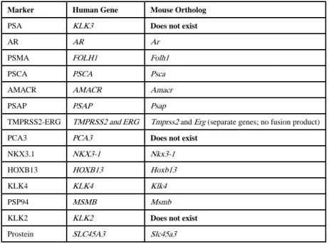
In terms of gene expression, mice do not express PSA, KLK2, or PCA3 (Table 3). Of the
kallikreins, only KLK4 has a murine ortholog
232. Mice express a PSCA ortholog, which is
70% similar to human
132. PSP94, PSMA, and PSAP are also expressed, and are specific to
the mouse prostate
233–235. Mice also express Hoxb13 independent of androgen, and this
gene has been used to create a GEMM of PCa
236,237. Nkx3.1 is another marker present in
mice, and its role in prostate development and tumorigenesis has been studied extensively in
mouse models
238–240. Mice also express an AMACR ortholog, though its role in murine
prostate biology is limited
241. It is unclear based on published literature if prostein is
expressed in the mouse prostate at the protein level, although RNA ISH has shown that the
Slc45a3 gene is expressed throughout developing tissue in mouse embryos
242. Mice express
both TMPRSS2 and ERG, although with no prostate specificity, and the TMPRSS2-ERG
fusion does not occur in mice because they never develop de novo PCa
243. Mice also express
AR; in fact, many of the androgen signaling paradigms have been discovered by studying
mouse or rat AR (see above section on AR). However, an important consideration is that AR
activity in mice might differ from human due to the amount of testosterone in either species
at any given time – it has been shown that a hormonally intact male mouse has
approximately as much circulating testosterone as an androgen-ablated male human
85,244.
Perhaps the best way to use mice as an in vivo model for rare cell studies is to inject
genetically labeled human or mouse cancer cells into the mice, harvest blood and/or BM at
specific time points, and then use the genetic marker for CTC/BM-DTC detection
29. It is
inefficient and less desirable to conduct rare cancer cell research in most GEMMs due to the
slow progression of the disease. However, some of the newer rapidly progressing PCa
models, especially those marked with fluorescent molecules, may allow for further study of
CTCs and DTCs in GEMMs
245. Some of the mouse PCa marker orthologs that exist could
be useful for detecting mouse CTCs/BM-DTCs with the intent to characterize and study
A
uthor Man
uscr
ipt
A
uthor Man
uscr
ipt
A
uthor Man
uscr
ipt
A
uthor Man
uscr
ipt
their roles. Ultimately, while mouse models have been invaluable to model prostate
development and disease, there is no substitute for detecting human prostate-specific
markers on prostate cancer cells in human blood or BM.
Discussion
Despite early detection and treatment advancements, PCa patients continue to have poor
outcomes largely due to bone metastasis. CTCs and BM-DTCs are the source of overt bone
metastases; therefore, these rare cells can offer important clinical insights, as well as a better
understanding of the biology underlying successful dissemination
12,13,246. Due to easier
sample access (blood versus BM), CTCs represent a cell population that will likely be more
clinically useful in real time. BM-DTCs, however, may represent a more biologically
important cell population because they have successfully disseminated. However, as
discussed, it is difficult to detect and accurately identify BM-DTCs due to their rarity and
the lack of sensitive and specific protein markers. While putative CTCs can generally be
found using epithelial markers in IF assays, BM-DTCs are more difficult to assess due to the
complex cellular heterogeneity of the BM relative to the blood, which includes
autofluorescent cell types and occasional cells that express certain epithelial markers
247,248.
While certain cancer-specific markers (e.g. Myc) might be expressed in rare cancer cells,
they are often also expressed in a variety of other cells in blood and BM. Therefore, we
propose that using prostate-specific markers could improve the accurate detection of rare
PCa cells in liquid biopsies.
Due to the sensitivity requirement of rare cell assays (detection level of one single cancer
cell in a field of millions of WBCs), new challenges have arisen with regard to the
specificity of putative prostate-specific markers. Several of the prostate-specific markers
described in this paper are used to help differentiate PCa tumors from other types of cancer,
particularly in the metastatic tissue setting. In rare cell assays, the use of RT-PCR and IF
(coupled with automated scanning microscopy)
29,249allows for highly sensitive detection of
RNA and protein, respectively. However, published reports about the specificity of these
putative prostate-specific markers were not focused on rare cell detection but rather
sectioned tissue, and thus were not as focused on confirming that every positively stained
cell was indeed of prostate origin. A protein that is considered sensitive and specific in a
tissue-based assay may not be considered as such in a rare cell assay. For example, if a BM
liquid biopsy containing ten million WBCs were to be stained for a putative cancer-specific
marker, and only 0.01% of WBCs expressed that marker, approximately 1,000 WBCs would
incorrectly be identified as a cancer cell using highly sensitive scanning techniques.
Therefore, putative PCa markers require rigorous testing in known control and patient
samples using rare cell-based assays, rather than tissue-based assays
250,251. RNA from
formalin-fixed CTCs or cells obtained via fluorescence activated cell sorting (FACS) or via
selection techniques and assess via RT-PCR for finite gene panels is one promising
methodology
33. New technologies, such as multiplexed ion beam imaging coupled with
mass cytometry (CyTOF) to determine the expression of a panel of approximately 100
markers at one time could be extremely useful to ascertain sensitivity and specificity of
marker in rare cells assays
252–254.
A
uthor Man
uscr
ipt
A
uthor Man
uscr
ipt
A
uthor Man
uscr
ipt
A
uthor Man
uscr
ipt
For IF-based assays, the selection of the detection antibody is particularly important, as
staining patterns and positivity can vary widely. Polyclonal antibodies are in general more
sensitive and have a higher probability of detection in a range of different conditions, but
they are generally less specific than the monoclonal antibodies
255. There are many other
factors that can influence the staining of an antibody, such as tissue processing, fixation
reagents and timing, antigen retrieval type and timing, microscope type, and automated
scanning settings
256–258. Proper training at each of these stages, as well as proper recording
and communication of protocols, is of utmost importance during the process of identifying
new markers for rare cell assays
259. Even if an antibody has been rigorously tested,
depending on the type of tissue and exact staining protocol involved, it can still result in
false positivity or negativity. For instance, NKX3.1 is present in the nucleus of prostate cells,
but can also stain in the cytoplasm of other tissues
187. Markers that only stain in the
cytoplasm, like PSA, might not be ideal markers for rare cell assays because diffuse false
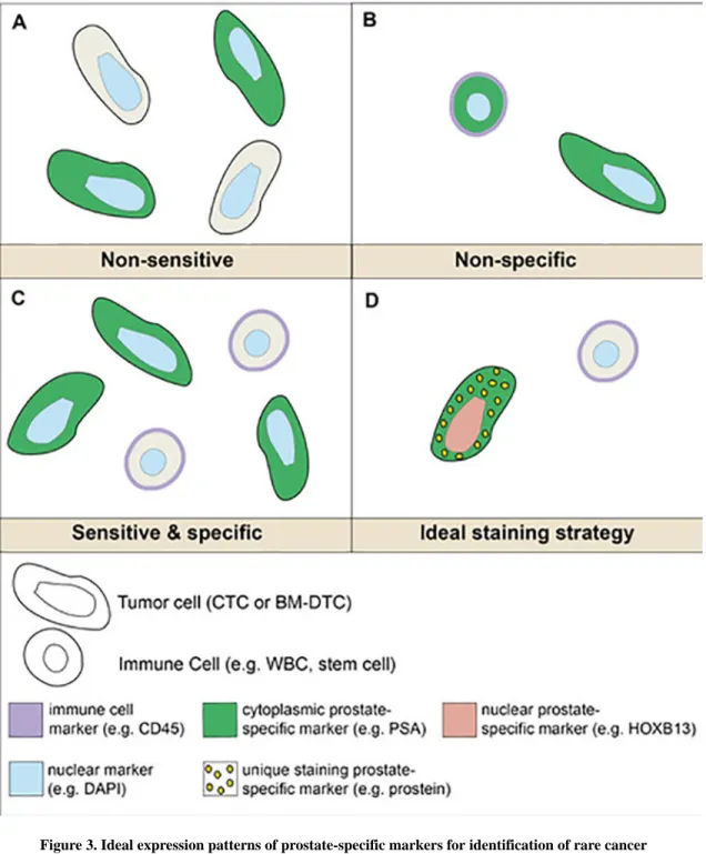
positive cytoplasmic staining is seen on occasion simply due to processing. Therefore, it
would be ideal to combine markers that have different staining patterns using multiplex
staining. For example, an ideal multiplex protocol might include a nuclear marker (e.g.
HOXB13), cytoplasmic marker (e.g. PSA), and a marker with a unique staining pattern (e.g.
prostein, which localizes to the Golgi apparatus) (FIG. 4). In this review, we have largely
focused on protein expression because IF can provide more information than other
techniques, such as RT-PCR. While RT-PCR is more sensitive in terms of its ability to detect
small amounts of RNA, it does not provide information about cellular heterogeneity in terms
of which cells express which RNA. IF can provide visual evidence of protein expression,
and in multiplex assays can provide expression information about multiple proteins on a
single cell. Given the fact that protein expression provides insight into function, IF-based
assays also have the advantage of being able to understand the role and clinical application
of detected cells. In addition, single cell picking techniques have improved to the point
where genomic and proteomic analyses can be performed at the single cell level
26,260–262.
Each prostate marker we have discussed in this article has a varying degree of specificity to
the prostate gland or PCa. Some, like PSA, prostein, HOXB13, and KLK2, appear to be
highly specific for prostate tissue, based on tissue-based assays. Others, like AR, PSAP,
PSCA, and PSMA are much less specific. In addition, some markers become aberrantly
expressed in a variety of cancers, even if they were not expressed in the corresponding
healthy tissue (e.g. PSA is occasionally found in lung cancer even though it is not expressed
in healthy lung tissue). However, we postulate that a prostate-specific marker only needs to
be specific to PCa cells in that any other cells that are present in a liquid biopsy do not
express the marker. This includes blood and BM cells such as all immune cells,
hematopoietic and mesenchymal stem cells, BM stromal cells, osteoclasts, and endothelial
cells, among others. This is based on the high unlikelihood that a PCa patient will have
cancer of another tissue, whereby even if a marker of interest is highly expressed in prostate
cells but also expressed in pancreatic cells, it would still be acceptable for use in a liquid
biopsy.
The sensitivity of the detection marker is also extremely important to ensure that every PCa
cell that is present in a blood or BM sample from a patient is identified. Since CTCs and
BM-DTCs are so rare, failing to detect only a few cells could have major clinical
A
uthor Man
uscr
ipt
A
uthor Man
uscr
ipt
A
uthor Man
uscr
ipt
A
uthor Man
uscr
ipt
implications. This means that every PCa cell that enters the bloodstream and/or BM would
ideally express the detection marker. Unfortunately, information to this degree is severely
lacking in the published literature. Most reports have determined the sensitivity of prostate
markers via IHC, where sensitivity is discussed in terms of the percentage of patients where
positive staining was observed. Instead, for rare cell assays, the number of PCa cells that are
detected with the marker out of a known total number of PCa cells present should be
determined. This may be impossible to assess in clinical samples, considering there is no
perfectly sensitive marker to our knowledge that would provide the true number of cancer
cells present in a sample. To overcome these obstacles, increasing the number of markers so
as to “catch” every cell would be helpful, as long as they are each highly specific. Even so,
for some less common types of PCa (e.g. neuroendocrine, small cell, or carcinoid), the
classic prostate markers like PSA or NKX3.1 will not be helpful
104. Instead, other markers
such as synaptophysin or chromogranin might be required to identify these cells
263.
An important concept to consider is that a marker does not need to be as sensitive or specific
if it is not being used for detection purposes. Once the CTC/BM-DTC is detected by highly
sensitive and specific marker(s), it does not matter if a marker being used to study biological
characteristics or to drive therapeutic decisions is also present on a non-PCa blood or BM
cell. For example, we have discussed AR as being a relatively non-prostate-specific marker,
as it is expressed in many other healthy tissues, including the BM. Therefore, we would not
recommend using AR to detect or identify PCa CTCs or BM-DTCs. However, the
expression of full-length AR or its variant form (AR-V7) has been shown to be clinically
informative as to whether to treat metastatic PCa patients with either taxanes or second line
hormonal therapy
19,21,107. This is an excellent example of the importance and applicable
range of using liquid biopsies and rare cell assays on liquid biopsies to directly impact
patient care.
Concluding Remarks
The aims of this review article were to emphasize the difficulties in accurately identifying
rare prostate CTCs or BM-DTCs with the commonly used epithelial markers, and the
subsequent need for prostate-specific biomarkers in the detection of these cells. While much
has been done to identify and quantify CTCs in the blood of cancer patients, much less has
been done in bone marrow to identify BM-DTCs. BM-DTCs are likely the “important
CTCs,” meaning they are responsible for lethal bone metastases, and therefore contain
biological characteristics required for successful dissemination. As rare cell assays need to
be exceptionally sensitive, it is crucial that sensitive and specific markers are used to
differentiate cancer cells from blood and BM cells, but unfortunately little is known about
candidate marker expression on PCa cells at an individual cell level. We have attempted to
compile an exhaustive list of published prostate-specific markers as a starting point for
determining which markers should be investigated further to be used for CTC/BM-DTC
detection in the future. Some markers, like AR and PSAP, are too non-specific to be used as
individual markers of PCa cells, while others, such as PSA, prostein, and HOXB13, hold
more promise as sensitive and specific markers. It is likely that multiple specific markers
will have to be combined to increase overall sensitivity. The goal of future studies must be to
consistently and reliably identify rare cancer cells using sensitive and specific markers.
A
uthor Man
uscr
ipt
A
uthor Man
uscr
ipt
A
uthor Man
uscr
ipt
A
uthor Man
uscr
ipt
Although this review has focused on PCa, the same strategies are applicable to rare cell
assays in any type of cancer.
Supplementary Material
Refer to Web version on PubMed Central for supplementary material.
Acknowledgments
Acknowledgements and funding
This work is supported by NCI grants U54CA143803, CA163124 CA093900, CA143055 as well as the Prostate Cancer Foundation, the Patrick C. Walsh Fund and a gift from the Stutt family. EEvdT is supported by the Cure for Cancer Foundation. KCV is supported by NCI grant F32CA206394.
References
1. Siegel RL, Miller KD, Jemal A. Cancer statistics, 2015. CA: a cancer journal for clinicians. 65:5– 29. DOI: 10.3322/caac.212542015; [PubMed: 25559415]
2. Han M, et al. Biochemical (prostate specific antigen) recurrence probability following radical prostatectomy for clinically localized prostate cancer. The Journal of urology. 169:517–523. DOI: 10.1097/01.ju.0000045749.90353.c72003; [PubMed: 12544300]
3. Mehra R, et al. Characterization of bone metastases from rapid autopsies of prostate cancer patients. Clinical cancer research : an official journal of the American Association for Cancer Research. 17:3924–3932. DOI: 10.1158/1078-0432.CCR-10-31202011; [PubMed: 21555375]
4. Ruppender NS, Morrissey C, Lange PH, Vessella RL. Dormancy in solid tumors: implications for prostate cancer. Cancer metastasis reviews. 32:501–509. DOI: 10.1007/s10555-013-9422-z2013; [PubMed: 23612741]
5. Lam HM, Vessella RL, Morrissey C. The role of the microenvironment-dormant prostate disseminated tumor cells in the bone marrow. Drug Discov Today Technol. 11:41–47. DOI: 10.1016/j.ddtec.2014.02.0022014; [PubMed: 24847652]
6. Mishra A, Shiozawa Y, Pienta KJ, Taichman RS. Homing of cancer cells to the bone. Cancer Microenviron. 4:221–235. DOI: 10.1007/s12307-011-0083-62011; [PubMed: 21826451] 7. Nguyen DX, Bos PD, Massague J. Metastasis: from dissemination to organ-specific colonization.
Nature reviews. Cancer. 9:274–284. DOI: 10.1038/nrc26222009; [PubMed: 19308067] 8. Mohler JL, et al. Prostate Cancer, Version 1.2016. J Natl Compr Canc Netw. 14:19–30.2016;
[PubMed: 26733552]
9. Li F, et al. Cell surface Thomsen-Friedenreich proteome profiling of metastatic prostate cancer cells reveals potential link with cancer stem cell-like phenotype. Oncotarget. 8:98598–98608. DOI: 10.18632/oncotarget.219852017; [PubMed: 29228713]
10. Cheung KJ, et al. Polyclonal breast cancer metastases arise from collective dissemination of keratin 14-expressing tumor cell clusters. Proceedings of the National Academy of Sciences of the United States of America. 113:E854–863. DOI: 10.1073/pnas.15085411132016; [PubMed: 26831077] 11. Alix-Panabieres C, Pantel K. Challenges in circulating tumour cell research. Nature reviews.
Cancer. 14:623–631. DOI: 10.1038/nrc38202014; [PubMed: 25154812]
12. Friedlander TW, et al. Detection and characterization of invasive circulating tumor cells derived from men with metastatic castration-resistant prostate cancer. International journal of cancer. Journal international du cancer. 134:2284–2293. DOI: 10.1002/ijc.285612014; [PubMed: 24166007]
13. Pantel K, Alix-Panabieres C. Real-time liquid biopsy in cancer patients: fact or fiction? Cancer research. 73:6384–6388. DOI: 10.1158/0008-5472.CAN-13-20302013; [PubMed: 24145355] 14. Gold B, Cankovic M, Furtado LV, Meier F, Gocke CD. Do circulating tumor cells, exosomes, and
circulating tumor nucleic acids have clinical utility? A report of the association for molecular
A
uthor Man
uscr
ipt
A
uthor Man
uscr
ipt
A
uthor Man
uscr
ipt
A
uthor Man
uscr
ipt
pathology. J Mol Diagn. 17:209–224. DOI: 10.1016/j.jmoldx.2015.02.0012015; [PubMed: 25908243]
15. Perakis S, Speicher MR. Emerging concepts in liquid biopsies. BMC Med. 15:75.2017; [PubMed: 28381299]
16. Zhang W, et al. Liquid Biopsy for Cancer: Circulating Tumor Cells, Circulating Free DNA or Exosomes? Cell Physiol Biochem. 41:755–768. DOI: 10.1159/0004587362017; [PubMed: 28214887]
17. de Bono JS, et al. Circulating tumor cells predict survival benefit from treatment in metastatic castration-resistant prostate cancer. Clinical cancer research : an official journal of the American Association for Cancer Research. 14:6302–6309. DOI: 10.1158/1078-0432.CCR-08-08722008; [PubMed: 18829513]
18. Scher HI, et al. Phenotypic Heterogeneity of Circulating Tumor Cells Informs Clinical Decisions between AR Signaling Inhibitors and Taxanes in Metastatic Prostate Cancer. Cancer research. 2017
19. Scher HI, et al. Nuclear-specific AR-V7 Protein Localization is Necessary to Guide Treatment Selection in Metastatic Castration-resistant Prostate Cancer. European urology. 71:874–882. DOI: 10.1016/j.eururo.2016.11.0242017; [PubMed: 27979426]
20. Scher HI, et al. Circulating tumor cell biomarker panel as an individual-level surrogate for survival in metastatic castration-resistant prostate cancer. Journal of clinical oncology : official journal of the American Society of Clinical Oncology. 33:1348–1355. DOI: 10.1200/JCO.
2014.55.34872015; [PubMed: 25800753]
21. Scher HI, et al. Association of AR-V7 on Circulating Tumor Cells as a Treatment-Specific Biomarker With Outcomes and Survival in Castration-Resistant Prostate Cancer. JAMA Oncol. 2:1441–1449. DOI: 10.1001/jamaoncol.2016.18282016; [PubMed: 27262168]
22. Kuske A, et al. Improved detection of circulating tumor cells in non-metastatic high-risk prostate cancer patients. Sci Rep. 6:39736.2016; [PubMed: 28000772]
23. Xu L, et al. The Novel Association of Circulating Tumor Cells and Circulating Megakaryocytes with Prostate Cancer Prognosis. Clinical cancer research : an official journal of the American Association for Cancer Research. 23:5112–5122. DOI: 10.1158/1078-0432.CCR-16-30812017; [PubMed: 28615267]
24. Amann R, Fuchs BM. Single-cell identification in microbial communities by improved fluorescence in situ hybridization techniques. Nat Rev Microbiol. 6:339–348. DOI: 10.1038/ nrmicro18882008; [PubMed: 18414500]
25. Yap TA, Lorente D, Omlin A, Olmos D, de Bono JS. Circulating tumor cells: a multifunctional biomarker. Clinical cancer research : an official journal of the American Association for Cancer Research. 20:2553–2568. DOI: 10.1158/1078-0432.CCR-13-26642014; [PubMed: 24831278] 26. Campton DE, et al. High-recovery visual identification and single-cell retrieval of circulating
tumor cells for genomic analysis using a dual-technology platform integrated with automated immunofluorescence staining. BMC cancer. 15:360.2015; [PubMed: 25944336]
27. Werner SL, et al. Analytical Validation and Capabilities of the Epic CTC Platform: Enrichment-Free Circulating Tumour Cell Detection and Characterization. J Circ Biomark. 4:3.2015; [PubMed: 28936239]
28. Nagrath S, et al. Isolation of rare circulating tumour cells in cancer patients by microchip technology. Nature. 450:1235–1239. DOI: 10.1038/nature063852007; [PubMed: 18097410] 29. Valkenburg KC, et al. A simple selection-free method for detecting disseminated tumor cells
(DTCs) in murine bone marrow. Oncotarget. 7:69794–69803. DOI: 10.18632/oncotarget. 120002016; [PubMed: 27634877]
30. Helo P, et al. Circulating prostate tumor cells detected by reverse transcription-PCR in men with localized or castration-refractory prostate cancer: concordance with CellSearch assay and association with bone metastases and with survival. Clin Chem. 55:765–773. DOI: 10.1373/ clinchem.2008.1179522009; [PubMed: 19233911]
31. O'Hara SM, et al. Multigene reverse transcription-PCR profiling of circulating tumor cells in hormone-refractory prostate cancer. Clin Chem. 50:826–835. DOI: 10.1373/clinchem. 2003.0285632004; [PubMed: 14988224]