Impedance Cardiography for
Demonstrating Procedural Efficacy of
Percutaneous Mitral Balloon Valvuloplasty
AABBSSTTRRAACCTT OObbjjeeccttiivvee:: Impedance Cardiography (ICG) method, facilitates measuring hemodynamic pa-rameters indirectly by recording thoracic impedance variations induced by cyclic changes in blood flow. The aim of this study is to evaluate alterations in hemodynamic parameters obtained by impedance car-diography (ICG) in addition to conventional echocardiographic and catheterization data after percuta-neous mitral balloon valvuloplasty (PMBV). MMaatteerriiaall aanndd MMeetthhooddss:: 18 patients with severe rheumatic mitral stenosis to whom PMBV had been performed were included in this study. Impedance cardiographic measurements were performed in addition to routine echocardiographic examination and invasive left atrial and pulmonary arterial pressure recordings before and after the procedure. Cardiac output was cal-culated with direct Fick method before PMBV. Average values of several measurements were used to ob-tain the most accurate results for atrial fibrillation patients in the study group. RReessuullttss:: Following successful PMBV, in impedance cardiographic evaluation an increase in cardiac output (4.69±1.46 and 5.68±1.3 l/min, before and after PMBV, respectively, p<0.001) and cardiac index (2.72±0.81 and 3.29±0.73 l/min/m2,
be-fore and after PMBV, respectively, p<0.001) an evident prolongation in ejection period (246.8±40.3 and 275.2±32.1 msec, before and after PMBV, respectively, p<0.001); and a statistically significant reduction in pre-ejection period (103.7±41.8 and 82.7±16.8 msec, before and after PMBV, respectively, p=0.033), and thoracic fluid content (34.3±11.2 and 27.7±5.31/kΩ, before and after PMBV, respectively, p=0.037) was observed. There was a strong correlation between preprocedural cardiac output calculations of ICG and direct Fick method (r:0.89, p<0.001). CCoonncclluussiioonn:: In addition to the conventional methods, serial record-ings of impedance cardiographic outputs may yield beneficial information for the assessment of PMBV ef-ficacy. As a non-invasive and easily applicable method, it might be utilized during in-hospital follow-up. KKeeyy WWoorrddss:: Cardiography, impedance; mitral valve stenosis; mitral valve annuloplasty
Ö
ÖZZEETT AAmmaaçç:: Empedans kardiyografi (EmKG), atımdan atıma değişen kan akımının oluşturduğu göğüs em-pedans değişikliklerini kaydederek hemodinamik parametrelerin dolaylı olarak ölçülmesine imkân tanıyan bir yöntemdir. Bu çalışmada, perkütan mitral balon valvuloplasti (PMBV) sonrası rutin olarak değerlendiri-len ekokardiyografi ve kateterizasyon verilerine ek olarak; EmKG ile elde edideğerlendiri-len hemodinamik göstergeler-deki değişimlerin incelenmesi amaçlandı. GGeerreeçç vvee YYöönntteemmlleerr:: Çalışmaya PMBV uygulanan 18 ciddi mitral darlığı hastası alındı. İşlem öncesi ve sonrasında, rutin ekokardiyografik inceleme ve invaziv sol atriyum ve pulmoner arter basınç kayıtlarına ek olarak EmKG ölçümleri yapıldı. PMBV öncesi kardiyak debi Fick yön-temiyle hesaplandı. Çalışma grubundaki atriyal fibrilasyon hastalarında ise doğruya en yakın sonucu elde ede-bilmek için tekrarlayan ölçümlerin ortalaması kullanıldı. BBuullgguullaarr:: Başarılı PMBV sonrası, EmKG değerlendirmesinde kardiyak debi (4,69±1,46; 5,68±1,3 l/dk; PBMV öncesi ve sonrası, sırasıyla p<0,001) ve kardiyak indekste (2,72±0,81; 3,29±0,73 l/dk/m2; öncesi ve sonrası, sırasıyla p<0,001) belirgin artış;
ejeksi-yon süresinde aşikar bir uzama (246,8±40,3; 275,2±32,1 ms; öncesi ve sonrası, sırasıyla p<0,001); ve pree-jeksiyon süresi (103,7±41,8; 82,7±16,8 ms; öncesi ve sonrası, sırasıyla p=0,033) ve torasik sıvı içeriğinde (34,3±11,2; 27,7±5,31/kΩ; öncesi ve sonrası, sırasıyla p=0,037) istatiksel olarak anlamlı bir azalma söz konusuydu. İşlem öncesi yapılan EmKG ölçümlerinde elde edilen kardiyak debi değerleri ile Fick yöntemiyle ölçülen değerler arasında güçlü bir korelasyon saptandı (r:0,89; p<0,001). SSoonnuuçç:: EmKG ölçümleriyle elde edilen verilerin takibi, PMBV’nin başarısını değerlendirmede diğer standart yöntemlere ek olarak faydalı bilgiler sunabilir. Non-invaziv ve kolay uygulanabilir olması dolayısıyla hastane içi takipte kullanılabilir. AAnnaahhttaarr KKeelliimmeelleerr:: Kardiyografi, impedans; mitral kapak darlığı; mitral kapak annuloplastisi TTuurrkkiiyyee KKlliinniikklleerrii JJ CCaarrddiioovvaasscc SSccii 22001155;;2277((33))::8811--77
Mehmet Onur OMAYGENÇ,a
Cem DOĞAN,b
Ruken Bengi BAKAL,c
Özkan CANDAN,d
Suzan HATİPOĞLU AKPINAR,e
Gamze BABÜR GÜLER,a
Cihangir KAYMAZ,c
Nihal ÖZDEMİRc aDepartment of Cardiology,
İstanbul Medipol University Faculty of Medicine, İstanbul
bClinic of Cardiology,
Malatya State Hospital, Malatya
cClinic of Cardiology
Kartal Koşuyolu Yüksek İhtisas Training and Research Hospital, İstanbul
dClinic of Cardiology,
Uşak State Hospital, Uşak
eClinic of Cardiology,
Edremit State Hospital, Balıkesir Ge liş Ta ri hi/Re ce i ved: 10.02.2015 Ka bul Ta ri hi/Ac cep ted: 23.11.2015
This study was presented as a poster at 23thNational Cardiology Congress, 19-23 October 2007, Antalya.
Ya zış ma Ad re si/Cor res pon den ce: Mehmet Onur OMAYGENÇ İstanbul Medipol University Faculty of Medicine,
Department of Cardiology, İstanbul, TÜRKİYE/TURKEY
doi: 10.5336/cardiosci.2015-44142
itral stenosis was diagnosed in 0.1% of participants at a population-based study (higher in developing countries due to increased prevalence of rheumatic heart disease) and percutaneous mitral balloon valvuloplasty (PMBV), if applicable, is the preferred option of managing severe mitral stenosis.1,2 Conventional
transthoracic and transesophageal echocardiogra-phy, invasive left atrial pressure, pulmonary arte-rial pressure and cardiac output measurements are widely used diagnostic tools to assess the success of PMBV.
In 1870, Fick described a method to estimate cardiac output in humans. Fick postulated that oxy-gen uptake in the lungs is entirely transferred to the blood. Therefore, cardiac output can be calcu-lated as the ratio between oxygen consumption (VO2) and arteriovenous difference in oxygen
(AVDO2).
Fick equation: CO= VO2 /AVDO2
This estimation is accurate when the hemody-namic status is sufficiently stable to allow constant gas diffusion during the mean transit time of blood through the lungs.3
Impedance cardiography (ICG) is designed to measure resistance of tissues to flow and alterations of these during dynamic processes like respiration and cardiac cycle. Thus it is possible to calculate fundamental parameters of hemodynamics by for-mulas.4 As mentioned above, ICG is a unique
method, fascilitating direct measurement of blood flow instead of utilizing pressure recordings.5There
are certain investigations readily performed with different concepts, validating ICG in various car-diovascular diseases like acute and chronic heart failure, acute myocardial infarction and valvu-lopathies.6-9
In this study we aimed to determine acute he-modynamic alterations before and after PMBV with ICG in severe mitral stenosis.
MATERIAL AND METHODS
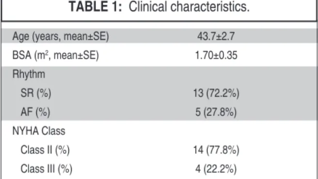
Eighteen consecutive female patients with severe mitral stenosis undergoing PMBV in Kartal Koşu-yolu Yuksek Ihtisas Training and Research
Hospi-tal were included. The mean age of our study pop-ulation was 43.7 ± 11.4 years and 5 patients had atrial fibrillation (Table 1). All individuals were evaluated with transthoracic and transesophageal echocardiography for clinical and anatomical eli-gibility before the procedure. Individuals with characteristics that may cause confusion for im-pedance measurements like left ventricular systolic dysfunction, severe pulmonary hypertension (mean pulmonary artery pressure >40 mmHg), ex-tremes of body weight and significant aortic valve insufficiency were excluded.
Impedance cardiographic measurements were performed before and 24 hours after the procedure. Planimetric mitral valve area (MVApln) and trans-valvular gradients were measured before and 24 hours after the procedure and also, left atrial and pulmonary arterial pressures were recorded just before and immediately after PMBV. Before the procedure cardiac output (CO) was measured inva-sively and calculated by using the Fick formula. Oral and written informed consents were received from all individuals and the study protocol was ap-proved by our institution’s local ethical committee.
BALLOON VALVULOPLASTY AND CARDIAC CATHETERISATION
Right common femoral artery and vein were used for vascular access. 6F pigtail catheters were used for arterial and venous blood sampling before the procedure and CO was calculated. Left atrial and pulmonary arterial pressure tracings were also recorded before and after the procedure using fluid filled 6F pigtail catheters. PMBV was performed by
Age (years, mean±SE) 43.7±2.7 BSA (m2, mean±SE) 1.70±0.35 Rhythm SR (%) 13 (72.2%) AF (%) 5 (27.8%) NYHA Class Class II (%) 14 (77.8%) Class III (%) 4 (22.2%)
TABLE 1: Clinical characteristics.
AF: Atrial Fibrillation; BSA: Body Surface Area; F: Female; M: Male; NYHA: New York Heart Association; SD: Standard Deviation; SR: Sinus Rhythm.
the transseptal approach using Inoue balloon (Toray Medical Industries, Houston, TX, USA) technique.
ECHOCARDIOGRAPHIC EXAMINATION
All patients underwent transthoracic echocardiog-raphy (TTE) before and 24 hours after the proce-dure. With TTE (GE, VingMed System Five, Horten, Norway and 3.5 MHz transducer) peak and mean diastolic transmitral gradients were measured by continuous-wave Doppler echocardiography. Estimated valve areas were verified by using the area trace method in all patients. Mitral regurgita-tion was categorized as absent, mild, moderate, or severe using a combination of qualitative and quan-titative parameters. Tricuspid regurgitation (TR) jet flow was assessed from the apical, subcostal, and parasternal views. Systolic pulmonary artery pres-sure (PAP) was meapres-sured with continuous-wave Doppler. The maximum peak TR velocity (V) recorded from any view was used to determine right ventricular systolic pressure with the simpli-fied Bernoulli equation (PAP = 4V2+ RAP). To
es-timate right atrial pressure, inferior vena cava diameter was measured from the subxiphoid long-axis view. Right atrial pressure was estimated using the caval respiratory index as described by Kircher et al.10Pulmonary artery systolic pressure was
esti-mated from the sum of the transtricuspid gradient and right atrial pressure.
IMPEDANCE CARDIOGRAPHIC MEASUREMENTS
The device used at this study (BioZ®
CardioDy-namics, San Diego, CA), uses electric current stim-ulations (electrical bioimpedance) for identification of thoracic or body impedance variations induced by cyclic changes in blood flow caused by each heart beat.
Eight ICG electrodes were placed on the carotid arteries and area where midaxillary line in-tersects with an imaginery line traversing apex at anatomical position. An automatic sphigmo-manometry was placed around right arm. Finally all of the cables for data input were attached to the monitor. Measurements were performed before and 24 hours after the procedure. In order to
elim-inate possible discrepancies caused by beat-to-beat variations in atrial fibrillation patients, five meas-urements were performed and average values were recorded. The values for CO, cardiac index (CI), ejection period (EP), preejection period (PEP), tho-racic fluid content (TFC), left cardiac workload (LCW), systemic vascular resistance (SVR) which had been displayed on the monitor were noted.
ICG utilizes two-dimensional wave forms to estimate hemodynamic status, principally cardiac output. First derivative measures fluid velocity and most of the parameters mentioned in the text are calculated from this data by specific indices. Sec-ond derivative measures acceleration and acts as a complementary entity. It also provides accurate coupling with cardiac cycle. Three major waves A, S and O waves correspond to atrial contraction, ventricular systole and mitral valve opening re-spectively. A wave is absent in atrial fibrillation and may show significant variation during frequent premature contractions. S wave principally, demonstrates cardiac output and larger S waves in-dicate increased CO. O waves can be barely visible even in healthy young individuals and they are mostly related to diastolic parameters.4,11
Extremes of body height and weight, aortic valve insufficency and respiratory artifacts are some other factors that may alter accurate data col-lection.4
STATISTICAL ANALYSIS
Baseline characteristics were expressed as mean ± standard error (SE) for continuous variables, and percentages for categorical ones. Due to the limited sample size (n=18), non-parametric tests were used to compare variables. Pre-test and post-test vari-ables within the study population were compared with the Wilcoxon signed rank test and the results were demonstrated on a table with the correspon-ding z and p values. The association between meas-urements of CO by direct Fick and ICG methods was tested by Spearman’s correlation analysis. The results of the analysis were shown with a graphic and the corresponding rho and p values. Accuracy of the cardiac output measured by Fick and ICG methods were examined by the Bland and Altman
analysis. A 2-tailed p value <0.05 was considered statistically significant. Statistical analysis was per-formed using MedCalc programme (MedCalc Soft-ware, Mariakerke, Belgium).
RESULTS
PMBV was performed by using Inoue balloon with transseptal approach and no complications were observed. Upon echocardiographic and invasive hemodynamic measures; PMBV might be consid-ered as efficacious for all study patients; namely sig-nificant decrease in left atrial pressure, invasive pulmonary artery pressure recordings and echocar-diographic gradients; and a rise in planimetric valve area were observed (Table 2). Only two patients were found to have moderate mitral regurgitation postprocedurally; others had trivial or mild regur-gitation.
In terms of impedance cardiographic meas-urements before and after the procedure; a signifi-cant increase of CO and CI; an evident
prolongation in EP; a shortened PEP and a signifi-cant decrease in TFC were observed. Reduction of LCW and SVR did not reach statistical significance (Table 3).
According to Spearman’s correlation; there was a strong relationship between cardiac output measurements obtained by ICG and direct Fick method (Spearman’s rho=0.89, p < 0.001). Correla-tions between the cardiac output measured by Fick and ICG were also summarized in Figure 1. The Bland-Altman plots revealed a good agreement of CO measured by direct Fick method and ICG (mean difference 0.69±1.09, 95% CI [0.153-1.237]) (Figure 2).
DISCUSSION
Impedance cardiography is a method, which had been used for experimental purposes in early 1960s; and several studies evaluating diagnostic value, val-idation, variability, cost-effectiveness and contri-bution to prognosis for diseases including coronary
Pre-PMBV Post-PMBV p value z score
MVApln (cm2, mean±SD) 1.14±0.03 1.81±0.04 <0.001 • -3.75
MEAN GRADIENT, (mmHg, mean±SD) 14.6±1.3 5.2±0.5 <0.001 • -3.73 LAP, (mmHg, mean±SD) 26.6±1.6 15.9±1.1 <0.001 • -3.73 PAPsystolic, (mmHg, mean±SD) 57.8±2.9 42±1.7 <0.001 • -3.63 PAPmean, (mmHg, mean±SD) 38.5±2.2 27.6±1.4 <0.001 • -3.73 PAPdiastolic, (mmHg, mean±SD) 26±2.2 18.1±1.5 0.001 • -3.47
TABLE 2: Echocardiographic valvular measurements and invasice left atrial and pulmonary artery pressure recordings
before and after the procedure.
LAP: Left atrial pressure; MVApln: Planimetric mitral valve area; PAP: Pulmonary arterial pressure; SD: Standard Deviation. • Statistical significance.
Pre-PMBV Post-PMBV p value z score
CO, (l/min, mean±SD) 4.69±0.35 5.68±0.33 <0.001 • -3.73 CI, (l/min/m2, mean±SD) 2.72±0,19 3.29±0.17 <0.001 • -3.73
EP, (msec, mean±SD) 246.8±9.5 275.2±7.6 <0.001 • -3.53 PEP, (msec, mean±SD) 103.7±9.9 82.7±3.4 0.003 • -2.98 TFC, (1/kΩ, mean±SD) 34.3±2.6 27.7±1.2 0.022 • -2.29 LCW, (kg m, mean±SD) 5.8±0.4 5.3±0.4 0.162 -1.4 SVR, (dynes sec/cm5, mean±SD) 1415.9±157.2 1256.8±88.7 0.396 -0.85
TABLE 3: Impedance cardiography measurements before and after the procedure.
CO: Cardiac output; CI: Cardiac index; EP: Ejection period; PEP: Pre-ejection period; SD: Standard Deviation; TFC: Thoracic fluid content; LCW: Left cardiac workload; SVR: Syste-mic vascular resistance.
artery disease, congestive heart failure, systemic hypertension, pulmonary hypertension and vari-ous valvular pathologies had been published since then.6,8,11-14
In the last decade, ICG had been utilized for monitoring heart failure and tailoring the therapy especially after combination with implantable de-vices.15-17
But the main purpose for utilization of ICG in most of the publications is to estimate cardiac out-put. At a relatively former article comparing CO measured by ICG with direct Fick method as a part of diagnostic cardiac catheterisation correlation
seems tremendous (r:0,93).18 At publications in
early 2000’s, it ranges between 0,76 to 0,89.19-21
Greenberg et al., evaluated the validity of BioZ®
CardioDynamics ICG device in heart failure pa-tients and found a close correlation between CO values obtained by ICG and thermodilution method.22At the largest randomised trial on heart
failure patients -the BIG study as a part of ESCAPE study- only a modest correlation was observed be-tween two techniques.23Results of our sample
pop-ulation affirm the strong relationship between invasive and ICG measurements of cardiac output. There are just a few studies investigating the role of ICG in valvular heart diseases. Aortic valve pathologies, especially significant regurgitation, are known to influence ICG waveforms and decreases the reliability of cardiac output measurements.24
Thus, patients with aortic insufficiency are invari-ably excluded in ICG studies. Significant aortic valve disease was also established as a specific contraindi-cation for PMBV and these patients had already been eliminated in the first step of patient selection. In a study comparing patients who have mi-tral regurgitation (MR) with healthy individuals, authors concluded that even in MR patients ICG can estimate CO reliably although degree of agree-ment was decreased.24
In comparison with healthy individuals, pa-tients with mitral stenosis are known to have lower stroke volume, consequently cardiac output and cardiac index values measured by ICG.7
Same group of authors published another re-port for mitral valve diseases and defined the restoration of O wave abnormalities after correc-tive surgery both for stenosis and regurgitation. They also stated that CO values were significantly lower preoperatively in mitral stenosis patients as compared with control group and normalization of these values were observed after surgery.25
As expected; in our sample population, an ev-ident increase in CO and related parameters at early stages were observed. And also, due to en-hanced flow passing through the expanded valvu-lar orifice, left ventricuvalvu-lar filling and ejection periods were altered.
FIGURE 1: Correlation between direct Fick and impedance cardiography
measurements of cardiac output.
CO: Cardiac output; ICG: Impedance cardiography; SD: Standard deviation.
FIGURE 2: Bland-Altman analysis of differences between cardiac output
measured by direct Fick and impedance cardiogarphy.
CO: Cardiac output; ICG: Impedance cardiography; SD: Standar deviation.
CO (b y F IC K) (l/ mi n)
CO (by ICG) (l/min)
1 2 3 4 5 6 7 6.2 6.0 5.8 5.6 5.4 5.2 5.0 4.8 4.6 Di ffe re nc e [ CO (F IC K) -C O( IC G) ] ( l/m in)
Mean of CO(FICK) and CO(ICG) (l/min)
3 4 5 6 7 +1.96 SD 2.8 Mean 0.7 -1.96 SD -1.4 4 3 2 1 0 -1 -2
LIMITATIONS
Main limitation of the study is definitely the size of the study population. Despite this disadvantage, statistically significant values were reached. Fick method was used for invasive determination of CO instead of thermodilution for timesaving. There-fore, concerning possible acute procedure-related changes in hemoglobin levels, measurement of CO indirectly by Fick method after the procedure was considered to be unreliable and was not per-formed.
Another issue is obtaining ICG data in atrial fibrillation patients. A clear signal recording may not be possible in these patients or when frequent premature beats exist. However, in a study com-paring ICG with inert gas rebreathing against echocardiographic examination for estimation of CO noninvasively, ICG was found to be superior and acceptable aggreement with echocardiographic measurements was observed.26We repeated the
ex-amination until we observed at least five clear waveforms for 5 patients with atrial fibrillation in
our sample group. We used average values of these serial recordings for statistical evaluation to over-come this predefined adversity.
CONCLUSION
This study defines alterations of the hemodynamic data obtained by ICG before and after PMBV. In addition to the echocardiographic findings and in-vasive measurements; serial recordings of ICG out-puts may yield beneficial and additional information for the assessment of PMBV efficacy.
To the best of our knowledge, hemodynamic changes with ICG after a percutaneous interven-tion to stenotic valve has not been investigated so far. This is the first study demonstrating the alter-ations of CO and other parameters calculated by ICG after successful PMBV.
A
Acckknnoowwlleeddggeemmeennttss
Authors would like to express their gratitude to Oguz Karaca MD for his contributions on statistical analysis and structural editing.
1. Nkomo VT, Gardin JM, Skelton TN, Gottdiener JS, Scott CG, Enriquez-Sarano M. Burden of valvular heart diseases: a population-based study. Lancet 2006;368(9540):1005-11. 2. Vahanian A, Alfieri O, Andreotti F, Antunes MJ,
Barón-Esquivias G, Baumgartner H, et al; ESC Committee for Practice Guidelines (CPG); Joint Task Force on the Management of Valvular Heart Disease of the European Society of diology (ESC); European Association for Car-dio-Thoracic Surgery (EACTS). Guidelines on the management of valvular heart disease (ver-sion 2012): the Joint Task Force on the Man-agement of Valvular Heart Disease of the European Society of Cardiology (ESC) and the European Association for Cardio-Thoracic Sur-gery (EACTS). Guidelines on the management of valvular heart disease (version 2012). Eur J Cardiothorac Surg 2012;42(4):S1-44. 3. Ultman JS, Bursztein S. Analysis of error in
the determination of respiratory gas exchange at varying FIO2. J Appl Physiol 1981;50(1): 210-6.
4. Sodolski T, Kutarski A. Impedance cardiogra-phy: a valuable method of evaluating haemo-dynamic parameters. Cardiol J 2007;14(2): 115-26.
5. Northridge DB, Findlay IN, Wilson J, Hender-son E, Dargie HJ. Non-invasive determination of cardiac output by Doppler echocardiogra-phy and electrical bioimpedance. Br Heart J 1990;63(2):93-7.
6. Brazdzionyte J, Macas A. Bland-Altman analy-sis as an alternative approach for statistical evaluation of agreement between two meth-ods for measuring hemodynamics during acute myocardial infarction. Medicina (Kau-nas) 2007;43(3):208-14.
7. Parulkar GB, Jindal GD, Padmashree RB, Haridasan GG, Dharani JB. Impedance car-diography in mitral valve disease. J Postgrad Med 1980;26(3):155-61.
8. Sharman DH, Gomes CP, Rutherford JP. Im-provement in blood pressure control with im-pedance cardiography-guided pharmacologic decision making. Congest Heart Fail 2004;10(1):54-8.
9. Strobeck JE, Silver MA. Beyond the four quad-rants: the critical and emerging role of imped-ance cardiography in heart failure. Congest Heart Fail 2004;10(2 Suppl 2):1-6.
10. Kircher BJ, Himelman RB, Schiller NB. Noninvasive estimation of right atrial
pres-sure from the inspiratory collapse of the in-ferior vena cava. Am J Cardiol 1990;66(4): 493-6.
11. Bour J, Kellett J. Impedance cardiography: a rapid and cost-effective screening tool for car-diac disease. Eur J Intern Med 2008;19(6): 399-405.
12. Treister N, Wagner K, Jansen PR. Repro-ducibility of impedance cardiography param-eters in outpatients with clinically stable coronary artery disease. Am J Hypertens 2005;18(2 Pt 2):44S-50S.
13. Ferrario C, Smith R; CONTROL Trial Investi-gators. Cost-effectiveness of impedance cardiography testing in uncontrolled hyper-tension. Am Heart Hosp J 2006;4(4):279-89. 14. Tonelli AR, Alnuaimat H, Li N, Carrie R,
Mubarak KK. Value of impedance cardiogra-phy in patients studied for pulmonary hyper-tension. Lung 2011;189(5):369-75. 15. Gielerak G, Piotrowicz E, Krzesiński P, Kowal
J, Grzęda M, Piotrowicz R. The effects of car-diac rehabilitation on haemodynamic parame-ters measured by impedance cardiography in patients with heart failure. Kardiol Pol 2011;69(4):309-17.
16. Khan FZ, Virdee MS, Hutchinson J, Smith B, Pugh PJ, Read PA, et al. Cardiac resyn-chronization therapy optimization using noninvasive cardiac output measurement. Pacing Clin Electrophysiol 2011;34(11): 1527-36.
17. Qiao Q, Hua W, Ding LG, Chen KP, Wang J, Wang FZ, et al. Single center experience with intrathoracic impedance monitoring. Chin Med J(Engl) 2011;124(14):2219-21.
18. Teo KK, Hetherington MD, Haennel RG, Greenwood PV, Rossall RE, Kappagoda T. Cardiac output measured by impedance car-diography during maximal exercise tests. Car-diovasc Res 1985;19(12):737-43.
19. Albert NM, Hail MD, Li J, Young JB. Equiva-lence of the bioimpedance and thermodilution methods in measuring cardiac output in hos-pitalized patients with advanced, decompen-sated chronic heart failure. Am J Crit Care 2004;13(6):469-79.
20. Drazner MH, Thompson B, Rosenberg PB, Kaiser PA, Boehrer JD, Baldwin BJ, et al. Comparison of impedance cardiography with invasive hemodynamic measurements in pa-tients with heart failure secondary to ischemic or nonischemic cardiomyopathy. Am J Cardiol 2002;89(8):993-5.
21. Sageman WS, Riffenburgh RH, Spiess BD. Equivalence of bioimpedance and thermodi-lution in measuring cardiac index after cardiac surgery. J Cardiothorac Vasc Anesth 2002;16 (1):8-14.
22. Greenberg BH, Hermann DD, Pranulis MF, Lazio L, Cloutier D. Reproducibility of imped-ance cardiography hemodynamic measures in clinically stable heart failure patients. Congest Heart Fail 2000;6(2):74-80.
23. Kamath SA, Drazner MH, Tasissa G, Rogers JG, Stevenson LW, Yancy CW. Correlation of impedance cardiography with invasive hemo-dynamic measurements in patients with
ad-vanced heart failure: the BioImpedance Car-dioGraphy (BIG) substudy of the Evaluation Study of Congestive Heart Failure and Pul-monary Artery Catheterization Effectiveness (ESCAPE) Trial. Am Heart J 2009;158(2):217-23.
24. van der Meer NJ, Vonk Noordegraaf A, Kamp O, de Vries PM. Noninvasive measurement of cardiac output: two methods compared in pa-tients with mitral regurgitation. Angiology 1999;50(2):95-101.
25. Parulkar GB, Jindal GD, Bhardwaj R, Suraokar S, Dharani JB. Impedance cardiog-raphy in mitral valve diseases. Indian Heart J 1985;37(1):37-42.
26. Osbak PS, Henriksen JH, Kofoed KF, Jensen GB. Non-invasive measurements of cardiac output in atrial fibrillation: inert gas rebreathing and impedance cardiography. Scand J Clin Lab Invest 2011;71(4):304-13.