Graphene Nanoreactors: Photoreduction of Prussian Blue in
Aqueous Solution
Silvia Nappini,
*
,†Alessia Matruglio,
‡,†,¶Denys Naumenko,
†,□Simone Dal Zilio,
†Marco Lazzarino,
†Frank.M. F. De Groot,
§Coskun Kocabas,
∥Osman Balci,
∥and Elena Magnano
*
,†,⊥†IOM-CNR, Laboratorio TASC, S.S. 14 - km 163.5, 34149 Basovizza, Trieste, Italy
‡University of Trieste, Graduate School of Nanotechnology, Piazzale Europa 1, 34127 Trieste, Italy
§Inorganic Chemistry and Catalysis, Debye Institute for Nanomaterials Science, Utrecht University, Universiteitsweg 99, Utrecht, Netherlands
∥Department of Physics, Bilkent University, 06800 Ankara, Turkey
⊥Department of Physics, University of Johannesburg, P.O. Box 524, Auckland Park, 2006 Johannesburg, South Africa
*
S Supporting InformationABSTRACT: Prussian dyes are characterized by interesting photo-magnetic properties due to the photoinduced electron transfer involved in the Fe oxidation and spin state changes. Ferromagnetic Prussian blue (PB) in contact with titanium dioxide (TiO2) can be reduced to paramagnetic Prussian white (PW) upon UV band gap excitation of TiO2. This process is promoted by the presence of a hole scavenger, such as water, fundamental to ensure the overall charge balance and the continuity of the process. In order to clarify the photoinduced reduction mechanism and the role of water, an innovative system of graphene nanobubbles (GNBs) filled with a PB aqueous solution was developed, enabling the application of electron spectroscopies to the liquid phase, up to now limited by the vacuum required to overcome
the short electron inelastic mean free path in dense medium. In this work GNBs formed on the photocatalytic substrate are able to act as “nanoreactors”, and they can control and take part in the reaction. The evolution of Fe L2,3 edge X-ray absorption spectra measured in total electron yield through the graphene membrane revealed the electron reduction from PB (FeIII−CN− FeII) to PW (FeII−CN−FeII) upon UV irradiation, shedding light on the photoinduced electron transfer mechanism in liquid
phase. The results, confirmed also by Raman spectroscopy, unequivocally demonstrate that the reaction occurs preferentially in aqueous solution, where water acts as hole scavenger.
■
INTRODUCTIONPrussian blue (PB) and its analogues (ferrocyanides) are characterized by interesting photomagnetic, electrocatalytic, and optical properties that can be mainly attributed to photoinduced electron transfer processes capable of changing the oxidation and spin state of Fe inside the complex structure. Thanks to these characteristics and the capability to tune the magnetic and optical properties by external stimuli, PB analogues have found promising applications for memory photomagnetic devices,1,2 electroactive layers in electro-chemistry,3,4 cathode materials for battery,5,6 and electrodes in biosensors.7−9 PB was also widely used as a pigment in different types of artwork of the 18th and 19th centuries, and, for this reason, it was the subject of several studies in the area of cultural heritage science to understand the fading process of the dye upon extended exposure to light.10,11
Prussian blue structure exists in two forms: “insoluble PB”, FeIII4[FeII(CN)6]3, and “soluble PB” or “Turnbull’s blue”,
KFeIII[FeII(CN) 6].
10,12
Prussian blue color is due to the intervalence charge transfer between low-spin FeII (LS-FeII−
C) and high-spin FeIII (HS-FeIII−N) centers. Discoloration of the soluble ferromagnetic PB ionic species [FeIII(FeII(CN)6)]+
under intense illumination is due to the reduction of FeIII−N to FeII−N centers which leads to the formation of the
para-magnetic colorless organometallic ion, well-known as Prussian white (PW) with formula [FeII(FeII(CN)
6)]2+.
Understanding the reduction mechanism of PB is important because it is the basic working principle of different biosensor
and ion detection systems based on PB film
electro-des.6,8,9,13−15 The photoinduced electron transfer is also responsible for the changes in the spin state of the metal centers of ferrocyanide compounds, leading to interesting photomagnetic properties which are widely used for several applications in thefield of photomagnetism.2,16For example, in Co−Fe Prussian blue, the electron excitation results in trapping the metastable high-spin state,16showing that the efficiency of Received: August 8, 2017
Revised: September 16, 2017 Published: September 19, 2017
In order to clarify the role of water and the mechanism of the photoinduced reduction of PB in an aqueous solution in contact with a TiO2catalyst, we investigated by soft X-ray
core-level spectroscopies the electronic evolution of Fe core core-levels. X-ray photoelectron spectroscopy (XPS) and X-ray absorption (XAS) are suitable techniques to study processes based on the transfer of electrons from a solid substrate to a solution or vice versa. The emitted photons and electrons with characteristic energies resulting from the direct excitation or relaxation of the core holes can be detected and analyzed to obtain information regarding the chemical environment, oxidation state, ligandfield strength, and charge transfer effects of the investigated system. Unfortunately, soft X-ray propagation and photoelectron detection require the ultra-high-vacuum (UHV) conditions, which hinder the application of soft X-ray techniques to the liquid−solid interfaces.
In the past decade substantial efforts have been devoted to the application of soft X-ray core-level spectroscopies to experiments under working conditions, such as ambient pressure photoelectron spectroscopy (AP-XPS)22−26 or the use of microfabricated liquid cells.27−34
Recentely, we have presented a novel, simple, and robust method to apply conventional electron spectroscopies to investigate the evolution of electrochemical or chemical reactions directly in their liquid environment.35 Graphene nanobubbles (GNBs)filled with the desired solution between a titanium dioxide TiO2(100) rutile single crystal and a
monolayer of graphene (Gr) were used to follow the electronic evolution during the thermal-induced reduction of an aqueous solution of FeCl3encased inside the GNBs.
The small thickness (only one atomic layer) and the high elasticity, mechanical strength, and impermeability of Gr provide the transparency to both photons and electrons required for the electronic characterization of the system placed under the Gr membrane.27,36,37
Here we did a step forward by using the GNB system for two purposes: it is a“sample holder” for electron spectroscopy from the liquid phase and a “reactor” that can control the process and take part in the reaction exploiting the photocatalytic properties of TiO2.20,38−40
A complete in situ spectroscopic study of the photoreduction mechanism of PB (soluble form, KFeIII[FeII(CN)
6]) in aqueous
solution encased in GNBs formed on a TiO2 catalyst is
presented. The photoreduction was induced by using a laser at 395 nm of wavelength, corresponding to the band gap of rutile (3.03 eV),41and the process was monitored after different UV exposure time. The reaction was followed in situ by core-level electron spectroscopy on the BACH beamline at Elettra
■
RESULTS AND DISCUSSIONAn array of GNBs filled with an aqueous solution of PB (10 mM) was obtained by transferring a CVD grown Gr layer onto a TiO2single crystal using a thermoplastic polymer as sacrificial layer (seeMethods). The formation of GNBs was described in detail in our previous work35and proved by several techniques, such as atomic force microscopy (AFM), Raman spectroscopy, XPS, and XAS. As previously described, the wettability of the bare TiO2 substrate, and thus the number of GNBs, was
modulated by oxygen plasma and thermal treatments up to 700 °C.42−44
Surface defects and adsorption of OH−groups on the crystal surface contribute to the formation of GNBs filled with an aqueous solution during the transfer protocol.
The presence of the aqueous solution inside GNBs was proved by measuring XPS O 1s spectra before the UV irradiation and after 45 min of irradiation with a laser tuned at 395 nm of wavelength and 15 mW of power (Figure 1a).
As previously reported for the GNBs filled with an aqueous solution,35 the O 1s peaks can be deconvoluted in four components. The component at 533.4 eV is related to liquid water and does not change after UV irradiation, demonstrating the sealing capability of the Gr membrane for the time scale of the experiment.27,28,45,46 The component at 532 eV can be associated with CO−OH, due to atmospheric contamination or polymer residuals, also observed on C 1s47(Figure 1b). The component at 531.2 eV can be related to hydroxyl radicals (OH) at the TiO2surface, andfinally, the component at 530.2 eV is related to oxygen deficiencies48or to bridging hydroxyls on the surface vacancies49of bulk TiO2; this latter component is very low due to the dense media interposed between the TiO2substrate and the Gr layer. XPS spectra of C 1s before and after 45 min of irradiation were also measured to check the Gr condition after the UV treatment. The obtained spectra are shown inFigure 1b, evidencing that the UV irradiation does not change Gr components. Before and after irradiation, C 1s spectra present a pronounced sp2 component at 284.6 eV associated with high-quality Gr, a sp3component at 285.3 eV
associated with defects in the lattice and grain boundaries, and weak C−OH and O−CO components at 286.5 and 288.6 eV due to atmospheric contamination and polymer residuals.
The irradiation does not introduce additional defects or damage into the Gr layer; indeed, a little decrease of the O− CO components is visible, indicating a cleaning effect of the UV laser in the presence of TiO2substrate.50,51
XPS spectra of Fe 2p are not reported in the paper because the oxidation state of iron can be analyzed with a higher
accuracy by XAS measurements. Indeed, Prussian blue photoreduction was examined by XAS at the Fe L2,3 edge measured in total electron yield (TEY) exploiting the high electrical conductivity of the Gr/liquid interface which enabled the measurement of spectra with unprecedented high signal/ noise ratio. Fe L2,3edge XAS corresponds to 2p→ 3d transition and provides a direct and sensitive measure of the fraction of Fe 3d orbitals hybridized with ligand 2p as a function of the metal valence and spin state. The sensitivity of the XAS line shape of Fe in a particular ligandfield allows a quantitative analysis of the valences of Fe atoms.
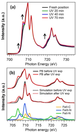
Indeed, XAS spectra can be accurately modeled by ligand field multiplet calculation enabling a direct assignment of each spectroscopic feature to a specific oxidation and spin state of Fe atoms in PB. InFigure 2a XAS spectra measured at different intervals of UV exposure time are reported: before the irradiation (black line), after 20 min (green line), after 40 min (blue line), and after 70 min (red line) of irradiation. Before irradiation the Fe L2,3-edge XAS spectrum has the
typical line shape expected for a mixed iron complex system such as PB,6,52 KFeIII[FeII(CN)6], which is a mixture of
low-spin FeIIcenters (LS-FeII−C) and high-spin FeIIIcenters
(HS-FeIII−N). Fe L-edge XAS spectra clearly show the progressive formation of reduced Fe species6 as a function of the UV exposure time: FeIII−CN−FeIIevolves gradually to FeII−CN− FeII, as shown by the intensity increase of the peaks at 707.15
and 720.3 eV as indicated inFigure 2a. In order to prove the reduction of HS-FeIII−N to HS-FeII−N centers, XAS spectra
were simulated by linear combination of the calculated spectral
components present in PB molecules before and after UV exposure. The simulated curves of each spectroscopic feature (LS-FeII−C, HS-FeIII−N, and HS-FeII−N) were calculated with the ligandfield multiplet approach (see Methods), and their linear combination was optimized to reproduce the exper-imental line shape of PB spectra reported inFigure 2b.
The calculations of the single spectral features were optimized by following those developed by Wang et al.6 and Hocking et al.52
The hopping parameters and back-bonding configuration energies were adjusted to reproduce the spectral components found in the literature6 and to model as best as possible the energy splittings of our experimental XAS spectra. The parameters optimized for the calculations are reported in
Table 1 in the Methods section. As expected, a linear combination of 50% LS-FeII−C and 50% HS-FeIII−N
reproduces quite well the line shape of the XAS spectrum of fresh PB. The best simulation of the XAS spectrum of PB irradiated 70 min with UV light was achieved by a linear combination of 15% HS-FeII−N, 35% HS-FeIII−N, and 50%
LS-FeII−C. Indeed, the calculation provides a direct verification that about 30% of FeIII−N atoms were reduced to FeII−N
species upon 70 min of UV irradiation. Despite the good
Figure 1.(a) XPS O 1s spectra of GNBs obtained before the UV irradiation (black curve) and after 45 min of irradiation (red curve) indicating the presence of water during the time scale of the experiment. (b) C 1s XPS spectra of GNBs before (black curve) and after 45 min of UV irradiation at 395 eV (red curve) show no additional defects in the Gr layer induced by UV exposure.
Figure 2. (a) Experimental XAS spectra at Fe L2,3 edge measured
before (black line) and after 20 min (green line), 40 min (blue line), and 70 min of UV irradiation (red line) of an aqueous solution of PB. (b) From top to bottom: experimental Fe L2,3edge XAS spectra of PB
before (black solid line) and after 70 min of UV irradiaton (red solid line); calculated spectra of PB before (black dashed line) and after laser exposure (red dashed line); simulated spectroscopic components of FeII−C (yellow line), FeII−N (green line), and FeIII−N (blue line)
atoms coordinated to cyano groups. The linear combination of the simulated spectral features (PB calculated spectra) indicates that 30% of FeIII−N atoms are reduced to FeII−N species after 70 min of UV
irradiation. The Journal of Physical Chemistry C
precursors are reported in Figure S-3 in the Supporting Information.
In order to be sure that the Fe reduction is due exclusively to the UV irradiation in our experimental conditions, but not to the possible damage induced by the X-rays,4,53the synchrotron light contribution was evaluated as well. A second fresh sample of GNBs filled with a 10 mM solution of PB was used as reference and exposed only to the synchrotron light for 45 min adopting the same experimental conditions used for XAS measurements (photon energy and beam flux) without laser irradiation. Figure 3 a reports the comparison between the spectrum measured before irradiation and the spectra collected after exposure to the UV laser for 45 min (bottom spectra) and to X-rays for 45 min (top spectra).
As visible inFigure 3a, upon UV irradiation the intensity of peaks at 707.15 and 720.3 eV increases, and the spectrum line shape approaches the one reported for reduced FeII−CN−FeII species typical of PW (as described before).6,52In the case of the sole X-ray irradiation, no evident change in the Fe L2,3edge
is visible after 45 min of exposure, confirming that the main factor responsible for the Fe reduction is the UV light exposure, whose energy corresponds to the band gap of TiO2.
The role of the aqueous environment was investigated as well to understand if the presence of water is necessary for the photoreduction process. Fe L2,3 edge XAS spectra of PB solution encased inside GNBs were compared with those acquired on a dry sample before and after laser exposure. The dry PB sample was prepared by drop-casting method on TiO2
rutile single crystal. Both samples were irradiated with the same UV laser for 20 min, a sufficient time to see Fe reduction (as previously observed in Figure 2 a). The obtained results are shown inFigure 3 b.
As widely discussed above, the PB solution inside GNBs on TiO2shows the clear sign of Fe reduction upon UV irradiation.
On the contrary, the dry PB sample drop casted on TiO2does not show any feature of reduced Fe. These results indicate that the water environment is a fundamental requirement for the photoinduced reduction of PB, and the GNB approach ensures this basic condition.
In light of the observed results, it is clear that the gradual photoinduced reduction of FeIII−CN−FeII species (PB) to FeII−CN−FeII species (PW) occurs preferentially under the
395 nm laser excitation energy and in the presence of an aqueous environment. Indeed, upon UV band gap excitation of rutile (Eg = 3.03 eV), the electrons of the valence band are
photoexcited to the conduction band of TiO2, generating both
holes and electrons in the valence and conduction bands, respectively.
The role of water is extremely important to allow the evolution of the reaction; in fact, water acts as a scavenger and reduces the generated holes in the valence band, preventing the electrons excited to the conduction band from being easily recombined with holes.54
These conditions are required to ensure a continuous supply of photoexcited electrons to the conduction band of TiO2,
which can be injected into the electronic states of PB to reduce FeIII−N to FeII−N atoms. The remaining holes in the valence band of TiO2 oxidize water molecules to produce protons, which are used to ensure the charge balance of the partial reduced dye.1 The proposed photoreduction mechanism is reported inFigure 4.
The process was further studied by Raman spectroscopy in liquid phase. Raman spectra of GNB samples (black and red lines in Figure 5a, corresponding to fresh and UV irradiated samples, respectively) show the characteristic features of Gr at
Figure 3.(a) XAS Fe L2,3edge spectra of an aqueous solution of PB in
GNBs exposed for 45 min to UV irradiation (red curve, bottom spectra) and for 45 min to X-rays irradiation (red curve, top spectra). (b) XAS Fe L2,3edge spectra measured after 20 min of UV exposure of
the following: a PB aqueous solution in GNBs/TiO2 (red curve,
bottom spectra) and a PB dryfilm on TiO2(red curve, top spectra).
The black spectra are measured before the irradiation. The photoreduction of Fe occurs only under UV light and in wet environment. No evident photoinduced effects are visible under X-rays exposure and in the absence of water.
1590 and 2695 cm−1which are ascribed to G and 2D peaks, respectively.55,56
G and 2D bands are not affected by UV illumination, and no defects are introduced within the exposure time, as also evident in Figure S-4 in theSupporting Information. Other features at 443.4 cm−1(Egrutile mode) and 610.8 cm−1(Agrutile mode)
are ascribed to TiO2 substrate, while the small components
between 200 and 700 cm−1can be assigned to Fe−C and Fe−N stretching and Fe−C−N and Fe−N−C bending modes11,57 typical of ferrocyanides, which are visible also on dry PB (blue line).
Finally, the characteristic CN vibrational peak around 2162 cm−1is ascribed to the FeII−CN−FeIIIA
g(symmetric)
stretching mode of PB.10,11
An expanded view of the CN vibrational component is presented in Figure 5 b, and all the spectra have been normalized to this peak for a comparative analysis.
On dry PB sample on glass (blue line), this mode is observed at 2160 cm−1, resulting downshifted by 2−3 cm−1with respect to PB solution in GNBs (red and black lines). This shift is most likely due to the presence of water or OH−ions, which can play an important role in the frequency shift of Agstretching mode. Thisfinding proves again the presence of aqueous solution in GNBs. The weak peak around 2098 cm−1is attributed to the Eg mode (asymmetric) of the FeII−CN−FeIII stretching
vibration,11 and its intensity remains unchanged upon UV illumination. Finally, the relative intensity of the shoulder at 2127 cm−1, corresponding to FeII−CN−FeIIstretching mode of CN group coordinated to two FeIIatoms, grows after 15 h of UV illumination (red line).
The higher intensity of this feature upon UV exposure proves an increase of reduced FeII cations, confirming the partial photoreduction of PB to PW in aqueous solution on a TiO2
substrate.
■
METHODSPrussian Blue Solution. An aqueous solution of the soluble form of PB (100 mM) was prepared by mixing at room temperature equal volumes of 100 mM solutions of K3 [Fe-(CN)6] (potassium ferri-cyanide, Sigma-Aldrich) and FeCl2·
4H2O (iron(II) chloride tetrahydrate, Sigma-Aldrich). The reaction is described by the chemicaleq 1:
+ → +
Fe ClII 2 K [Fe (CN) ]3 III 6 KFe [Fe (CN) ]III II 6 2KCl (1)
The final PB solution was diluted 10 times to avoid the formation of aggregates, but the concentration of Fe was chosen high enough to have a good XAS signal. A picture of the starting and the final solutions of K3[Fe(CN)6] and
KFeIII[FeII(CN)
6] is shown in Figure S-1 in the Supporting
Information.
Graphene Nanobubbles Fabrication. The Gr layers were provided by the Department of Physics of Bilkent University in Ankara, Turkey. Graphene was synthesized on copper foils by chemical vapor deposition (CVD) according to the procedure described elsewhere.58,59 The copper foils were placed in a quartz chamber and heated to 1000°C under flow of hydrogen
Figure 4.Proposed photoreduction mechanism of PB: upon UV band gap excitation of TiO2(Eg= 3.03 eV) the electrons (e−) of the valence
band (VB) are promoted to the conduction band (CB) of TiO2,
generating both holes (h+) and e−in the VB and CB, respectively. H2O acts as a scavenger to reduce the generated h+ in the VB,
preventing the e−of CB from being easily recombined with h+. The photoexcited e− can be injected into the electronic states of PB to reduce FeIII−N to FeII−N atoms. The remaining h+in the VB of TiO2
are used to oxidize the H2O molecules and produce H3O+ions to
ensure the charge balance of the reduced PB.
Figure 5.(a) Raman spectra of dry PB on glass (blue line), freshly prepared GNBsfilled with PB aqueous solution on TiO2(black line)
and after 15 h of UV exposure (red line). (b) Zoomed region of Raman CN stretching mode of PB. The vibrational peak around 2162 cm−1on PB solution in GNBs (black and red lines) results up-shifted by 2−3 cm−1 with respect to dry PB (blue line) due to the
presence of water. The intensity of the FeII−CN−FeIIstretching
mode component at 2127 cm−1grows after 15 h of UV illumination (red line), while the peak around 2098 cm−1of the FeII−CN−FeIII
stretching vibration remains unchanged. The higher intensity of the FeII−CN−FeIIfeature upon UV exposure proves the formation of
new FeIIspecies, thus the photoreduction of PB in aqueous solution
on a TiO2substrate.
Dry samples were also prepared by drop-casting method in order to analyze the role of water.
Electron Spectroscopies. XPS and XAS measurements of the samples were carried out on the CNR BACH beamline at Elettra Sincrotrone in Trieste (Italy).60,61
XPS spectra of C 1s and O 1s core levels were recorded at 596 eV of photon energy. A VG-Scienta R3000 hemispherical analyzer,62working with an overall energy resolution of 0.2 eV, was used. All spectra were fitted by a Gaussian function after the prior subtraction of a Shirley background to account for the inelastic photoelectrons. XAS spectra at the L2,3 edge of Fe
before and after the UV laser (15 mW, 395 nm) exposure were measured in TEY collecting the currentflowing through the Gr membrane.
The laser (PS-LBX, 395 nm, 15 mW, Oxxius) was placed on a laser board, and the light was impinged on the samples through an optical window mounted on the experimental chamber of BACH beamline at 22.5° from the sample surface normal.
Raman Spectroscopy. Raman measurements were per-formed in the reflection geometry on an inverted optical microscope (Axiovert 200, Zeiss) coupled with a 750 mm long spectrometer (Shamrock SR-750, Andor Technology plc). CW laser with the excitation wavelength of 532 nm (Cobolt Samba, 50 mW, bandwidth 1 MHz) was used as an excitation source. A laser beam was reflected by 45° oriented beamsplitter (532 nm RazorEdge Dichroic), directed to the inverted microscope and then focused on the mounted sample by 100× air objective (NA 0.8, EC Epiplan), resulting in a diameter of laser spot of around 0.35μm. The laser power on the sample was controlled by variable neutral densityfilter (NDC-50C-4M, Thorlabs) and kept at 1 mW to avoid temperature-induced effects. The Raman scattered light was collected by the same objective and sent to the spectrometer by the same optical path, dispersed by a diffractive grating of 600 lines mm−1, andfinally analyzed using TE-cooled EMCCD (Newton DU971-UVB, Andor Technol-ogy plc). The Rayleigh scattered light was blocked by the long-pass edge filter (532 nm RazorEdge) at the entrance of spectrometer. The acquisition time in all experiments was 60 s. Simulations. Ligandfield multiplet simulations were carried out exploiting the multiplet model implemented by Thole63
and Cowan64 and by using the CTM4XAS 5.5 program
developed by Stavitski and de Groot65 which includes spin− orbit coupling, Coulomb interactions, and crystal field effects. Atomic Slater integrals were used, and all spectra were broadened by a Lorentzian with a half-width of 0.2(0.3) eV and by a Gaussian with a sigma value of 0.3 eV. Metal-to-ligand and ligand-to-metal charge transfer (MLCT and LMCT) were
N components expected in PB and PW species are reported in
Table 1. The best simulation of high-spin state FeII−N was achieved using a single configuration (3dN) and without the use of mixing parameters.
Since the presence of additional components due to PB precursors cannot be completely excluded, extra components of FeIII−C and FeII−Cl ascribed to FeCl
2 and K3[FeIII(CN)6]
were simulated and reported in Figure S-3 together with the corresponding parameters in Table S-1 (Supporting Informa-tion).
■
CONCLUSIONThis work sheds light on the photoinduced reduction mechanism of a PB dye in aqueous solution in the presence of TiO2catalyst. The role of the solvent was analyzed, and the photochemical reaction was monitored for the first time by using conventional electron spectroscopy methods in UHV conditions. GNBs were employed to follow in situ and in operando the evolution of the electronic properties of the sample in liquid environment. Thanks to the possibility of filling “quasi” transparent to photons and electrons GNBs with liquids, the well-known UV-induced reduction of an aqueous solution of PB catalyzed by band gap excitation of TiO2was
followed by XAS measurements.
XAS spectra at the L2,3 edge of Fe showed the gradual
evolution from PB (FeIII−CN−FeII) to PW (FeII−CN−FeII), confirmed also by ligand field multiplet calculation, and O 1s XPS measurements proved the presence of water inside GNBs before and after UV irradiation.
Our data suggest that photoexcited electrons promoted to the conduction band of TiO2 are used by PB to reduce HS-FeIII−N to HS-FeII−N species. The results clearly demonstrate
that this process requires the presence of water as scavenger to reduce the generated holes in the valence band of rutile; otherwise, the excited electrons could be easily recombined with holes, stopping the reduction of Fe species. The results were further confirmed by Raman measurements of PB solution in GNBs, evidencing changes of the CN vibrational stretching modes as a function of the Fe oxidation state.
In conclusion, thanks to the GNB approach, the mechanism of photocatalytic reduction of PB in aqueous solution was finally investigated by electron spectroscopy. This paper proves again the robustness and reliability of GNBs as a valuable method to study the evolution of a large variety of physical− chemical processes in liquid phase, with a particular attention to liquid/solid electron transfer in catalysis, energy conversion processes, photocatalysis, water splitting, and so on.
■
ASSOCIATED CONTENT*
S Supporting InformationThe Supporting Information is available free of charge on the
ACS Publications websiteat DOI:10.1021/acs.jpcc.7b07898. Preparation of PB (pictures of the starting solution K3[Fe(CN)6] and thefinal solution KFeIII[FeII(CN)
6]);
Raman and SEM of “as-grown” Gr on Cu foil; LFM
calculation of PB precursors (FeCl2 and K3[Fe(CN)6])
and PB components; Raman spectra of G and 2D peaks of GNBs and a dryfilm of PB (PDF)
■
AUTHOR INFORMATION Corresponding Authors *E-mail:[email protected]. *E-mail:[email protected]. ORCID Silvia Nappini: 0000-0002-4944-5487 Marco Lazzarino:0000-0003-1077-6569 Frank.M. F. De Groot:0000-0002-1340-2186 Coskun Kocabas: 0000-0003-0831-5552 Present Addresses¶CERIC-ERIC (Central European Research Infrastructure Consortium), S.S. Fourteen Km 163.5, 34149 Basovizza, Trieste, Italy.
□Elettra-Sincrotrone Trieste S.C.p.A, S.S. Fourteen Km 163.5, 34149 Basovizza, Trieste, Italy.
Author Contributions
S.N. and E.M. planned and executed electron spectroscopy experiments and evaluated the data. S.N. drafted the manu-script. E.M. actively participated in planning the manumanu-script. S.N. and A.M. prepared PB solutions. A.M. prepared GNBs filled with liquid and helped with data acquisition. F.D.G actively helped in ligandfield multiplet analysis. D.N. executed Raman characterization and analysis. M.L. and S.D.Z. gave general advisory and actively participated in planning the experiments. C.K. and O.B. provided the Gr layers grown on Cu foils. All authors have given approval to thefinal version of the manuscript.
Notes
The authors declare no competingfinancial interest.
■
ACKNOWLEDGMENTSThis work was supported by CNR and the Italian Ministry of Education MIUR through the national grants FIRB Futuro in Ricerca 2012 RBFR128BEC “Beyond graphene: Tailored C-layers for novel catalytic materials and green chemistry”, progetto premiale ABNANOTECH and FIRB RBAP11ET-KA_003. We are thankful to Dr. Federica Bondino for constructive feedback on the work. The technical support of Federico Salvador and Paolo Bertoch is acknowledged.
■
ABBREVIATIONSGr, graphene; GNB, graphene nanobubble; PB, Prussian blue; PW, Prussian white; XPS, X-ray photoemission spectroscopy; XAS, X-ray absorption spectroscopy; AFM, atomic force microscopy; SEM, scanning electron microscopy; UHV, ultrahigh vacuum; BE, binding energy; TEY, total electron yield; CVD, chemical vapor deposition
■
REFERENCES(1) Yamamoto, T.; Saso, N.; Umemura, Y.; Einaga, Y. Photo-reduction of Prussian Blue Intercalated into Titania Nanosheet Ultrathin Films. J. Am. Chem. Soc. 2009, 131, 13196−13197.
(2) Aguilà, D.; Prado, Y.; Koumousi, E. S.; Mathonière, C.; Clérac, R. Switchable Fe/Co Prussian Blue Networks and Molecular Analogues. Chem. Soc. Rev. 2016, 45, 203−224.
(3) Itaya, K.; Uchida, I.; Neff, V. D. Electrochemistry of Polynuclear Transition Metal Cyanides: Prussian Blue and Its Analogues. Acc. Chem. Res. 1986, 19, 162−168.
(4) Risch, M.; Stoerzinger, K. A.; Regier, T. Z.; Peak, D.; Sayed, S. Y.; Shao-Horn, Y. Reversibility of Ferri-/Ferrocyanide Redox during Operando Soft X-Ray Spectroscopy. J. Phys. Chem. C 2015, 119, 18903−18910.
(5) Neff, V. D. Some Performance Characteristics of a Prussian Blue Battery. J. Electrochem. Soc. 1985, 132, 1382−1384.
(6) Wang, L.; Song, J.; Qiao, R.; Wray, L. A.; Hossain, M. A.; Chuang, Y.-D.; Yang, W.; Lu, Y.; Evans, D.; Lee, J.-J.; et al. Rhombohedral Prussian White as Cathode for Rechargeable Sodium-Ion Batteries. J. Am. Chem. Soc. 2015, 137, 2548−2554.
(7) Malinauskas, A.; Ruzgas, T.; Gorton, L. Electrochemical Study of the Redox Dyes Nile Blue and Toluidine Blue Adsorbed on Graphite and Zirconium Phosphate Modified Graphite. J. Electroanal. Chem. 2000, 484, 55−63.
(8) Ricci, F.; Palleschi, G. Sensor and Biosensor Preparation, Optimisation and Applications of Prussian Blue Modified Electrodes. Biosens. Bioelectron. 2005, 21, 389−407.
(9) Ricci, F.; Gonçalves, C.; Amine, A.; Gorton, L.; Palleschi, G.; Moscone, D. Electroanalytical Study of Prussian Blue Modified Glassy Carbon Paste Electrodes. Electroanalysis 2003, 15, 1204−1211.
(10) Gervais, C.; Languille, M.-A.; Réguer, S.; Gillet, M.; Pelletier, S.; Garnier, C.; Vicenzi, E. P.; Bertrand, L. Why Does Prussian Blue Fade? Understanding the Role(s) of the Substrate. J. Anal. At. Spectrom. 2013, 28, 1600−1609.
(11) Samain, L.; Gilbert, B.; Grandjean, F.; Long, G. J.; Strivay, D. Redox Reactions in Prussian Blue Containing Paint Layers as a Result of Light Exposure. J. Anal. At. Spectrom. 2013, 28, 524−535.
(12) Fading and Colour Change of Prussian Blue: Methods of Manufacture and the Influence of Extenders, Technical Bulletin, National Gallery, London https://www.nationalgallery.org.uk/technical-bulletin/kirby_saunders2004(accessed Jan 10, 2017).
(13) Itaya, K.; Shibayama, K.; Akahoshi, H.; Toshima, S. Prussian-blue-modified Electrodes: An Application for a Stable Electrochromic Display Device. J. Appl. Phys. 1982, 53, 804−805.
(14) Karyakin, A. A. Prussian Blue and Its Analogues: Electro-chemistry and Analytical Applications. Electroanalysis 2001, 13, 813− 819.
(15) Agrisuelas, J.; Bueno, P. R.; Ferreira, F. F.; Gabrielli, C.; García-Jareño, J. J.; Gimenez-Romero, D.; Perrot, H.; Vicente, F. Electronic Perspective on the Electrochemistry of Prussian Blue Films. J. Electrochem. Soc. 2009, 156, P74−P80.
(16) Sato, O.; Iyoda, T.; Fujishima, A.; Hashimoto, K. Photoinduced Magnetization of a Cobalt-Iron Cyanide. Science 1996, 272, 704−705. (17) Escax, V.; Bleuzen, A.; Cartier Dit Moulin, C.; Villain, F.; Goujon, A.; Varret, F.; Verdaguer, M. Photoinduced Ferrimagnetic Systems in Prussian Blue Analogues C(I)XCo4[Fe(CN)6]y (C(I) = Alkali Cation). 3. Control of the Photo- and Thermally Induced Electron Transfer by the [Fe(CN)6] Vacancies in Cesium Derivatives.
J. Am. Chem. Soc. 2001, 123, 12536−12543.
(18) Taguchi, M.; Yagi, I.; Nakagawa, M.; Iyoda, T.; Einaga, Y. Photocontrolled Magnetization of CdS-Modified Prussian Blue Nanoparticles. J. Am. Chem. Soc. 2006, 128, 10978−10982.
(19) Jiang, G.; Lin, Z.; Chen, C.; Zhu, L.; Chang, Q.; Wang, N.; Wei, W.; Tang, H. TiO2 Nanoparticles Assembled on Graphene Oxide Nanosheets with High Photocatalytic Activity for Removal of Pollutants. Carbon 2011, 49, 2693−2701.
(20) Fujishima, A.; Zhang, X.; Tryk, D. A. Heterogeneous Photocatalysis: From Water Photolysis to Applications in Environ-mental Cleanup. Int. J. Hydrogen Energy 2007, 32, 2664−2672. The Journal of Physical Chemistry C
Interface. Sci. Rep. 2015, 5, 9788.
(25) Favaro, M.; Drisdell, W. S.; Marcus, M. A.; Gregoire, J. M.; Crumlin, E. J.; Haber, J. A.; Yano, J. An Operando Investigation of (Ni−Fe−Co−Ce)Ox System as Highly Efficient Electrocatalyst for Oxygen Evolution Reaction. ACS Catal. 2017, 7, 1248−1258.
(26) Favaro, M.; Yang, J.; Nappini, S.; Magnano, E.; Toma, F. M.; Crumlin, E. J.; Yano, J.; Sharp, I. D. Understanding the Oxygen Evolution Reaction Mechanism on CoOx Using Operando Ambient-Pressure X-Ray Photoelectron Spectroscopy. J. Am. Chem. Soc. 2017, 139, 8960−8970.
(27) Kolmakov, A.; Dikin, D. A.; Cote, L. J.; Huang, J.; Abyaneh, M. K.; Amati, M.; Gregoratti, L.; Günther, S.; Kiskinova, M. Graphene Oxide Windows for in Situ Environmental Cell Photoelectron Spectroscopy. Nat. Nanotechnol. 2011, 6, 651−657.
(28) Kraus, J.; Reichelt, R.; Günther, S.; Gregoratti, L.; Amati, M.; Kiskinova, M.; Yulaev, A.; Vlassiouk, I.; Kolmakov, A. Photoelectron Spectroscopy of Wet and Gaseous Samples through Graphene Membranes. Nanoscale 2014, 6, 14394−14403.
(29) Yulaev, A.; Guo, H.; Strelcov, E.; Chen, L.; Vlassiouk, I.; Kolmakov, A. Graphene Microcapsule Arrays for Combinatorial Electron Microscopy and Spectroscopy in Liquids. ACS Appl. Mater. Interfaces 2017, 9, 26492−26502.
(30) Guo, J.; Tong, T.; Svec, L.; Go, J.; Dong, C.; Chiou, J.-W. Soft-x-Ray Spectroscopy Experiment of Liquids. J. Vac. Sci. Technol., A 2007, 25, 1231−1233.
(31) Bora, D. K.; Glans, P.-A.; Pepper, J.; Liu, Y.-S.; Du, C.; Wang, D.; Guo, J.-H. An Ultra-High Vacuum Electrochemical Flow Cell for in Situ/Operando Soft X-Ray Spectroscopy Study. Rev. Sci. Instrum. 2014, 85, 043106.
(32) Guo, H.; Strelcov, E.; Yulaev, A.; Wang, J.; Appathurai, N.; Urquhart, S.; Vinson, J.; Sahu, S.; Zwolak, M.; Kolmakov, A. Enabling Photoemission Electron Microscopy in Liquids via Graphene-Capped Microchannel Arrays. Nano Lett. 2017, 17, 1034−1041.
(33) Yuk, J. M.; Park, J.; Ercius, P.; Kim, K.; Hellebusch, D. J.; Crommie, M. F.; Lee, J. Y.; Zettl, A.; Alivisatos, A. P. High-Resolution EM of Colloidal Nanocrystal Growth Using Graphene Liquid Cells. Science 2012, 336, 61−64.
(34) Wang, C.; Qiao, Q.; Shokuhfar, T.; Klie, R. F. High-Resolution Electron Microscopy and Spectroscopy of Ferritin in Biocompatible Graphene Liquid Cells and Graphene Sandwiches. Adv. Mater. 2014, 26, 3410−3414.
(35) Nappini, S.; Matruglio, A.; Naumenko, D.; Zilio, S. D.; Bondino, F.; Lazzarino, M.; Magnano, E. Graphene Nanobubbles on TiO2for
in-Operando Electron Spectroscopy of Liquid-Phase Chemistry. Nanoscale 2017, 9, 4456−4466.
(36) Kolmakov, A.; Gregoratti, L.; Kiskinova, M.; Günther, S. Recent Approaches for Bridging the Pressure Gap in Photoelectron Microspectroscopy. Top. Catal. 2016, 59, 448−468.
(37) Kraus, J.; Böbel, M.; Günther, S. Suppressing Graphene Nucleation during CVD on Polycrystalline Cu by Controlling the Carbon Content of the Support Foils. Carbon 2016, 96, 153−165.
(38) Martyanov, I. N.; Klabunde, K. J. Photocatalytic Purification of Water and Air over Nanoparticulate TiO2. In Nanoscale Materials in
Array Films. Nanoscale Res. Lett. 2014, 9, 621.
(43) Zhang, K.-X.; Wang, W.; Hou, J.-L.; Zhao, J.-H.; Zhang, Y.; Fang, Y.-C. Oxygen Plasma Induced Hydrophilicity of TiO2 Thin Films. Vacuum 2011, 85, 990−993.
(44) Jung, C.-K.; Bae, I.-S.; Song, Y.-H.; Boo, J.-H. Plasma Surface Modification of TiO2 Photocatalysts for Improvement of Catalytic Efficiency. Surf. Coat. Technol. 2005, 200, 1320−1324.
(45) Tissot, H.; Gallet, J.-J.; Bournel, F.; Olivieri, G.; Silly, M. G.; Sirotti, F.; Boucly, A.; Rochet, F. The Electronic Structure of Saturated NaCl and NaI Solutions in Contact with a Gold Substrate. Top. Catal. 2016, 59, 605−620.
(46) Ketteler, G.; Yamamoto, S.; Bluhm, H.; Andersson, K.; Starr, D. E.; Ogletree, D. F.; Ogasawara, H.; Nilsson, A.; Salmeron, M. The Nature of Water Nucleation Sites on TiO2(110) Surfaces Revealed by Ambient Pressure X-Ray Photoelectron Spectroscopy. J. Phys. Chem. C 2007, 111, 8278−8282.
(47) Yang, D.; Velamakanni, A.; Bozoklu, G.; Park, S.; Stoller, M.; Piner, R. D.; Stankovich, S.; Jung, I.; Field, D. A.; Ventrice, C. A., Jr.; et al. Chemical Analysis of Graphene Oxide Films after Heat and Chemical Treatments by X-Ray Photoelectron and Micro-Raman Spectroscopy. Carbon 2009, 47, 145−152.
(48) Chu, D.; Younis, A.; Li, S. Direct Growth of TiO 2 Nanotubes on Transparent Substrates and Their Resistive Switching Character-istics. J. Phys. D: Appl. Phys. 2012, 45, 355306.
(49) Xing, M.; Shen, F.; Qiu, B.; Zhang, J. Highly-Dispersed Boron-Doped Graphene Nanosheets Loaded with TiO2 Nanoparticles for
Enhancing CO2Photoreduction. Sci. Rep. 2015, 4, 6341.
(50) Jia, Y.; Gong, X.; Peng, P.; Wang, Z.; Tian, Z.; Ren, L.; Fu, Y.; Zhang, H. Toward High Carrier Mobility and Low Contact Resistance: Laser Cleaning of PMMA Residues on Graphene Surfaces. Nano-Micro Lett. 2016, 8, 336−346.
(51) Banerjee, S.; Dionysiou, D. D.; Pillai, S. C. Self-Cleaning Applications of TiO2 by Induced Hydrophilicity and
Photo-catalysis. Appl. Catal., B 2015, 176, 396−428.
(52) Hocking, R. K.; Wasinger, E. C.; de Groot, F. M. F.; Hodgson, K. O.; Hedman, B.; Solomon, E. I. Fe L-Edge XAS Studies of K4[Fe(CN)6] and K3[Fe(CN)6]: A Direct Probe of Back-Bonding. J. Am. Chem. Soc. 2006, 128, 10442−10451.
(53) Gervais, C.; Languille, M.-A.; Moretti, G.; Réguer, S. X-Ray Photochemistry of Prussian Blue Cellulosic Materials: Evidence for a Substrate-Mediated Redox Process. Langmuir 2015, 31, 8168−8175.
(54) Dung, D.; Ramsden, J.; Graetzel, M. Dynamics of Interfacial Electron-Transfer Processes in Colloidal Semiconductor Systems. J. Am. Chem. Soc. 1982, 104, 2977−2985.
(55) Ferrari, A. C. Raman Spectroscopy of Graphene and Graphite: Disorder, Electron−phonon Coupling, Doping and Nonadiabatic Effects. Solid State Commun. 2007, 143, 47−57.
(56) Ferrari, A. C.; Basko, D. M. Raman Spectroscopy as a Versatile Tool for Studying the Properties of Graphene. Nat. Nanotechnol. 2013, 8, 235−246.
(57) Barsan, M. M.; Butler, I. S.; Fitzpatrick, J.; Gilson, D. F. R. High-Pressure Studies of the Micro-Raman Spectra of Iron Cyanide Complexes: Prussian Blue (Fe4[Fe(CN)6]3), Potassium Ferricyanide
(K3[Fe(CN)6]), and Sodium Nitroprusside (Na2[Fe(CN)5(NO)]· 2H2O). J. Raman Spectrosc. 2011, 42, 1820−1824.
(58) Salihoglu, O.; Balci, S.; Kocabas, C. Plasmon-Polaritons on Graphene-Metal Surface and Their Use in Biosensors. Appl. Phys. Lett. 2012, 100, 213110.
(59) Li, X.; Cai, W.; An, J.; Kim, S.; Nah, J.; Yang, D.; Piner, R.; Velamakanni, A.; Jung, I.; Tutuc, E.; et al. Large-Area Synthesis of High-Quality and Uniform Graphene Films on Copper Foils. Science 2009, 324, 1312−1314.
(60) Zangrando, M.; Finazzi, M.; Paolucci, G.; Comelli, G.; Diviacco, B.; Walker, R. P.; Cocco, D.; Parmigiani, F. BACH, the Beamline for Advanced Dichroic and Scattering Experiments at ELETTRA. Rev. Sci. Instrum. 2001, 72, 1313−1319.
(61) Zangrando, M.; Zacchigna, M.; Finazzi, M.; Cocco, D.; Rochow, R.; Parmigiani, F. Polarized High-Brilliance and High-Resolution Soft X-ray Source at ELETTRA: The Performance of Beamline BACH. Rev. Sci. Instrum. 2004, 75, 31−36.
(62) Drera, G.; Salvinelli, G.; Åhlund, J.; Karlsson, P. G.; Wannberg, B.; Magnano, E.; Nappini, S.; Sangaletti, L. Transmission Function Calibration of an Angular Resolved Analyzer for X-Ray Photoemission Spectroscopy: Theory vs Experiment. J. Electron Spectrosc. Relat. Phenom. 2014, 195, 109−116.
(63) Thole, B. T.; van der Laan, G.; Fuggle, J. C.; Sawatzky, G. A.; Karnatak, R. C.; Esteva, J.-M. 3d. Phys. Rev. B: Condens. Matter Mater. Phys. 1985, 32, 5107−5118.
(64) Cowan, R. D. The Theory of Atomic Structure and Spectra; University of California Press: Berkeley, CA, 1981.
(65) Stavitski, E.; de Groot, F. M. F. The CTM4XAS Program for EELS and XAS Spectral Shape Analysis of Transition Metal L Edges. Micron 2010, 41, 687−694.