nv
estiga
tion
Percutaneous Laser Disc Decompression:
Retrospective Analysis of 197 Cases and Review of
The Literature
Perkütan Lazer Disk Dekompresyonu: 197 Olgunun Retrospektif Analizi
ve Literatürün Taranması
Yahya Cem ERBAs1, serhat PUsAT2, Ersin ERDOGAN3
1Bilgi Hospital, Department of Neurosurgery, Ankara, Turkey
2Etimesgut Military Hospital, Department of Neurosurgery, Ankara, Turkey 3Ufuk University, School of Medicine, Department of Neurosurgery, Ankara, Turkey
Corresponding Author: Serhat PUSAt / E-mail: [email protected]
ABSTRACT
AIm: Percutaneous laser disc decompression (PLDD) is a one of the well-known minimal invasive treatment methods of disc herniations. The aim of this study is to present our clinical experience and to show the benefits of this technique.
mAterIAl and methOds: A total of 197 patients, who met the criteria of PLDD, underwent treatment between 2007 and 2009. The data of the patients was reviewed retrospectively. Among them, 107 (54.3 %) patients were male and 90 were female with a mean age of 46.34 years (ranged between 23 and 86 years). Seventy-two patients underwent one level PLDD, 112 (56.8 %) patients two levels PLDD and 13 patients three levels PLDD procedures. The mean follow-up time was 42 months.
results: Among the 72 patients, the level of PLDD was L3-L4 in 4 patients, L4-L5 in 39 patients and L5-S1 in 29 patients. L4-L5 and L5-S1 levels were the most common 2-level PLDD locations in 71 patients. Twenty-five (12.7 %) patients underwent microsurgical discectomy after PLDD. The procedure was repeated in 3 patients. Discitis secondary to possible thermal injury occurred in 2 (0.1%) patients and this complication was improved with conservative treatment.
COnClusIOn: PLDD is a safe and effective procedure in the treatment of discogenic pain if the patient met the selection criteria. However, this technique is not an alternative to open surgery.
KeywOrds: Percutaneous laser disc decompression, Discogenic back pain, Minimal invasive
ÖZ
AmAÇ: Perkütan lazer disk dekompresyonu (PLDD) disk hernilerinin tedavisinde iyi bilinen minimal invaziv bir yöntemdir. Çalışmanın amacı PLDD konusunda klinik tecrübemizi paylaşmak ve bu tekniğin faydalarını göstermektir.
yÖntem ve GereÇler: 2007 ile 2009 yılları arasında kriterlere uyan 197 hastaya PLDD uygulanmıştır. Bu 197 hastanın verileri retrospektif olarak incelendi. Olguların 107 (% 54,3)’si erkek ve 90’ı kadın olup, ortalama yaş 46,34’dü (23-86 yaş arası). Yetmiş iki hastaya tek seviye, 112 (% 56,8) hastaya iki seviye ve 13 hastaya üç seviye PLDD uygulandı. Ortalama takip süresi 42 ay idi.
BulGulAr: 72 hasta içinde, 4 hastada L3-L4 seviyesine, 39 hastada L4-L5 seviyesine ve 29 hastada L5-S1 seviyesine PLDD uygulandı. L4-L5 ve L5-S1 seviyesi en sık 2-seviye PLDD uygulanan seviyedir ve 71 hastaya uygulanmıştır. Yirmibeş (% 12,7) hastaya PLDD’den sonra mikrocerrahi yöntemle diskektomi uygulanmıştır. Üç hastada prosedür tekrar uygulanmıştır. Muhtemel termal hasara sekonder diskitis 2 (% 0,1) hastada izlenmiştir ve bu komplikasyon konservatif yöntemle düzelmiştir.
sOnuÇ: Perkütan lazer disk dekompresyonu hasta kriterlere uyduğu takdirde güvenli ve etkin bir tedavi yöntemidir. Ancak bu teknik hiçbir zaman açık cerrahiye alternatif değildir.
AnAhtAr sÖZCÜKler: Perkütan lazer disk dekompresyonu, Diskojenik bel ağrısı, Minimal invaziv
INTRODUCTION
Sixty to eighty percent of the adults suffer from back pain at least once in their life (12, 13). Back pain is the second common cause of admission to the hospital after upper respiratory tract infection and it is in the first rank among the diseases that cause sick leave (1, 13). 75-85% of all acute back pain improves in the first 2 months without any treatment, but
15-20% of the cases back pain becomes chronic despite the medical treatment (1, 22, 25). Discogenic pain is responsible for chronic back pain in 40% of the patients (13). Open surgery and spinal instrumentation were the conventional treatment methods of discogenic pain for many years. The rate of recurrence is 2-5% and the rate of clinical success in the early postoperative period is 95-98% after open surgery (8, 9). Fibrosis secondary to inadequate physiotherapy in
the postoperative period and secondary to epidural wound healing causes back and leg pains and reduces the success of surgery to 80% in the late follow-up period (8, 9).
Minimal invasive techniques for back pain are more popu-lar today because knowledge on the spinal anatomy has in-creased, clinical outcomes of conventional procedures have proven unsatisfactory, and imaging techniques have signifi-cantly improved (28). Percutaneous procedures used in pa-tients with back pain are prolotherapy (sclerotherapy), facet joint corticosteroid injection, medial branch blocks, intradis-cal corticosteroid injection, radiofrequency denervation, in-tradiscal electrothermal therapy (IDET), epidural steroid injec-tion, trigger point injecinjec-tion, adhesiolysis, nucleoplasty and percutaneous laser disc decompression (PLDD) (7, 27). In this paper, we tried to discuss our experience on the PLDD procedures and to present our clinical results of this minimal invasive technique in patients with discogenic back pain.
MATERIAL and METHODS
Written informed consent about the treatment protocol was obtained from all patients before the procedure. A total of 197 (0.02%) patients among the 79979 patients underwent PLDD treatment between 2007 and 2009. Of the 79979 patients, 9541 patients presented to neurosurgery, 29360 patients presented to physical therapy, 19550 patients presented
to neurology and 21528 patients presented to orthopedics outpatient clinics. All patients had back pain and the selection of the patients was made according to the indications as follow (14);
1. Leg pain is worse than back pain
2. Disc protrusion in MRI and no sequestrated disc 3. Chronic back pain longer than 3 months 4. Failure of non-invasive treatment methods 5. No neurological deficit
6. No segmental instability
7. Preservation of more than 75% of the disc height 8. No psychogenic component
Magnetic resonance imaging (MRI) was performed in all patients in order to detect the level of disc herniation. This imaging was also used for the differential diagnosis. The patients who had neurological deficit, instability or spinal tumor in the preoperative radiological examinations did not undergo PLDD for back pain. All procedures were performed by the same surgeon and preoperative demographic features of the patients were recorded. The procedure was performed under local anesthesia and with C-arm fluoroscopy guidance (Figure 1). Multi Diode PL 3DTM 980 nm Laser (INTERmedic) and
Menchetti’s handpiece with a 21 G needle were used for the PLDD (Figure 2A-C). Laser power was 8 –12 W, the exposure time was 500 – 600 ms and the pause was 2–2.5 sec with this technique. During the procedure, anteroposterior and lateral lumbosacral plain x-rays were obtained in all patients to verify the level of PLDD (Figure 3A-C). The mean follow-up time was 42 months (range 24 to 72 months). The visual analogue scale (VAS) was used to grade back pain in pre-procedure and post-procedure periods and each patient completed a VAS from 0 to 10 with 0 representing no pain and 10 representing unbearable pain. Postoperative complications and its management were also reviewed in this retrospective study.
RESULTS
Of the 197 patients, 107 (54.3%) were male and 90 were female with a mean age 46.34 years (ran023 to 86 years). The average pre-procedure VAS score was 6.3 (range 4 to 7). Among the 72 patients, the level of PLDD was L3-L4 in 4 patients, L4-L5 in 39 (54.2%) patients and L5-S1 in 29 patients. L4-L5 and
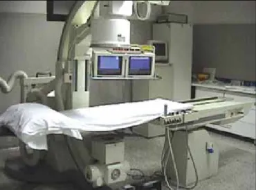
Figure 1: The operating room design and C-arm fluoroscopy for
PLDD procedure.
Figure 2: The laser source (A) and handpiece (B,C) for PLDD are shown in this picture.
L5-S1 levels were the most 2-level PLDD in 71 patients. This procedure was performed in 13 patients as 3-level at L3-L4, L4-L5 and L5-S1. The average post-procedure VAS score at the end of the first week was 7.9 (range 6 to 10). There was an improvement in the post-procedure period based on VAS scores. A total of 25 (12.7%) patients (9 female and 16 male) underwent microsurgical discectomy until the end of 2014. PLDD procedure was repeated in 3 patients (1 female and 2 male) because of the recurrence of the symptoms. Discitis secondary to possible thermal injury occurred in 2 (0.1%) patients (1 female and 1 male) and this complication improved with conservative treatment. There was no other complication secondary to PLDD procedure.
DISCUSSION
We documented the results of 197 patients who underwent PLDD for discogenic lumbar back pain. Most of the patients were male and 2-level PLDD was the most commonly used procedure. Single level PLDD was most frequently performed at the L4-L5 level. The rate of open surgical treatment was 12.7% among the 197 patients. Discitis was the only complication of our series. It was observed in 2 patients and treated conservatively.
Laser is the abbreviation of Light Amplification by Stimulated Emission of Radiation and in Turkish language it is abbreviated as “Laser”. PLDD was first performed in 1986 by Choy in Europe and in 1991 this procedure received approval of FDA in the United States. Choy (5) reported that intradiscal YAG laser application causes evaporation of nucleus pulposus and provides 75% clinical success (5). It is well known that the different doses of Laser affect cell proliferation, motility and secretion (21). Laser types, which are currently in use for the musculoskeletal system, are:
1. UV laser (Excimer) 2. Apparent laser (Argon) 3. IR laser (Ion resonance)
Apparent laser is well absorbed by hemoglobin and produces its effects by production of heat in the tissue (15, 17). This effect results in apoptosis by activation of oxygen in the cell nucleus. UV laser disrupts molecular connections without heat production (21).
The current indications of PLDD are discogenic pain, radicular pain, lumbar spinal stenosis and herniated lumbar discs (2, 6, 17, 26). Brouwer et al. (4) recently compared the results of PLDD and open conventional microdiscectomy in patients with lumbar disc herniation and radicular pain. They concluded that a strategy of PLDD, followed by surgery if necessary, resulted in non-inferior outcomes compared with surgery. Lee and Kang suggested percutaneous endoscopic laser annuloplasty for lumbar discogenic pain and they emphasized that Ho:YAG laser provides favorable outcomes for carefully selected groups of patients with DLBP (16). This technique is also suitable for thoracic disc herniations (11). The duration of application and energy requirements in PLDD varies according to the wavelength of the laser that is used in the procedure (15, 17). The selection of the patient and sufficient conservative treatment are important before the application of PLDD (17, 26). The patient selection criteria for PLDD are previously reported in the literature as follows (14): leg pain is worse than back pain, disc protrusion in MRI and no sequestrated disc, chronic back pain longer than 3 months, failure of non-invasive treatment methods, no neurological deficit, no segmental instability, preservation of more than 75% of the disc height, and no presence of psychogenic component. We also used these criteria for our patients in PLDD procedures. In our series, we used the same criteria with Kim (14) and we obtained satisfactory results with this technique. These criteria are therefore important in order to obtain better clinical results.
Phillips and Lauryssen reported that intradiscal procedures should be in the upper steps of the discogenic pain management algorithm and should be considered before the open surgical options (23). Intradiscal procedures were also suggested before the open surgical procedures in another article (18). Singh et al. performed a systematic review of lumbar laser spine surgery in the literature and they identified a total of 2447 patients from published studies, 72% of whom fulfilled the criteria for improvement. They concluded that level II-2 evidence (U.S. Preventive Services Task Force) existed for percutaneous laser decompression for short (less than one year) and long-term (more than one year) pain relief (24). Zileli and Ozer also pointed out the importance of PLDD in patients with back pain (28). In our series, the main complaint of our patients is back pain that is not improved with medical
Figue 3: Pictures show the application of procedure (A) and intraprocedural anteroposterior (B) and lateral (C) lumbosacral graphies.
4. Brouwer PA, Brand R, van den Akker-van Marle ME, Jacobs WC, Schenk B, van den Berg-Huijsmans AA, Koes BW, van Buchem MA, Arts MP, Peul WC: Percutaneous laser disc decompression versus conventional microdiscectomy in sciatica: A randomized controlled trial. Spine J 15(5):857-865, 2015
5. Choy D: Percutaneous laser disc decompression (PLDD). J Clin Laser Med Surg 16: 325-333, 1988
6. Duarte R, Costa JC: Percutaneous laser disc decompression for lumbar discogenic radicular pain. Radiologia 54(4):336-341, 2012 (in Spanish)
7. Erdine S: Interventional techniques in pain management. ANKEM 16(3): 182-184, 2002 (in Turkish)
8. Erdine S, Ozyalcin NS, Cimen A: Percutaneous lumbar nucleoplasty. Ağrı 2:17-22, 2005 (in Turkish)
9. Gangi A, Dietemann JL, Mortazavi R, Pfleger D, Kauff C, Roy C: CT-guided interventional procedures for pain management in the lumbosacral spine. RadioGraphics 18: 621-633, 1998 10. Grönemeyer DHW, Buschkamp H, Braun M, Schirp S,
Weinsheimer PA, Gevargez A: Image-guided percutaneous laser disk decompression for herniated lumbar disks: A 4-year follow-up in 200 patients. J Clin Laser Med Surg 21(3): 131-138, 2003
11. Haufe SM, Mork AR, Pyne M, Baker RA: Percutaneous laser disc decompression for thoracic disc disease: Report of 10 cases. Int J Med Sci 7(3):155-159, 2010
12. Izci Y, Apaydin O, Ozdem T, Cerrahoglu K: The bony malformations of lumbosacral junction and sacrum: Case reports and literature review. Tohoku J Exp Med 201(4):277-281, 2003
13. Izci Y, Taskaynatan MA: Management of lower back pain in young Turkish recruits. Mil Med 169(10):824-828, 2004 14. Kim PS: Nucleoplasty. Tech Reg Anesthesia Pain Man 8: 46-52,
2004
15. Knight M, Goswami A: Lumbar percutaneous KTP532 wavelength laser disc decompression and disc ablation in the management of discogenic pain. J Clin Laser Med Surg 20(1): 9-13, 2002
16. Lee SH, Kang HS: Percutaneous endoscopic laser annuloplasty for discogenic low back pain. World Neurosurg 73(3):198-206, 2010
17. McMillan MR, Patterson PA, Parker V: Percutaneous la-ser disc decompression for the treatment of discogenic lumbar pain and sciatica: A preliminary report with 3-month follow-up in a general pain clinic population. Photomed La-ser Surg 22(5): 434-438, 2004
18. Menchetti PPM, Bini W: Percutaneous treatment in lumbar disc herniation. In: Minimally Invasive Surgery of the Lumbar Spine. Springer Verlag, 2014: 83-105
19. Menchetti PPM, Canero G, Bini W: Percutaneous laser discectomy: Experience and long term follow-up. Acta Neurochir Suppl 108: 117-121, 2011
treatment and we therefore decided to perform PLDD in order to provide a minimal invasive treatment option to the patients.
PLDD has been used in more than 50000 patients all over the world (20). Menchetti et al. reported a large series of 900 patients who underwent percutaneous laser discectomy with 5 year follow-up. Outcomes were measured using VAS and Macnab criteria (19). The VAS improved from 8.5 to 3.4. 68% reported good or better outcomes according to Macnab criteria. Grönemeyer et al. reported that success rate of PLDD was 74% among 200 patients with disc herniation over a 4 year follow-up period (10). Brat et al. investigated the radiological results of PLDD in 2003 (3). They analysed changes in disc herniation and its native intervertebral disc at a mean follow-up of 7.5 months after PLDD in asymptomatic patients. In this study, the main observations at MRI are appearance of a high signal on T2 weighted images in the hernia in 59%, shrinking of the hernia in 66% and overall stability of disc height. In our series, lumbar disc herniation was present in all patients before the PLDD, but 25 patients underwent microsurgical discectomy after the procedure because of the increase in disc herniation associated with worsening clinical condition. All of these 25 patients were improved after open surgery. Discitis possibly secondary to thermal injury was observed in 2 patients and improved with conservative management. There are 2 limitations of this study. Firstly, the number of patients was too low in order to obtain statistically significant results. Secondly, analysis of patient satisfaction was not performed in our series because of the difficulties of including an adequate number of patients for statistical comparison.
CONCLUSION
PLDD provides satisfactory results in patients with appropri-ate indications. This procedure is not an alternative to conven-tional surgical techniques. However, there aremany studies showing the effectiveness of PLDD. This procedure is in the second step after medical treatment and physical therapy in the management algorithm of discogenic back pain and should be used in patients who meet the criteria of PLDD.
REFERENCES
1. Andersson GB: Epidemiological features of chronic low-back pain. Lancet 354: 581-585, 1999
2. Black W, Fejos AS, Choy DS: Percutaneous laser disc de-compression in the treatment of discogenic back pain. Photomed Laser Surg 22(5):431-433, 2004
3. Brat HG, Bouziane T, Lambert J, Divano L: Changes in disc herniation after CT-guided Percutaneous Laser Disc Decompression (PLDD): MR findings. Proc. SPIE 5610, Laser Florence 2003: A Window on the Laser Medicine World 54:2004
25. Taskaynatan MA, Izci Y, Ozgul A, Hazneci B, Dursun H, Kalyon TA: Clinical significance of congenital lumbosacral malformations in young male population with prolonged low back pain. Spine (Phila Pa 1976) 30(8):E210-E213, 2005 26. Tassi GP: Comparison of results of 500 microdiscectomies and
500 percutaneous laser disc decompression procedures for lumbar disc herniation. Photomed Laser Surg 24(6): 694-697, 2006
27. Yucel A: Algologic approaches to the back pain. Turk J Phys Med Rehab 5 (Special issue): 1-10, 1998 (in Turkish)
28. Zileli M, Ozer AF: Perkutan lazer disk dekompresyonu. In: Zileli M, Ozer AF (eds), Omurga ve Omurilik Cerrahisi. Vol: 3. İzmir: İntertıp Yayınevi, 2014: 1751
20. Menchetti PPM, Longo L: Percutaneous Laser Diode Disc Decompression (PL3Dtm) - 500 Consecutive Cases Laser Florence 2004 Abs, Suppl. Laser in Medical Science, Springer Publ., London
21. Moore P, Ridgway TD, Higbee RG, Howard EW, Lucroy MD: Effect of wavelength on low-intensity laser irradiation-stimulated cell proliferation in vitro. Laser Surg Med 36(1): 8-12, 2005
22. Nachemson AL: Newest knowledge of low back pain: A critical look. Clin Orthop 279: 8-20, 1992
23. Phillips FM, Lauryssen C: Intradiscal therapy. In: International Society for the Advancement of Spine Surgery (ed). The Lumbar Intervertebral Disc. Thieme, USA, 2010: 181
24. Singh VL, Manchikanti RM, Benyamin SH, Hirsch JA: Percutaneous lumbar laser disc decompression: A systematic review of current evidence. Pain Physician 12(3):573-588, 2009