21
7tepeklinik
Clinical evaluation of
107 anterior teeth
restored with
direct nanofilled
resin composite: up to
32 months
Nanofil rezin
kompozitlerle restore
edilmiş 107
anterior dişin klinik
olarak
değerlendirilmesi: 32
aylık takip
Assist. Prof. Tuğba Toz Akalın
Istanbul Medipol University, Faculty of Dentistry, Department of Restorative Dentistry, Istanbul
Assist. Prof. Mahmut Kuşdemir
Istanbul Medipol University, Faculty of Dentistry, Department of Restorative Dentistry, Istanbul
Assist. Prof. Harika Gözükara Bağ
Inonü University, School of Medicine, Department of Biostatistics, Malatya
Prof. Mutlu Özcan
Dental Materials Unit, Center for Dental and Oral Medicine, Clinic for Fixed and Removable Prostho-dontics and Dental Materials Science, University of Zurich, Switzerland
Received: 18 April 2016 Accepted: 23 June 2016
DOI: 10.5505/yeditepe.2016.87587
Corresponding author:
Assist. Prof. Dr. Tuğba Toz Akalın
Department of Restorative Dentistry, Istanbul Medipol University
Atatürk Bulvarı 34083, Fatih, Unkapanı, Istanbul, Turkey
Tel: +905433242554 Fax: +902125210426 Email: [email protected]
SUMMARY
Aim: This study evaluated the clinical performance of a
nano-fill resin composite in Class III and IV cavities.
Materials and Methods: One hundred and seven Class III
and Class IV restorations were performed with a nanofill resin composite. Restorations were evaluated using the modified USPHS/FDI criteria. The changes were analyzed using McNe-mar and Marginal Homogeneity and Kaplan-Meier tests.
Results: Fourteen absolute failures were encountered
result-ing in a survival rate of 86.8% (Kaplan-Meier). Nanofill resin composite showed acceptable clinical performance up to 32 months of service.
Conclusion: However color stability and retention decreased
at the end of two year fol-low up instead of fractures.
Key words: Adhesion, class III cavities, nanofilled composite
resin
ÖZET
Amaç: Bu çalışmanın amacı, nanofil bir rezin kompozitin
klinik performansının sınıf 3 ve sınıf 4 kavitelere yapılan resto-rasyonlar ile değerlendirilmesidir.
Gereç ve Yöntem: 107 adet sınıf 3 ve sınıf 4 restorasyon
nanofil rezin kompozit ile re-store edildi ve daha sonra mod-ifiye USPHS/FDI kriterleri ile değerlendirildi. Sonuçlar McNe-mar ve Marjinal Homojenite ve Kaplan-Meier testleri ile analiz edildi.
Bulgular: 14 adet restorasyonda başarısızlık tespitiyle birlikte
%86,8 lik bir restorasyon başarı oranı yakalandı. Nanofil rezin kompozitin 32 aylık periyotta kabul edilebilir bir klin-ik başarı gösterdiği tespit edildi.
Sonuç: 2 yıllık takip sonunda renk stabilitesi ve retansiyonun
azaldığı belirlendi.
Anahtar kelimeler: Adezyon, class III kaviteler, nanofil
kom-pozit rezin
INTRODUCTION
One of the most significant contributions to dental technolo-gy has been the introduction of adhesive dentistry. Regard-ing the adhesive restorative materials from the time they were introduced to dental clinicians, a significant evolution was observed1 and with this evolution these materials have gained the advantage of preserving sound tooth tissue and providing acceptable esthetic.2 Minimal invasive treatments avoid reaching an early end of a “tooth’s lifecycle” and there-fore they are more preferable. During the replacement of decayed or missing tooth tissue, dentists achieve excellent esthetic results with direct res-in composites with a variety of colors and effects.3
Microfilled resin composites were developed to eliminate the rough surface characteristic of macrofilled materials. These restoratives have worse mechanical properties instead of improved handling and polishing properties. In an effort to maintain the advantages of both conventional and
22
7tepeklinik
filled resins, hybrid resin composites were introduced to cli-nicians. These materials have a smoother surface char-acteristic due to smaller particular ingredients.4 Further efforts in filler technology resulted in microhybrid com-posites named as universal comcom-posites which are used for both anterior and posterior areas.5
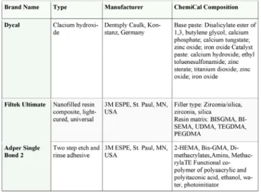
Apart from conventional resin composites, nanofill and/or nanohybrid adhesive restora-tive materials represent the state of the art in terms of filler ingredient and have a sim-ilar or slightly better performance.6 In addition to the ad-vantages of nano fill and/or nanohy-brid resin composites such as strength, low wear, and polishability these mate-rials general-ly has different shades, allowing the natural dental tissue reproduction in an efficient way, with the stratification technique.1 Nanofill resin composites are composed of both nanomer and nanoclusters, where-as nanohybrid is a hybrid resin composite with nanofill-er in a prepolymnanofill-erized fillnanofill-er (PPF) form.7 Filtek Ultimate Universal Restorative (3M ESPE, St.Paul, MN, USA) is a nanofill resin composite largely used in the daily practice. The material contains Bis-GMA, urethane dimethacrylate (UDMA), triethylene glycol di-methacrylate (TEGDMA), and Bis-EMA resins. The inorganic filler loading is about 78.5 % by weight (63.3 % by volume). The including fillers are a combination of silica, zirco-nia and aggregated zir-conia/silica cluster fillers. The dentin, enamel, and body shades that have been used in the study have an average cluster particle size of 0,6–10ų.8
The knowledge that is necessary for the decision of re-storative material by clinician is reserved from the results of in vitro and in vivo literature. Even though in vitro evalu-ations have the advantage of high reproducibility, in vivo literature is necessary for the final evaluation of a restor-ative material due to factors which can not be simulated in vitro conditions.9 In daily practice, an evidence-based approach and clinical observations are becoming more expected of dentists. Clinical evaluations of direct adhe-sive restorations showed good wear resistance and ad-aptation to tooth tissues; however they also have a high replacement rate due to seconder caries and low color stability.10 Although there were various clinical evalua-tions of direct posterior composite restoraevalua-tions,11-13 reli-able data about the clinical performance of direct ante-rior restorations is rare. Thus the aim of this study was to evaluate the clinical performance of a nano hybrid resin composite (Fil-tek Ultimate) in Class III and IV cavities.
MATERIALS AND METHODS
The brands, manufacturers and chemical composition of the materials used in this study are listed in Table 1.
Study Design
Patients who have at least two anterior teeth with de-cay and met the inclusion criteria were included in this
study which was performed between January-2013 and October-2014. Participants recruited for this study were referred from the surrounding local gen-eral practices. Before entering the trial, all patients were provided with informed consent form approved by the ethical commit-tee of the university institutional review board (10840098-54). Information was given to each patient regarding the alternative treatment options.
Table 1: The brand, type, manufacturer, and chemical composition of the main materials used in this study
Patient inclusion/exclusion criteria
The inclusion and exclusion criteria were as follows: Inclusion Criteria:
1. Good general health.
2. Having at least two Class III and/or Class IV carious le-sions or existing defective res-torations, including proxi-mal surfaces in permanent maxillary anterior teeth, which were asymptomatic.
Exclusion Criteria:
1. Absence of adjacent and antagonist teeth.
2. Severe periodontal diseases and poor oral hygiene. 3. Symptoms of pulpitis, such as spontaneous pain or sensitivity to pressure.
Tooth preparation
One operator with experience in adhesive dentistry, more than 12 years since graduation, placed a total of 107 res-torations in 34 patients (18 female, 16 male; mean age 35.4±13.5 years old, range: 18-57 years old) with the help of a dental assistant. The teeth were cleaned and the col-or was determined by the a custom composite sample col-or Vita Shade Guide (VITA Zahnfabrik, Germany). After the shade selection preparations were cut un-der local anes-thesia if it was necessary. The cavity design (restricted to the elimination of carious tissue or defective restorations) was prepared using diamond and stainless steel burs (Diatech, Heerbrugg, Switzerland) and all buccal enamel cavosurface margins were beveled. All preparations were performed as adhesive-only cavities. The pulp tissue was
23
7tepeklinik
protected with calcium hydroxide (Dycal, Dentsply De-trey; Konstanz, Germany) in deeper cavities when when the remaining dentin on the cavity floor was close to the pulp. Glass ionomer liner (Glass-Liner, Willmann&Pein Gmbh, Hamburg, Germany) was ap-plied to cover the calcium hydroxide; therefore, most of the dentin surface was left ex-posed for adhesion.
Restorative procedure
The teeth were restored esthetically with a nano hybrid resin composite using etch-and rinse technique. Isolation was achieved with cotton rolls and suction instead of rub-ber dam and retraction cords (Ultrapak, Ultradent, USA) were used to minimize crevicular fluid flow. To ensure optimal adhesive bonding, a two-step etch and rinse ad-hesive sys-tem (Adper Single Bond 2, 3M ESPE, St.Paul, MN, USA), which is included the etching procedure with phosphoric acid (3M ESPE, St.Paul, MN, USA) was used. Enamel surfaces were conditioned for 30 seconds while dentin surfaces were conditioned for 15 seconds with phosphoric etching gel. After etching, the cavities were water rinsed thoroughly for 30 s with water, and after-wards clorhexidine antibacterial solution (Con-sepsis, Ultradent South Jordan, UT, USA) to remove the acidic agent and the teeth were dried by gently air blowing. Adhesive was applied with a disposable brush and tooth sur-faces were scrubbed with brushing motion for 20 s. This application was repeated second-ly with a new drop of adhesive with 20 s waiting period in between the coats. The surfac-es were dried with gentle air blowing and then medium at least 5 s and light cured for 20s (Gui-lin Woodpecker Medical Instrument Co., Ltd, China) ac-cording to the manufactur-er’s instructions.
The nanofill resin composite (Filtek Ultimate) was used for the composite build-ups due to its good handling property and shade matching. The resin composite was placed and polymerized using incremental layering tech-nique as dentin, body and enamel composite to simulate natural tooth color and translucency. Restorations were light-cured for 40 sec-onds each from facial and lingual directions. Particular attention was given to the con-tour-ing of the apical finish line of the restorations. After po-lymerization checking the oc-clusion, finishing and pol-ishing of the restorations took place with finpol-ishing burs, polish-ing discs Opti Disc (Kerr Corporation, CA, USA) and rubbers HiLuster PLUS Polishing System (Kerr Cor-poration, CA, USA).
Evaluation
Two calibrated observers different from the operator evaluated the restorations at base-line, at 6 months, and at final recall. For maximum validity, both examiners were cali-brated by using the recommended web- based
train-ing and calibration tool.14 Restorations were evaluated, according to the modified United States Public Health Service (USPHS) criteria or FDI criteria.15,16 According to these criteria, clinically excellent restorations were scored as 1, clinically good restorations as 2, clinically sufficient restorations as 3, and clinically unsatisfactory and clini-cally poor restorations as 4 and 5, respectively. The res-torations were inspected visually with a dental mirror and probe. Caries, chipping, debonding, fractures, and severe discoloration were considered as absolute fail-ures. Pa-tients were instructed to call the researchers in the event of any kind of failure. In case of an unfavorable event, such as chipping of direct adhesive restorative ma-terials, the date of the event was recorded. If it was impos-sible to identify the exact date of a defect, or the defect was detected only during the recall observation, the date of the unfavorable event was accepted as the date when the dentist observed the damage. All data were recorded by typing directly into an anonymous database.
When a complete restoration was lost or was impossible to repair, or the restarted tooth required root canal treat-ment, the restoration was deemed a failure. When less damaging events occurred, such as minor composite fractures, chipping fractures, marginal gaps, caries, or color or surface deterioration, the restorations were re-paired, polished, and the type of unfavorable event was documented in the patients’ record. These cases were de-fined as survival cases. Restorations having no failure or unfavorable event were classi-fied as a success.
Statistical analysis
Survival analyses were performed with statistical soft-ware program (SPSS 21,0; SPSS Inc, Chicago, IL, USA) using Kaplan-Meier test to obtain the survival rates in re-lation to observation time. P values less than 0,05 were considered to be statistically significant in all tests. To compare changes in the quality parameters between ob-servations, the marginal homogeneity test was used.
Results
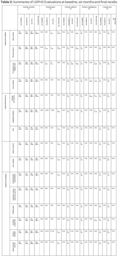
Results of the clinical evaluations of the 107 restorations at baseline, 6 months, 1 year and final recalls are shown in Table 2. In total, no drop-out was experienced yielding to the evaluation of 107 direct anterior restorations. The mean observation time was 22.4 ± 7 months with a min-imum observation period of 9.63 months and maxmin-imum 32.10 months. Of these 107 direct restorations 3 resto-rations were located in the mandible and 104 in the max-illa and 48 restorations were Class III and 79 restorations were Class IV.
Most of the restorations were scored as clinically excel-lent at baseline and six-month re-calls. Secondary car-ies and endodontic complications were not detected in
24
7tepeklinik
any of the teeth. At the 1-year recalls, 106 restorations (99.06%) restorations were classified as suc-cessful.
Table 2: Summaries of USPHS Evaluations at baseline, six months and final recalls.
One restoration (0.9%) was retreated due to the chipping at the margins and rec-orded as failure. One (0.9%) res-torations showed unfavorable event (chip fracture not affecting the marginal integrity) was repolished and ac-cepted as survival case. During the follow-up periods, 92 (86.8%) restorations were accepted as clinically accept-able; 18 (16.9%) were repaired and they were accepted as survival cases. Fourteen (13.2%) resto-rations were classified as failures, due to color mismatches, chipping and fractures and were retreated and recorded as failure (Table 3). The data for survival without failure and
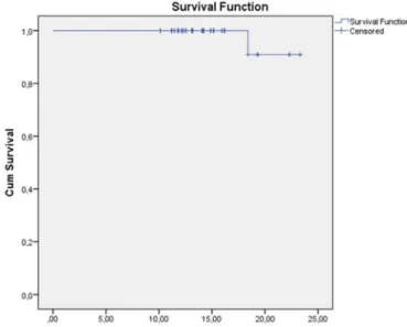
unfa-vorable events are shown in a Kaplan-Meier plot in Figure 1 and 2, with a sur-vival rate, after 1 year of 99.1 and at up to 32 months, of 86.8. The mean survival time (± standard error) was 30.1±0.6 months [95%C.I.=(28.97-31.23)].
Table 3: The distribution of failures according to the evaluated USPHS/FDI criteria.
The clinical evaluations of the 107 restorations at base-line, 6 months, and final recalls are shown in Table 2. The statistically significant differences were observed for the criteria of color stability at six month (p=0,016). Seven restorations showed minor deviations in translucency and scored as clinically good. At one year recall except the failed restoration three restorations showed deterio-ration in color; two of them were highly translucent while the other was more darker (p=0.012). At final recalls the statistically significant differences were observed for the criteria of surface luster (p<0,001), surface staining (p<0.001), color stability and translucency (p=0,010), fracture and retention (p=0.008) and marginal adaptation (p<0,001). For the criteria of surface luster 14 restorations were observed as slightly dull. However they were not noticeable from speaking distance. Mi-nor marginal and surface staining were observed at 15 restorations and
25
7tepeklinik
they were re-moved by repolishing. These restorations were accepted as survival. Three restorations were too darker that they were corrected by repair and accepted as failure. Eight restora-tions were scored as clinically unacceptable for the criteria of “fracture of material and
retention”; six of them showed chipping that damages
marginal quality while bulk frac-tures were observed at two restorations. Larger irregularities with the necessi-ty of repair were observed at the other two restorations. These restorations were also accepted as fail-ure.
Figure 1: Kaplan-Meier plot with the survival rate showed the survival without fail-ure and unfavorable event after 1 year.
Figure 2: Kaplan-Meier plot with the survival rate showed the survival without fail-ure and unfavorable event after up to 32 months.
DISCUSSION
Resin composites have gained popularity as restorative materials due to their esthetic and adhesive properties since their introduction to dentistry5 and currently, ac-cepted as the first choice to restore both anterior and posterior teeth.17 There are several journals observ-ing the clinical behavior of posterior resin composite resto-rations in the literature. These journals usually revealed that posterior resin restorations might present low annual
failure rates and long-lasting survival.12,18 with failure rea-sons such as secondary caries and frac-ture.18,19 Despite the extended usage of resin composites in anterior resto-rations, there is a lack of evidence from clinical observa-tions regarding the performance of anterior restora-observa-tions especially in long term.
In the present investigation, a two-step etch and rinse adhesive system was used to ensure optimal adhesive bonding after the etching of enamel surfaces for 30 sec-onds with phos-phoric etching gel. The marginal quality of the restorations were observed generally suc-cessful at final recalls due to the etch and rinse adhesive proce-dure applied in the study. Some studies reported that etch&rinse adhesives or additional etching exhibited higher percentages of gap free margins in enamel after thermo-mechanical loading when com-pared to two-step self-etch adhesives.20 Ermis et al.21 also mentioned in their clinical study that additional etching of the enamel mar-gins improved the marginal quality of res-torations bond-ed with this adhesive system.
The most obvious reason for failures in anterior resto-rations are directly or indirectly relat-ed to the esthetic ap-pearance of a tooth or restoration, while secondary caries is seldom the reason for replacement and endodontic complications are limited. Anterior restora-tions gener-ally behave differently from posterior restorations. The restoration loss of ante-rior region is more present than in posterior teeth and esthetic appearance plays an obvi-ous role in the desire of the patient to have a restoration replaced.22 In our study the patients are satisfied with the esthetically appearance of their final restorations. Fail-ures at final recalls were generally related with the loss of retention or fracture or bad marginal quality. Only 3 resto-rations were retreated due to the criteria of color stability and translucency. The perception of aesthetics itself may vary among individuals according to their age, educa-tional level, and environment to which they are exposed. The higher patient satis-faction scores in our clinical ob-servations can be related with these aspects.
The available literature is still insufficient to determine the longevity of direct anterior adhesive restorations. Many variables, like the size, location and type of restoration23 and the patient’s dentition24 affect the longevity of com-posite restorations. In anterior teeth, Class IV restorations involving the incisal edge are subjected to high mastica-tory loads, with fracture as a possible clinical outcome over time.17 The lack of mechanical retention in most Class IV restorations may lead to a greater challenge to the tooth-restoration bonded interface.25 van Dijken and Pallesen26 found a higher prevalence of failure in Class IV restorations performed with different materials in bruxers, indicating that over-loading in individuals with occlusal disturbances may increase the mechanical stresses in
26
7tepeklinik
the restorations, making them more prone to fracture. Lucarotti et al.27 reported that in-volvement of the incisal angle in incisors and canine teeth resulted in an associ-ated reduc-tion in median survival time. In our clinical observation we performed 48 Class III and 79 Class IV restorations. This repair protocol can be accepted as suf-ficiently flexible to allow for improving the shape, surface luster, or marginal staining even after long clinical peri-ods.
At final recalls 8 restorations were retreated due to the chipping, fracture and loss of res-torations and all of them were Class IV restorations. These scores can be related with the high masticatory loads that the restorations were exposed. It seems that failure behavior in anterior resto-rations is different from posterior teeth, with less second-ary caries pre-sent.22 No secondary caries was observed for any of the restorations.
Spinas28 reported that, despite repairs of restorations, in-terventions like repolishing were alternative procedures that could be repeated several times as necessary. The survival rate in our study provides evidence that minimal repair procedures, such as repolishing, can be accepted as an important factor in prolonging the clinical success of direct adhesive resto-rations. Daily clinical experiences show that minor unfavorable events are generally easy to repair.16 Reuses et al.29 observed microfilled and hy-brid anterior restorations and ob-served higher marginal discoloration for the microfilled resin composite. They accepted that hybrid resin composites performed well as an anterior restorative material. Microfill restorations were polishable but were weak because of their relative-ly low filler content. Thus, since then, the particle size of the conventional composites has been reduced through further grinding to produce more advanced restorative materials. Microhybrid resin composites were generally accepted as universal composites, with these materials used for most anterior and posterior regions. When com-bined, these materials improved strength and polishabil-ity characteristics.5 Narhi et al.30 evaluated the clinical behavior of microhybrid anterior restorations after 1 year and found the performance of the restora-tions to be clin-ically acceptable, similar with the clinical observations of Peumans et al.31,32
One of the biggest innovations could be the develop-ment of nanofill and nanohybrid res-in composites. Nano-hybrid properties, such as flexure strength and modulus, tend to be similar to those of microhybrids; however, they can be considered, as a group, to be in the lower range of the microhybrids, with both being superior to previ-ous microfills.5 Similar to the preceding resin composites, nanofill anterior restorations showed acceptable clinical behavior in our in vivo evaluations after up to 32 months.
CONCLUSIONS
Within the limitations of the current design and recall pe-riod, the following could be con-cluded:
1. Direct Class III and IV restorations made of nanofilled
resin composite presented 86.8% survival.
2. Repair and maintance protocols were practiced to
pro-long the restoration lifetime al-ready after 6 months to im-prove marginal adaptation, surface staining and lusture and morphology which deteriorated in some cases up to clinical service time of 32 months.
Acknowledgement
The authors acknowledge dental assistants Miss B. Yildiz from Istanbul Medipol University, School of Dentistry, De-partment of Restorative Dentistry Clinics, for organizing the recalls of the patients.
Conflict of interest
The authors did not have any commercial interest in any of the materials used in this study.
REFERENCES
1. de Carvalho LD, Machado RG, Lopes GC, de Andrada
MC. Nanofilled composite restorations with different ad-hesives strategies: clinical cases. Case Rep Dent 2012: 969627.
2. Loguercio AD, Lorini E, Weiss RV, Tori AP, Picinatto CC,
Ribeiro NR, Reis A A 12-month clinical evaluation of com-posite resins in class III restorations. J Adhes Dent 2007; 9: 57-64.
3. Pontons-Melo JC, Furuse AY, Mondelli J A direct
com-posite resin stratification tech-nique for restoration of the smile. Quintessence Int 2011; 42: 205-211.
4. van der Veen HJ, Pilon HF, Henry PP Clinical
perfor-mance of one microfilled and two hybrid anterior com-posite resins. Quintessence Int 1989; 20: 547-550.
5. Ferracane JL Resin composite--state of the art. Dental
Mater 2011; 27: 29-38.
6. Can Say E, Yurdaguven H, Yaman BC, Ozer F. Surface
roughness and morphology of resin composites polished with two-step polishing systems. Dent Materials J 2014; 33: 332-342.
7. Senawongse P, Pongprueksa P.J Surface roughness of
nanofill and nanohybrid resin composites after polishing and brushing. J Esthet Restor Dent 2007; 19: 265-273.
8. Prodan DA, Gasparik C, Mada DC, Miclăuş V, Băciuţ M,
Dudea D Influence of opacity on the color stability of a nanocomposite Clin Oral Invest 2015; 19: 867-875.
9. Geitel B, Kwiatkowski R, Zimmer S, Barthel CR, Roulet
JF, Jahn KR Clinically con-trolled study on the quality of class III, IV and V composite restorations after two years. J Adhes Dent 2004; 6: 247-253.
10. van Dijken JW Longevity of new hybrid restorative
27
7tepeklinik
materials in class III cavities. Eur J Oral Sci 1999; 107: 215-219.
11. Manhart J, Chen HY, Hickel R Clinical evaluation of
the posterior composite Quixfil in class I and II cavities: 4-year follow-up of a randomized controlled trial. J Adhes Dent 2010; 12: 237-243.
12. Da Rosa Rodolpho PA, Donassollo TA, Cenci MS,
Loguercio AD, Moraes RR, Bronkhorst EM, Opdam NJ, Demarco FF 22-Year clinical evaluation of the perfor-mance of two posterior composites with different filler characteristics. Dent Mater 2011; 27:955-963.
13. Lempel E, Toth A, Fabian T, Krajczar K, Szalma J
Retro-spective evaluation of poste-rior direct composite resto-rations: 10-Year findings. Dent Mater 2015; 31: 115-122.
14. Hickel R, Peschke A, Tyas M, Mjör I, Bayne S, Peters
M, Hiller KA, Randall R, Vanherle G, Heintze SD FDI World Dental Federation – clinical criteria for the evalua-tion of direct and indirect restorations. Update and clinical ex-amples. J Adhes Dent 2010; 12: 259-272.
15. Hickel R, Roulet JF, Bayne S, Heintze SD, Mjor IA,
Pe-ters M, Rousson V, Randall R, Schmalz G, Tyas M, Vanher-le G Recommendations for conducting controlVanher-led clinical studies of dental restorative materials. Int Dent J 2007; 57: 300-302.
16. Frese C, Schiller P, Staehle HJ, Wolff D Recontouring
teeth and closing diastemas with direct composite build-ups: a 5-year follow-up. J Dent 2013; 41:979-985.
17. Baldissera RA, Correa MB, Schuch HS, Collares K,
Na-scimento GG, Jardim PS, Moraes RR, Opdam NJ, Demar-co FF Are there universal restorative Demar-composites for an-te-rior and posterior teeth? J Dent 2013; 41:1027-1035.
18. Opdam NJ, van de Sande FH, Bronkhorst E, Cenci MS,
Bottenberg P, Pallesen U, Gaengler P, Lindberg A, Huys-mans MC, van Dijken JW. Longevity of posterior compos-ite restorations: a systematic review and meta-analysis. J Dent Res 2014; 93: 943-949.
19. Demarco FF, Correa MB, Cenci MS, Moraes RR,
Op-dam NJ Longevity of posterior composite restorations: not only a matter of materials. Dent Mater 2012; 28: 87-101.
20. Frankenberger R, Tay FR Self-etch vs etch-and-rinse
adhesives: effect of thermo-mechanical fatigue loading on marginal quality of bonded resin composite resto-rations. Dental Mater 2005; 21: 397-412.
21. Ermis RB, Temel UB, Cellik EU, Kam O Clinical
perfor-mance of a two-step self-etch adhesive with additional enamel etching in Class III cavities. Oper Dent 2010; 35: 147-155.
22. Demarco FF, Collares K, Coelho-de-Souza FH, Correa
MB, Cenci MS, Moraes RR, Opdam NJ. Anterior compos-ite restorations: A systematic review on long-term survival and reasons for failure. Dent Mater 2015; 31: 1214-1224.
23. Qvist V, Strom C 11-year assessment of Class-III resin
restorations completed with two restorative procedures. Acta Odontol Scand 1993; 51: 253-262.
24. Hunter B Survival of dental restorations in young
pa-tients. Community Dent Oral Epidemiol 1985; 13: 285-287.
25. Heintze SD, Rousson V, Hickel R Clinical
effective-ness of direct anterior restora-tions-a meta-analysis. Dent Mater 2015; 31: 481-495.
26. van Dijken JW, Pallesen U Fracture frequency and
longevity of fractured resin com-posite, polyacid-mod-ified resin composite, and resin-modpolyacid-mod-ified glass ionomer cement class IV restorations: an up to 14 years of fol-low-up. Clin Oral Invest 2010; 14: 217-222.
27. Lucarotti PS, Holder RL, Burke FJ Outcome of direct
restorations placed within the general dental services in England and Wales (Part 1): variation by type of resto-ration and re-intervention. J Dent 2005; 33: 805-815.
28. Spinas E Longevity of composite restorations of
trau-matically injured teeth. Am J Dent 2004; 17: 407-411.
29. Reusens, B, D’Hoore W & Vreven J In vivo comparison
of a microfilled and a hybrid minifilled composite resin in Class III restorations: 2-year follow-up Clin Oral Invest 1999; 3: 62-69.
30. Närhi TO, Tanner J, Ostela I, Narva K, Nohrström T,
Tirri T & Vallittu PK Anterior Z250 resin composite resto-rations: one-year evaluation of clinical performance. Clin Oral Invest 2003; 7: 241-243.
31. Peumans M, Van Meerbeek B, Lambrechts P &
Van-herle G The 5-year clinical per-formance of direct com-posite additions to correct tooth form and position. I. Es-thetic qualities. Clin Oral Invest 1997; 1: 12-18.
32. Peumans M, Van Meerbeek B, Lambrechts P &
Van-herle G The 5-year clinical per-formance of direct com-posite additions to correct tooth form and position. II. Marginal qualities. Clin Oral Invest 1997; 1: 19-26.