© 2015 by the Texas Heart ® Institute, Houston Volume 42, Number 4, 2015 400 http://dx.doi.org/10.14503/THIJ-14-4381
Chondromyxoid Fibroma
of the Sternum in a
63-Year-Old Woman
Primary chondromyxoid fibroma is a benign bone tumor. Its localization in the sternum is quite rare; we found only 6 relevant reports. We report our diagnosis and treatment of a chondromyxoid fibroma in the sternum of a 63-year-old woman. The patient underwent subtotal sternectomy and chest-wall reconstruction with use of a titanium rib bridge system and Prolene mesh. The patient’s clinical course was uneventful, and she had no local recur-rence 41 months postoperatively. Our review herein of the 6 previous cases reveals that our patient is the oldest thus far to have been diagnosed with a sternal chondromyxoid fibroma. (Tex Heart Inst J 2015;42(4):400-2)
C
hondromyxoid fibroma (CMF) is a benign bone tumor that constitutes 0.5% to 1% of primary bone neoplasms.1 It typically occurs in the metaphyses oflong bones such as the tibia; its localization in the sternum is extremely rare. We present a case of primary CMF of the sternum, and we review the few previously reported cases.
Case Report
In November 2010, a 63-year-old-woman was admitted to our hospital with chest pain and a 10-year history of swelling of the sternum. She had an irregular, palpable mass (diameter, 10 cm) in the upper two thirds of the sternum (Fig. 1). A radio-graph revealed a bone tumor in the sternum. A computed tomogram (CT) showed an osteolytic lesion with discrete calcific deposits in the sternal bone marrow (Fig. 2). Positron emission tomography (PET) of the mass with 18F-fluorodeoxyglucose
(18F-FDG) contrast medium revealed a maximal standardized uptake value (SUV
max) of 5.7. Upon histologic analysis, a biopsy specimen obtained with use of an ultrasonographically guided Tru-Cut® needle (CareFusion Corporation; Waukegan,
Ill) yielded a benign tumor. The tumor had a slightly lobular architecture and was
Case
Reports
Dalokay Kilic, MD Alper Findikcioglu, MD Merih Tepeoglu, MD Cigdem Vural, MDKey words: Bone
neo- plasms/diagnosis/sur-gery; chest pain/etiology; chondroma/radiography/ surgery; fibroma/diagnosis; fluorodeoxyglucose F18/ diagnostic use; prostheses and implants; reconstructive surgical procedures/meth-ods; sternum/pathology/ surgery; thoracic neoplasms/ diagnosis/surgery; treatment outcome
From: Departments of
Tho-racic Surgery (Drs. Findikcio-glu and Kilic) and Pathology (Drs. Tepeoglu and Vural), Baskent University Faculty of Medicine, 06490 Ankara, Turkey
Address for reprints:
Dalokay Kilic, MD, Depart-ment of Thoracic Surgery, Baskent University Faculty of Medicine, Ankara Teach-ing and Medical Research Center, 5 Sokak No:48, Kat 0, Bahcelievler, 06490 Ankara, Turkey
E-mail:
Fig. 1 Intraoperative photograph
Texas Heart Institute Journal Chondromyxoid Fibroma of the Sternum 401
composed of a myxoid matrix populated by stellate and spindle cells, with moderate nuclear enlargement and eosinophilic cytoplasmic processes (Fig. 3). The diag-nosis was CMF.
We performed a subtotal sternectomy and resected the mass with a clear surgical margin. Rigid sternal re-construction was achieved with use of the Stratos
Strasbourg Thoracic Osteosyntheses System (MedX-pert GmbH; Eschbach, Germany). After the ribs were prepared laterally to the margin of resection, the rib clips and connecting bar of the Stratos were bent to
proper shape and fitted to reconstruct the chest defect. The patient’s clinical course was uneventful, and she had no local recurrence as of March 2014.
Fig. 2 Computed tomogram shows an osteolytic lesion with
discrete calcification in the bone marrow of the upper sternum.
Fig. 3 Photomicrograph shows moderate nuclear enlargement
and prominent eosinophilic cytoplasmic processes (H & E, orig. ×200).
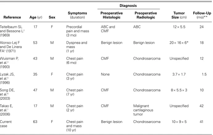
TABLE I. Reports of Chondromyxoid Fibroma of the Sternum
Diagnosis
Symptoms Preoperative Preoperative Tumor Follow-Up
Reference Age (yr) Sex (duration) Histologic Radiologic Size (cm) (mo)**
Teitelbaum SL 17 F Precordial ABC and ABC 12 × 5.5 24
and Bessone L2 pain and mass CMF
(1969) (3 mo)
Alonso-Lej F 53 M Dyspnea and Benign lesion Benign lesion 20 × 16 × 6* 18
and De Linera mass
FA3 (1971) (1 yr)
Wuisman P, 43 M Chest pain CMF Chondrosarcoma Unspecified 12
et al.4 (6 mo)
(1993)
Lyzak JS, 35 F Chest pain None Chondrosarcoma 3.7 × 1.7 1.5
et al.5 (3 yr)
(1996)
Song DE, 47 M Chest pain CMF Chondrosarcoma 8 × 5.5 × 3 10
et al.6 (7 yr)
(2003)
Takao E, 17 M Chest pain CMF Malignant Unspecified 42
et al.7 (2 yr) cartilaginous
(2006) tumor
Current 63 F Chest pain Benign lesion Chondrosarcoma 10 × 9 × 5 41
case and mass
(10 yr)
ABC = aneurysmal bone cyst; CMF = chondromyxoid fibroma; F = female; M = male *Tumor (size, 24 × 18 × 14 cm) had spread to the mediastinum.
Volume 42, Number 4, 2015 402 Chondromyxoid Fibroma of the Sternum
Discussion
To our knowledge, this is the 7th case of primary sternal CMF to be reported in the English-language medical literature (Table I).2-7 The patients’ ages have ranged
from 17 to our patient’s 63 years.
Clinically, the predominant symptom is chest pain, as in our patient. Radiologically, CMF most often pre-sents as a sharply demarcated, lytic lesion with scalloped margins.8 Some authors whose patients had a primary
sternal CMF suspected a chondrosarcoma or malignant cartilaginous tumor preoperatively (Table I) because of cortical destruction and expansion shown on CT.4-7 Our
patient’s CT showed an osteolytic lesion with discrete calcification in the bone marrow of the upper sternum. Low-grade chondrosarcoma, like CMF, can have a pre-dilection for low 18F-FDG uptake, so the differential
diagnosis of primary bone malignancies can be con-founding. Factors that might contribute to the lower avidity include a high proportion of acellular gelatinous matrix with respect to cellular density, and lower mi-totic rates than those in higher-grade tumors.9 In our
patient’s mass, FDG uptake evaluated by means of PET had an SUV max of 5.7. Because of the radiologic find-ings, we performed a biopsy in order to narrow the dif-ferential diagnosis.
Primary sternal tumors include chondroma, chondro-sarcoma, and osteochondroma. Particular caution is required to differentiate chondrosarcoma from CMF. Because of the presence of pleomorphic cells with hy-perchromatic nuclei, the histologic diagnosis of CMF might be difficult and result in an erroneous diagno-sis of chondrosarcoma.2 The preoperative histologic
specimens were evaluated correctly in some previous cases.2,4,6,7 In our patient, the preoperative histologic
di-agnosis was a benign lesion.
En bloc resection with clear margins is satisfactory treatment for CMF. As in our patient, no recurrence has been reported (Table I).
Chest-wall reconstruction with use of rigid material is necessary in cases of wall resections larger than 5 cm. In our patient, we used the Stratos, which has
rigid material composed of moldable titanium bars and rib clips. Gonfiotti and colleagues10 used a Stratos
for sternal reconstruction after subtotal sternectomy for primary tumors in one patient. We have used this graft in 5 subsequent sternectomy patients and prefer it when large resections of the chest wall include the entire sternum, cartilage, and medial ends of the clavicle. All our patients thus treated were extubated in the early postoperative period, and paradoxical respiration was not observed during a mean follow-up period of 28 ± 9 months (range, 18–41 mo). In addition, we have used Prolene mesh, to avoid lung herniation.
In our experience, the Stratos adapts progressively
and well to each required sternal shape, because of
the flexibility of the titanium bars and the absence of dead parietal spaces. We think that the Stratos plus
Prolene mesh should be considered in the reconstruc-tion of large chest-wall defects (including the sternum), because it results in minimal morbidity, is easy to use, prevents paradoxical respiration, and provides a good cosmetic appearance, as in our patient with CMF.
References
1. Campanacci M. Chondromyxoid fibroma. In: Bone and soft tissue tumors: clinical features, imaging, pathology and treat-ment. 2nd ed. Padova (Italy): Piccin Nuova Libraria S.p.A.; 1999. p. 265-77.
2. Teitelbaum SL, Bessone L. Resection of a large chondromyx-oid fibroma of the sternum: report of the first case and review of the literature. J Thorac Cardiovasc Surg 1969;57(3):333-40.
3. Alonso-Lej F, De Linera FA. Resection of the entire sternum and replacement with acrylic resin. Report of a case of giant chondromyxoid fibroma. J Thorac Cardiovasc Surg 1971;62 (2):271-80.
4. Wuisman P, Scheld H, Tjan T, Roessner A, Blasius S, Vestring T, Winkelmann W. Chondromyxoid fibroma of the sternum. Case report. Arch Orthop Trauma Surg 1993;112(5):255-6. 5. Lyzak JS, Gurley J, Boyle C, Dixon L, Olak J.
Chondromyx-oid fibroma of the sternum. Skeletal Radiol 1996;25(5):489-92.
6. Song DE, Khang SK, Cho KJ, Kim DK. Chondromyxoid fibroma of the sternum. Ann Thorac Surg 2003;75(6):1948-50.
7. Takao E, Morioka H, Yabe H, Anazawa U, Morii T, Horinou-chi H, et al. Chondromyxoid fibroma of the sternum. J Tho-rac Cardiovasc Surg 2006;132(2):430-1.
8. Mirra JM. Bone tumors: clinical, radiologic, and pathologic correlations. Vol 1. Philadelphia: Lea & Febiger; 1989. p. 623-48.
9. Makis W, Ciarallo A, Lisbona R. Chondromyxoid fibroma of the rib mimics a chondrosarcoma on 18F-FDG PET/CT. Acta Radiol 2011;52(5):554-6.
10. Gonfiotti A, Santini PF, Campanacci D, Innocenti M, Fer-rarello S, Caldarella A, Janni A. Malignant primary chest-wall tumours: techniques of reconstruction and survival. Eur J Cardiothorac Surg 2010;38(1):39-45.