ORIGINAL ARTICLE
Vanishing white matter disease with different faces
Gülay Güngör1&Olcay Güngör2 &Seda Çakmaklı3&Hülya Maraş Genç4
&Hülyaİnce5&Gözde Yeşil6
&Cengiz Dilber7& Kürşad Aydın8
Received: 15 June 2019 / Accepted: 28 July 2019
# Springer-Verlag GmbH Germany, part of Springer Nature 2019 Abstract
Purpose The goal of this study was to better understand vanishing white matter (VWM) disease, which is one of the most common hereditary white matter disorders, and its relationship to radiologic features, genetic analyses, and clinical findings. Methods We performed a study on 11 patients to describe the clinical and neuroimaging features of VWM. Patients were grouped into“infantile,” “early childhood,” and “juvenile” based on their onset age. EIF2B1–5 genes encoding five subunits of eukaryotic translation initiation factor 2B (eIF2B) were analyzed in all patients with clinically suspected VWM disease. Results In brain magnetic resonance imaging (MRI), all patients showed white matter abnormalities with various degrees. The initial clinical presentation in five of patients was ataxia, with severe refractory epilepsy in three patients. In children with infantile-onset VWM, a rapid deterioration of motor function was detected, and the frequency of epilepsy was higher. Two patients showed manifestations of end-stage VWM disease, and one of them had chronic subdural hematoma. One of our patients and his father were diagnosed with Brugada syndrome. Sequencing of the exons and exon-intron boundaries of the EIF2B1–5 genes revealed mutations in the genes EIF2B5 (5 cases), EIF2B3 (3 cases), and EIF2B4 (2 cases). We also found a novel mutation in one patient: c.323_325delGAA in the EIF2B1 gene.
Conclusions In this study, in addition to classical clinical and radiological findings, we wanted to emphasize that we may be confronted with refractory epilepsy (early infancy), cardiac problems, and intracranial complications that may occur in advanced stages.
Keywords Brugada syndrome . EIF2B gene . Epilepsy . MRI . Vanishing white matter (VWM)
Introduction
VWM disease is one of the most common hereditary white matter disorders. It is also known as childhood ataxia accom-panied by central hypomyelination [1]. The disease typically manifests itself by progressive neurological deterioration caused by certain triggers like infections and trauma in a completely healthy child [2,3]. VWM disease may be seen in all age groups, including antenatal, infantile, early child-hood, juvenile, and adult, and shows a wide phenotypic vari-ation [4,5]. Early childhood type is the most common type of VWM. As far as we know, more than 250 cases and 150 mutations of EIF2B1–5 have been reported in the literature [6,7]. MRI shows significant widespread abnormalities on T1 and T2 in almost all the cerebral white matter. It is also ac-companied by progressive cystic degeneration and a rarefac-tion leading to complete disappearance of white matter. There are genetic studies showing that there is a relationship between molecular mutations in five genes (eIF2B1–5) encoding the five subunits of eukaryotic translation initiation factor eIF2B * Olcay Güngör
1
Department of Radiology, Faculty of Medicine, Pamukkale University, Denizli, Turkey
2
Department of Pediatric Neurology, Faculty of Medicine, Pamukkale University, 20100 Denizli, Turkey
3 Department of Medical Genetics, Necip Fazıl City Hospital,
Kahramanmaras, Turkey
4
Department of Pediatric Neurology, Ümraniye Training and Research Hospital,İstanbul, Turkey
5 Department of Pediatric Neurology, Faculty of Medicine, Medical
Park University, Samsun, Turkey
6
Department of Medical Genetics, Faculty of Medicine, Bezmialem Vakıf University, İstanbul, Turkey
7
Department of Pediatric Neurology, Faculty of Medicine, Medipol University,İstanbul, Turkey
8 Department of Pediatric Neurology, Marash Life Hospital,
Kahramanmaraş, Turkey
(eIF2Ba-ε) with VWM disease [3]. In our study, we aimed to stress that children with VWM may present with resistant epilepsy, different intracranial pathologies or cardiac arrhyth-mias. Therefore, we wanted to emphasize the importance of the contribution of MR imaging to the diagnostic process and adding electrocardiographic imaging to patients with suspected cardiac pathology.
Materials and methods
VWM patients were taken from the pediatric neurology de-partment between 2013 and 2018. We have retrospectively studied all cases, and VWM was diagnosed on the basis of clinical findings, neuroimaging, and genetics tests. We cate-gorized our patients as infantile (onset before 2 years of age), early childhood VWM (onset age 2–6 years), and juvenile (onset later than 6 years of age) types [3,8,9]. Eleven patients were followed for 1–5 years. During that period, clinical signs, episodic attacks (progression of neurological deterioration), radiological images, and genetic analysis were examined. Patients’ follow-ups were performed via telephone calls, home visits, and routine outpatient clinic visits.
Patients’ progression of motor deterioration, seizures, and episodic aggravation (chronic progressive or rapid) were assessed. All triggering conditions, such as infections and mi-nor head trauma, have been reported. The evaluation of motor function was classified according to the Gross Motor Function Classification System (GMFCS) which was leveled between I and V [10].
Genotype analysis
Next generation sequencing analysis (Miseq-İllumina Inc.) of the coding exons and exon-intron boundaries of the EIF2B1–5 genes were performed in all of our patients except for patient 5. We screened EIF2B5 gene firstly based on the high muta-tion rate reported by previous studies. When no mutamuta-tions were found in the EIF2B5 gene, we continued to analyze the remaining four genes (EIF2B1–4). Patient 5 was diagnosed through next generation sequencing analysis using TruSight One Panel (Illumina Inc., San Diego, California).
Analysis of brain MRI
All brain MRI examinations were performed with 1.5T. MRI protocols included axial T1-weighted image, axial/sagittal T2-weighted image, and axial T2 fluid-attenuated inversion re-covery (FLAIR) weighted image. Additionally, some patients were examined with coronal T2 FLAIR, axial diffusion-weighted imaging (DWI), and apparent diffusion coefficient (ADC). Having a hypointense appearance on T1-weighted images and hyperintense on T2-weighted images was defined
as white matter involvement. Rarefaction was described as hypointensity on both T1WI and T2 FLAIR images. Diffusion limitation was indicated by high signals in DWI.
Results
A total of 11 patients (5 female and 6 male) with VWM dis-ease were included in the study. Age of symptom onset was 35.18 ± 19.13 months, and the current mean age was 75.66 ± 28.92 months. Of all patients, 36.3% (4/11) had infantile-onset disease and 63.6% (7/11) had early childhood-onset disease. Case numbers 1 and 2 were maternal first cousins, and cases 7 and 8 were paternal first cousins. Case numbers 1, 4, and 8 had a history of parental consanguineous marriage. The elderly brother of a case with infantile onset had died at the age of 7 months due to an unknown cause. The siblings of the other cases were alive and healthy. The follow-up duration was 32.73 ± 16.42 months. Five of our cases presented with gait disturbance, two with convulsions, two with neuromotor de-lay, one with speech disturbance, and one with dystonia. Seven (63.6%) out of 11 patients had a progressive chronic course while 36.3% (4/11) showed an acute and rapidly pro-gressive course. Rapid or propro-gressive motor deterioration was more common with the infantile (75%, 3/4) type than the early childhood (14.2%, 1/7) type. Children having a chronic pro-gressive course suffered 1 to 5 attacks of deterioration (mean 1.13 episodes). Episodic attacks were caused by an upper respiratory tract infection at least once in every patient, pneu-monia was the culprit in 3 children, urinary tract infection in 2 children, and mild head trauma in 2 children. Among the surviving children, 27.2% (3/11) could independently walk (GMFCSI-II) while (27.2%, 3/11) children lost their ability to walk (GMFCSIV-V). Patient 5 and patient 6 died at the age of 2.5 and 3 respectively. Neuromotor delay preceding disease onset was more common in infantile patients than the early childhood cases. Two of our patients with infantile onset presented with convulsions which were controlled by a triple antiepileptic drug regimen. Other patients with infantile onset started having convulsions 2 months after they had been diagnosed, and their convulsions were controlled by a dual antiepileptic drug regimen. Two of our patients with early childhood type had convulsions which started after the second year of follow-up. Mainly, they were tonic or myoclonic seizures. Antiepileptics used were valproate, topiramate, levetiracetam, and lamotrigine. Other rare signs that became manifest in some of our cases were optic atrophy and macrocephaly. Gene analysis revealed eIF2B5 (5 cases), eIF2B3 (3 cases), eIF2B4 (2 cases), and eIF2B1 (1 case) mutations (Table 1). A mutation (in the SCN5A gene) that is consistent with Brugada syndrome was found in patient 5.
Discussion
Our study demonstrated a higher infantile-onset epilepsy rate compared with childhood-onset epilepsy. Moreover, it was not an expected finding that some of our infantile-onset epilepsy patients had convulsions as the initial symptom and they were diagnosed with VWM disease while etiologies for epilepsy syndromes were investigated. Epileptic seizures are common in patients with VWM, and in 30% of infancy and childhood cases, epileptic seizures occur 1–2 years after the onset of the disease [9]. Our infantile patients presenting with convulsions were brought under control by triple antiepileptic therapy (re-sistant epilepsy). Such seizures are easily controlled by med-ications. Zhou et al. [11] reported an epilepsy rate of 37.5%, Turón-Viñas et al. [12] 42.8%, and Zhang et al. [8] reported an epilepsy rate of 50%. In all those studies, time to onset of epilepsy was at least 1–2 years after diagnosis; control of seizures with antiepileptic drugs is generally successful.
VWM disease is a genetic disorder with autosomal reces-sive transmission [12]. One of our patients had an elderly brother that died during the investigation of neuromotor de-velopment delay before the diagnosis has been established. As the prevalence of consanguineous marriage is high in our re-gion, the risk of having genetic disorders is equally high. Three of our cases had a family history of consanguineous marriage.
In VWM patients, developmental delay and motor deteri-oration are inevitable consequences in the long-term
follow-up. Fogli et al. [4,13] demonstrated by a Kaplan-Meier scale that 95% of infantile patients and 76% of early childhood patients would develop severe disability within a 10-year fol-low-up period. In a study led by Zhang et al. [8], 21.7% of the subjects walked independently at 3-year follow-up, which de-creased to 9.1% at 5 years and zero at 8-year follow-up. Among our cases with 1–5 years of follow-up, none of the ones with infantile onset could gain the ability to walk. Three of 7 patients with childhood onset lost their ability to walk. Our follow-up times were shorter; had they been longer, we could have detected loss of ability to walk at higher rates.
In Cree leukoencephalopathy, which is a phenotypic vari-ant of VWM disease that manifests at the age of 3–9 months and has a rapidly fatal course, 100% of patients die by the age of 2 years [14,15]. A study led by Zhang et al. [8], 79% of patients with infantile onset died at 1.2 years and 14% of patients with early onset died at 2.8 years. Half of our patients with infantile-onset disease died at a 1.7-year follow-up. In those two patients (patients 5 and 6), the disease had no rap-idly progressive course and both died after the age of 2.
Every episodic attack causes neuromotor impairment to worsen; however, the incidence of such an attack frequency is unclear. There is usually a history of infection or trauma at the time of symptom onset [12]; all our patients had a similar history. Labauge [16] reported that episodic attacks emerged in 38% of adult VWM patients. Turón-Viñas et al. [12] report-ed that a chronic progressive course with episodic attacks was observed in 16 of 21 patients while 5 patients had a rapidly Table 1 Characteristics of
EIF2B1–5 mutations in 11 patients
No Gene cDNA Protein Type Exons Mutation IDRS (https://www.ncbi.nlm. nih.gov/SNP/) 1 EIF2B5 943C>G R315G M Exon 7 Reported rs113994063 2 EIF2B5 943C>G R315G M Exon 7 Reported rs113994063 3 EIF2B5 943C>G R315G M Exon 7 Reported rs113994063 4 EIF2B5 943C>G R315G M Exon 7 Reported rs113994063 5 EIF2B4 1151G>A R384Q M Exon
10
Reported rs113994034 6 EIF2B4 1091G>A R384Q M Exon
11
Reported rs113994034 7 EIF2B3 674G>A R225Q M Exon
7
Reported rs113994024 8 EIF2B3 674G>A R225Q M Exon
7
Reported rs113994024 9 EIF2B3 674G>A R225Q M Exon
7 Reported rs113994024 10 EIF2B1 323_ 325delG-AA 108delR ID Exon 4 Novel No rs
11 EIF2B1 806G>A R269Q M Exon 6
Reported rs113994057
progressive course. Zhang et al. [8] reported that episodic attacks occurred in 90% of patients with infantile disease and 71.4% of early childhood disease. This study demonstrat-ed that episodic aggravations were more common in infantile and early childhood onset VWM. However, it is still unclear why this disease can lead to an acute or chronic course.
Schiffmann and van der Knaap [3,17] defined some MRI-based radiological criteria for VWM diagnosis. There is a homogenous T2-weighted and FLAIR hyperintense signal and T1-weighted hypointense signal in cerebral white matter. This neuroimaging finding remains constant throughout the disease course. Nevertheless, there may be signs of cystic degeneration showing CSF-like signal intensity that causes progressive rarefaction and complete loss of nearly whole ce-rebral white matter [4,12,18] (Fig. 1a–j). Moreover, other criteria include relative preservation of temporal lobes and the absence of cysts in cerebellar white matter and parenchy-mal contrast enhancement [3]. In early childhood brain, MRI is strongly recommended for clinical diagnosis of VWM [3, 19].
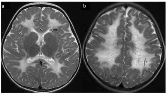
Other imaging patterns may be seen especially in the newborn and early infantile forms as well as in the early stages of other forms. Selective involvement in the inner edge of the corpus callosum may be a clue in the early stages of the disease (Fig. 2a). Additionally, symmetrical central tegmental tract hyperintensity is a prominent sign at disease onset but it lacks specificity [19]. In contrast, MRI features in infantile patients are not so informative for diagnosis. In the early disease stages, white matter
“may not be vanishing” and repeat radiological studies may be done [20].
The tigroid appearance of the white matter, which is caused by the protection of perivascular nerve fibers, can also be seen in VWM, metachromatic leukodystrophy and other disorders [21] (Fig.2b). Furthermore, neither cranial nor spinal nerves play a role in differential diagnosis of imaging studies for VWM.
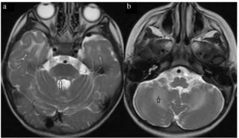
Brain MRI is characterized by extensive and symmetrical involvement of white matter. Also, internal capsules, subcor-tical fibers, and outer part of the corpus callosum are spared [3, 11,19]. FLAIR images show markedly hypointense areas in deep white matter regions probably corresponding to cavita-tions [22] (Fig.3a, b). A band-like pattern may be found in rarified and cystic white matter. Brain stem and cerebellar white matter can also be involved, but cystic degeneration has not been reported [23] (Fig.4a, b). Additionally, variable cerebellar atrophy primarily affecting the vermis and involve-ment of pontine teginvolve-mentum tracts are prominent [20].
Dissociation of the U fibers may be present to a variable degree and is better seen in T1-weighted images. Even at the end stage, when almost all white matter becomes cystic, it is noteworthy that atrophy, if present, is usually of mild degree. These abnormalities of white matter are unique and they are completely different from other neuropsychiatric disorders in which the white matter abnormalities on brain and cerebellum are seen diffuse and homogeneous [12].
Proton MR spectroscopy is normal at the onset, but it is typical that it gradually features a CSF-like spectrum [12].
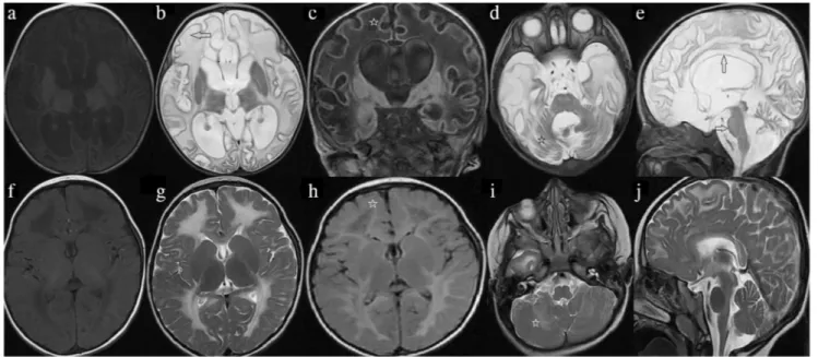
Fig. 1 Brain MRIs of infantile (patient 6, a–e) and early childhood (patient 10, f–j). VWM patients images were taken at 8 months of age and were taken at 4 years of age, respectively. Axial T1-weighted and T2-weighted images show a more extensive white matter involvement (arrow) in the infantile patient (a, b, f, g). Also, the infantile patient shows early subcortical rarefaction (stars) in the coronal and axial T2
FLAIR images (c, h). Cerebellar white matter regions (stars) were related in infantile and early childhood cases in the axial T2-weighted images (d, i). Brain stem was only involved (white arrow) in the infantile patient axial and sagittal T2-weighted image (d, e, i, j). Additionally, there is marked atrophy of the corpus callosum (black arrow) in the infantile patient on the sagittal T2-weighted image (e, j)
DWI properties in VWM have been rarely described [22,24]. Several studies have suggested that diffusion limitation is seen in regions with higher cell density [25] or that this effect is secondary to cytotoxicity or myelin edema [26]. We are of the opinion that it is difficult to comment on to identify if the effect is transient because of the limited existence of follow-up brain MRI studies in our patients (Fig.5). We presume that hyperintensities in DWI may possibly occur in parallel to acute aggravation episodes under stress.
While carrying out clinical or MRI follow-up of late-stage patients, one should also be careful about possible intracranial complications that may occur. One of our infantile-onset cases developed a subdural hematoma (Fig.6). As in this example, since MRI findings are not very specific in the end stages of the disease, it is very difficult to diagnose VWM at this stage. Leukoencephalopathy with VWM (OMIM # 603896) is a rare autosomal recessive transmitted disorder caused by mu-tations in any of the five genes: EIF2B1, EIF2B2, EIF2B3, EIF2B4, and EIF2B5. These genes are localized to chromo-somes 12q24.3, 14q24, 1p34.20, 2p23.3, and 3q27. EIF2B1–
5 genes encode five subunits of eukaryotic factor 2B (eIF2B): EIF2Bα, EIF2Bβ, EIF2Bγ, EIF2Bδ, and EIF2Bε. eIF2B as-sumes a task in the initiation of mRNA translation. Under stress conditions, protein synthesis is inhibited via eIF2B in-activation [27].
EIF2B5 is the most frequently mutated gene; mutations in EIF2B5 are detected in 57% of the patients. Mutation rates of the other genes are EIF2B4 (17%), EIF2B2 (15%), EIF2B3 (7%), and EIF2B1 (4%) respectively [12]. In this study, we had five patients with mutations in EIF2B5 (45.4%), two pa-tients with mutations in EIF2B4 (18.1%), three papa-tients with mutations in EIF2B3 (27.2%), and one patient with a mutation in EIF2B1 (9.1%). We did not find any mutation in the EIF2B2 gene. In our cohort, the most prevalent mutation is in the EIF2B5 gene which is compatible with previous reports although our sample size is too small to reach any conclusions.
Fogli et al. [13] investigated 93 patients with VWM dis-ease, 64% of the patients had mutations affecting the EIF2B5 gene. They observed that the most frequent mutation in Fig. 2 Axial T2-weighted images
of patient 1and patient 4. a There is hyperintensity on the inner rim of the corpus callosum (arrow). b In another patient, there are radially oriented stripes called tigroid pattern within abnormally hyperintense deep cerebral white matter (arrow)
Fig. 3 Axial FLAIR images of different patients (9 and 3) with VWM. a There are confluent parietal subcortical areas of hyperintensity (star) with periventricular cystic
degeneration (arrow) (patient 9). b In another patient, an axial FLAIR image shows parietal subcortical cystic formation (arrow) that occurred in advanced stage (patient 3)
EIF2B5 was the R113H substitution [13]. In this study, we had five patients with mutations in EIF2B5 (45.4%); four of them were carrying R315G mutation; one patient had R269Q mutation. Two of the patients were siblings (patients 1 and 2), and two of them were distantly related individuals (patients 3 and 4). We did not find R113H mutation in any of our patients. A novel homozygous mutation in the EIF2B1 gene was detected in patient 10: c.323_325delGAA (p.108delR). Confirmation and segregation analysis of parents performed by Sanger sequencing revealed that both parents were hetero-zygous. The deletion in the EIF2B1 gene was neither found in Exome Aggregation Consortium nor 1000 Genomes
databases. The prediction score in PROVEAN (http:// provean.jcvi.org/index.php) [28] was found to be“− 12.00” (deleterious). According to Human Splicing Finder (http:// www.umd.be/HSF/) [29], HSF score was“− 39.68” showing potential alteration of splicing. Mutation taster (http://www. mutationtaster.org/) [30] prediction was disease-causing. The affected nucleotide seemed to be conserved among species with a phyloP score of 3.058 and phastCons score of 0.992.
Most of the mutations reported in VWM are point mutations (88%), followed by deletions and insertions (10%). Splice site mutations are found in only 2% of the cases [31]. All of the mutations we detected in our patients were missense except for Fig. 5 DWI findings of an
infantile patient 11. There are initial MRIs at 12 months of age (a–f). On the initial DWI images (a–c), there are hyperintensities in the central and subcortical white matter, internal capsule, corpus callosum, and medulla (arrows). On the ADC images (d–f), hypointensities were seen in the same regions (arrows)
Fig. 4 Axial T2-weighted images of the patient 2. a There are bilateral pontine tegmental hyperintense lesions (arrow). b There is symmetrical T2 hyperintensity in the deep white matter of the cerebellum (star)
the novel in frame deletion in the EIF2B1 gene. Both homozy-gous and compound heterozyhomozy-gous mutations were stated in the literature; the inheritance pattern of the disease is autosomal recessive [6]. In our cohort of patients, all of the mutations in the EIF2B1–5 genes were in the homozygous state.
Patient 5 was diagnosed through next generation sequenc-ing analysis ussequenc-ing TruSight One Panel (Illumina Inc.). We found c.1151G>A mutation in the EIF2B4 gene and a hetero-zygote c.2893C>G mutation (p. R965G) in the SCN5A gene. This mutation was reported before in Brugada syndrome [32]. The patient’s father had also a heterozygous c.2893C>G mu-tation in the SCN5A gene, but the mother’s mumu-tation analysis was normal. We detected this mutation incidentally in our patient and her father. We referred the family to the cardiology
department for further examination. While his father’s electro-cardiography (ECG) findings were consistent with Brugada syndrome, our patient’s ECG findings were normal (Fig. 7). They did not have any cardiac symptoms. This syndrome should be closely monitored clinically because it is an inherited disease with an increased risk of sudden cardiac death [33]. To our knowledge, this is the first report of a patient with VWM disease and Brugada syndrome.
Limitations
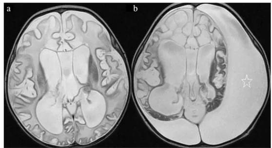
1- The number of infantile-onset patients was low, and we lacked juvenile-onset patients. Therefore, the compari-sons between the disease groups became inadequate. Fig. 6 Axial T2-weighted
images(a, b) of the patient 6. The first brain MRI at the age of 8 months shows cystic degeneration (cerebrospinal fluid–like signal intensity) that causes progressive rarefaction and complete loss of nearly whole cerebral white matter. The image obtained at the age of 15 months exhibits that subdural hematoma (star) developed in both hemispheres, with the left one being more diffuse
Fig. 7 Father’s
electrocardiography of the patient 5 with Brugada syndrome. There is a ST elevation (concave) in the V1, V2, and V3
2- As the disease portends a worse prognosis and lacks ther-apy, follow-up appointments were not regular. Therefore, motor function and cognitive process assessments could not be performed in detail.
3- With an increased number of patients, it would be wise to group patients on the basis of age at symptom onset. As more cases are gathered, we will conduct more detailed analyses and more comprehensive follow-ups.
Conclusion
VWM disease shows a quite wide range of phenotypic varia-tions. It affects all age groups. Especially, the contribution of MR imaging to the diagnosis process in early childhood is more valuable than infantile patients. We suggest that our study is noteworthy of remembering by documenting that the initial man-ifestation was convulsion followed by resistant epilepsy and by detecting the Brugada syndrome in a family for the first time. Author contributions GG, OG, and SÇ were involved in the design of this study. OG, Hİ, CD, KA, and HMG contributed to the analysis of the data. GG, OG, and SÇ contributed to the writing the manuscript. All authors critically reviewed and approved the final manuscript.
Compliance with ethical standards
Conflict of interest The authors declare that they have no conflict of interest.
Ethical approval This article does not contain any studies with human participants performed by any of the authors.
Informed consent For this type of study, formal consent is not required.
References
1. Bugiani M, Boor I, Powers JM, Scheper GC, van der Knaap MS (2010) Leukoencephalopathy with vanishing white matter: a re-view. J Neuropathol Exp Neurol 69(10):987–996
2. van der Knaap MS, Fogli A, Boespflug-Tanguy O, Abbink TEM, Schiffmann R (1993-2019) Childhood ataxia with central nervous sys-tem hypomyelination / vanishing white matter. In: Adam MP, Ardinger HH, Pagon RA, Wallace SE, Bean LJH, Stephens K, Amemiya A (eds) GeneReviews®. University of Washington, Seattle, WA 3. van der Knaap MS, Pronk JC, Scheper GC (2006) Vanishing white
matter disease. Lancet Neurol 5(5):413–423.https://doi.org/10. 1016/S1474-4422(06)70440-9
4. Fogli A, Dionisi-Vici C, Deodato F, Bartuli A, Boespflug-Tanguy O, Bertini E (2002) A severe variant of childhood ataxia with cen-tral hypomyelination/vanishing white matter leukoencephalopathy related to EIF21B5 mutation. Neurology 59(12):1966–1968 5. van der Knaap MS, van Berkel CG, Herms J, van Coster R,
Baethmann M, Naidu S, Boltshauser E, Willemsen MA, Plecko B, Hoffmann GF, Proud CG, Scheper GC, Pronk JC (2003) eIF2B-related disorders: antenatal onset and involvement of
multiple organs. Am J Hum Genet 73(5):1199–1207.https://doi. org/10.1086/379524
6. Maletkovic J, Schiffmann R, Gorospe JR, Gordon ES, Mintz M, Hoffman EP, Alper G, Lynch DR, Singhal BS, Harding C, Amartino H, Brown CM, Chan A, Renaud D, Geraghty M, Jensen L, Senbil N, Kadom N, Nazarian J, Yuanjian F, Zuyi W, Hartka T, Morizono H, Vanderver A (2008) Genetic and clinical heterogeneity in eIF2B-related disorder. J Child Neurol 23(2):205– 215.https://doi.org/10.1177/0883073807308705
7. Proud CG (2012) Vanishing white matter: the next 10 years. Future Neurol 7(1):81–92
8. Zhang H, Dai L, Chen N, Zang L, Leng X, Du L, Wang J, Jiang Y, Zhang F, Wu X, Wu Y (2015) Fifteen novel EIF2B1-5 mutations identified in Chinese children with leukoencephalopathy with vanishing white matter and a long term follow-up. PLoS One 10(3):e0118001.https://doi.org/10.1371/journal.pone.0118001
9. van der Knaap MS, Barth PG, Gabreels FJ, Franzoni E, Begeer JH, Stroink H, Rotteveel JJ, Valk J (1997) A new leukoencephalopathy with vanishing white matter. Neurology 48(4):845–855
10. Gunel MK, Mutlu A, Tarsuslu T, Livanelioglu A (2009) Relationship among the Manual Ability Classification System (MACS), the Gross Motor Function Classification System (GMFCS), and the functional status (WeeFIM) in children with spastic cerebral palsy. Eur J Pediatr 168(4):477–485.https://doi. org/10.1007/s00431-008-0775-1
11. Zhou L, Zhang H, Chen N, Zhang Z, Liu M, Dai L, Wang J, Jiang Y, Wu Y (2018) Similarities and differences between infantile and early childhood onset vanishing white matter disease. J Neurol 265(6):1410–1418.https://doi.org/10.1007/s00415-018-8851-6
12. Turon-Vinas E, Pineda M, Cusi V, Lopez-Laso E, Del Pozo RL, Gutierrez-Solana LG, Moreno DC, Sierra-Corcoles C, Olabarrieta-Hoyos N, Madruga-Garrido M, Aguirre-Rodriguez J, Gonzalez-Alvarez V, O’Callaghan M, Muchart J, Armstrong-Moron J (2014) Vanishing white matter disease in a spanish population. J Cent Nerv Syst Dis 6:59–68.https://doi.org/10.4137/JCNSD. S13540
13. Fogli A, Schiffmann R, Bertini E, Ughetto S, Combes P, Eymard-Pierre E, Kaneski CR, Pineda M, Troncoso M, Uziel G, Surtees R, Pugin D, Chaunu MP, Rodriguez D, Boespflug-Tanguy O (2004) The effect of genotype on the natural history of eIF2B-related leu-kodystrophies. Neurology 62(9):1509–1517
14. Alorainy IA, Patenaude YG, O’Gorman AM, Black DN, Meagher-Villemure K (1999) Cree leukoencephalopathy: neuroimaging find-ings. Radiology 213(2):400–406
15. Fogli A, Wong K, Eymard-Pierre E, Wenger J, Bouffard JP, Goldin E, Black DN, Boespflug-Tanguy O, Schiffmann R (2002) Cree leukoencephalopathy and CACH/VWM disease are allelic at the EIF2B5 locus. Ann Neurol 52(4):506–510.https://doi.org/10. 1002/ana.10339
16. Labauge P, Horzinski L, Ayrignac X, Blanc P, Vukusic S, Rodriguez D, Mauguiere F, Peter L, Goizet C, Bouhour F, Denier C, Confavreux C, Obadia M, Blanc F, de Seze J, Fogli A, Boespflug-Tanguy O (2009) Natural history of adult-onset eIF2B-related disorders: a multi-centric survey of 16 cases. Brain 132(Pt 8):2161–2169.https://doi.org/10.1093/brain/awp171
17. Schiffmann R, van der Knaap MS (2009) Invited article: an MRI-based approach to the diagnosis of white matter disorders. Neurology 72(8):750–759.https://doi.org/10.1212/01.wnl. 0000343049.00540.c8
18. Tedeschi G, Schiffmann R, Barton NW, Shih HH, Gospe SM Jr, Brady RO, Alger JR, Di Chiro G (1995) Proton magnetic resonance spectroscopic imaging in childhood ataxia with diffuse central ner-vous system hypomyelination. Neurology 45(8):1526–1532 19. van der Lei HD, Steenweg ME, Barkhof F, de Grauw T, d’Hooghe
M, Morton R, Shah S, Wolf N, van der Knaap MS (2012) Characteristics of early MRI in children and adolescents with
vanishing white matter. Neuropediatrics 43(1):22–26.https://doi. org/10.1055/s-0032-1307456
20. Wilson CJ, Pronk JC, Van der Knaap MS (2005) Vanishing white matter disease in a child presenting with ataxia. J Paediatr Child Health 41(1–2):65–67.https://doi.org/10.1111/j.1440-1754.2005. 00540.x
21. Singh RR, Livingston J, Lim M, Berry IR, Siddiqui A (2017) An unusual neuroimaging finding and response to immunotherapy in a child with genetically confirmed vanishing white matter disease. Eur J Paediatr Neurol 21(2):410–413.https://doi.org/10.1016/j. ejpn.2016.08.012
22. Patay Z (2005) Diffusion-weighted MR imaging in leukodystro-phies. Eur Radiol 15(11):2284–2303.https://doi.org/10.1007/ s00330-005-2846-2
23. Barros SR, Parreira SCR, Miranda AFB, Pereira AMB, Campos NMP (2018) New insights in vanishing white matter disease: iso-lated bilateral optic neuropathy in adult onset disease. J Neuroophthalmol 38(1):42–46. https://doi.org/10.1097/WNO. 0000000000000565
24. Ding XQ, Bley A, Ohlenbusch A, Kohlschutter A, Fiehler J, Zhu W, Lanfermann H (2012) Imaging evidence of early brain tissue degeneration in patients with vanishing white matter disease: a multimodal MR study. J Magn Reson Imaging 35(4):926–932.
https://doi.org/10.1002/jmri.23517
25. van der Lei HD, Steenweg ME, Bugiani M, Pouwels PJ, Vent IM, Barkhof F, van Wieringen WN, van der Knaap MS (2012) Restricted diffusion in vanishing white matter. Arch Neurol 69(6): 723–727.https://doi.org/10.1001/archneurol.2011.1658
26. Van Haren K, van der Voorn JP, Peterson DR, van der Knaap MS, Powers JM (2004) The life and death of oligodendrocytes in
vanishing white matter disease. J Neuropathol Exp Neurol 63(6): 618–630.https://doi.org/10.1093/jnen/63.6.618
27. Pronk JC, van Kollenburg B, Scheper GC, van der Knaap MS (2006) Vanishing white matter disease: a review with focus on its genetics. Ment Retard Dev Disabil Res Rev 12(2):123–128.https:// doi.org/10.1002/mrdd.20104
28. Venter JC (2019)http://provean.jcvi.org/index.php. J Creig Venter. Accessed 14/5/2019
29. LEVY N (2019) http://www.umd.be/HSF/. Aix Marseille Universite. Accessed 14/5/2019
30. Schwarz JM, Cooper DN, Schuelke M, Seelow D (2014) MutationTaster2: mutation prediction for the deep-sequencing age. Nat Methods 11(4):361–362.https://doi.org/10.1038/nmeth. 2890
31. Scali O, Di Perri C, Federico A (2006) The spectrum of mutations for the diagnosis of vanishing white matter disease. Neurol Sci 27(4):271–277.https://doi.org/10.1007/s10072-006-0683-y
32. Priori SG, Napolitano C, Gasparini M, Pappone C, Della Bella P, Brignole M, Giordano U, Giovannini T, Menozzi C, Bloise R, Crotti L, Terreni L, Schwartz PJ (2000) Clinical and genetic hetero-geneity of right bundle branch block and ST-segment elevation syndrome: a prospective evaluation of 52 families. Circulation 102(20):2509–2515
33. Iglesias DG, Rubín J, Pérez D, Morís C, Calvo D (2019) Insights for stratification of risk in Brugada syndrome. Eur Cardiol 14(1): 45–49.https://doi.org/10.15420/ecr.2018.31.2
Publisher’s note Springer Nature remains neutral with regard to jurisdic-tional claims in published maps and institujurisdic-tional affiliations.