Contents lists available atScienceDirect
Carbohydrate Polymers
journal homepage:www.elsevier.com/locate/carbpol
One-step green synthesis of antibacterial silver nanoparticles embedded in
electrospun cyclodextrin nano
fibers
Asli Celebioglu, Fuat Topuz, Zehra Irem Yildiz, Tamer Uyar
⁎Institute of Materials Science & Nanotechnology, UNAM-National Nanotechnology Research Center, Bilkent University, 06800 Ankara, Turkey
A R T I C L E I N F O Keywords: Cyclodextrin Nanofibers Electrospinning Silver nanoparticles Green synthesis Antibacterial A B S T R A C T
Antibacterial electrospun nanofibers based on cyclodextrin (CD) and silver nanoparticles (Ag-NPs) were pro-duced by solution electrospinning from aqueous and DMF solutions using different Ag contents. CD molecules acted as the reducing agent and catalyzed the formation of Ag-NPs. The nanofibers with smaller diameters were observed for thefibers generated from DMF solutions than those produced from aqueous solutions. TEM and STEM analyses revealed the Ag-NPs (∼2–5 nm depending on solvent-type and Ag loading) in nanofibers, while FTIR and surface enhanced Raman scattering (SERS) analyses showed the apparent frequency shift of OH stretching band and the enhancement of Raman bands of CD molecules with the incorporation of the Ag-NPs. The polycrystalline structure of the Ag-NPs was shown by XRD and SAED analyses over {111}, {200}, {220} and {311} planes. The nanofibers showed significant inhibition against the growth of Escherichia coli and Staphylococcus aureus owing to the antibacterial activity of the Ag-NPs.
1. Introduction
Antibacterial materials have taken considerable interest in targeting pathogenic bacteria in daily life and protecting human health (Li, Chen, Zhao, & Urmila, 2015). Particularly, pathogenic bacteria like Escher-ichia coli (E. coli) and Staphylococcus aureus (S. aureus) can lead to life-threatening diseases, e.g., clinical mastitis (Bannerman et al., 2004). Although most types of E. coli are considered as harmless and com-monly found in the lower intestine, some types can lead to various intestinal and extraintestinal illnesses through virulence factors, such as yersiniabactin, aerobactin, flagellin and intimin (Kaper, Nataro, & Mobley, 2004). Particularly, the pathotypes of E. coli can provoke dis-eases like dysentery (Chanter, Hall, Bland, Hayle, & Parsons, 1986) and diarrhoea (Levine, 1987). On the other hand, S. aureus was listed as one of high-priority pathogens by the World Health Organization (WHO). In this regard, various drug molecules (Ruszczak & Friess, 2003), anti-biotics (Williamson, Maroudas, & Wilkie, 1971), nanoparticles (Wang, Hu, & Shao, 2017), quaternary ammonium compounds (Makvandi, Jamaledin, Jabbari, Nikfarjam, & Borzacchiello, 2018), metal ions (Du, Niu, Xu, Xu, & Fan, 2009), a water-soluble antibacterial polysaccharide extracted from dandelion and its derivative (Lin, Zhu, Li et al., 2018), essential oils (thyme (Lin, Zhu, Thangaraj, Abdel-Samie, & Cui, 2018), and tea tree oil (Cui, Bai, & Lin, 2018)), clove oil/chitosan nano-particles (Cui, Bai, Rashed, & Lin, 2018), nisin-loaded nanoparticles
(Cui, Wu, Li, & Lin, 2017), and cinnamon essential oil/beta-cyclodex-trin (β-CD) proteoliposomes (Lin, Dai, & Cui, 2017) were exploited to inhibit bacterial growth and destroy the cellular structure of micro-organisms. Apart from the above biocides, silver nanoparticles (Ag-NPs) have also shown high antibacterial activity, and therefore, were in-corporated into different material systems, including hydrogels (González-Sánchez et al., 2015;Lustosa et al., 2017), nanogels (Qasim, Udomluck, Chang, Park, & Kim, 2018), and coatings (Zhao et al., 2011). They were also incorporated into electrospunfiber systems to generate antibacterial nanofibrous materials. In this regard, various Ag-loaded electrospun fibers based on a polymer and Ag precursors were pre-viously reported (Patel, Li, Wang, Zhang, & Wei, 2007; Rujitanaroj, Pimpha, & Supaphol, 2008;Son, Youk, Lee, & Park, 2004;Xu et al., 2006).
The formation of Ag-NPs through the reduction of Ag(II) can take place in the presence of a reducing agent, such as polyols, or by heat treatment. In this context, Hong et al. reported poly(vinyl alcohol) (PVA)fibers containing Ag-NPs, which were prepared by the electro-spinning of PVA/silver nitrate (AgNO3) and subsequent heat treatment (Hong, Park, Sul, Youk, & Kang, 2006). Zhuang et al. produced Ag-NPs containing chitosan/gelatin nanofibers, where the nanoparticles sizing between 1 and 5 nm were synthesized using a microcrystalline chitosan as the reducing agent (Zhuang, Cheng, Kang, & Xu, 2010). An inter-esting study on the use of solvent as a reducing agent was reported by
https://doi.org/10.1016/j.carbpol.2018.12.008
Received 19 September 2018; Received in revised form 30 November 2018; Accepted 6 December 2018
⁎Corresponding author.
E-mail address:[email protected](T. Uyar).
Carbohydrate Polymers 207 (2019) 471–479
Available online 06 December 2018
0144-8617/ © 2018 Elsevier Ltd. All rights reserved.
Zhang and co-workers (Shi et al., 2011). The authors used formic acid as the reducing agent for AgNO3 and solvent for the dissolution of nylon-6 to obtain electrospunfibers loaded with Ag-NPs sizing between 2 and 4 nm. Li et al. reported the synthesis of Ag-NPs with the help of a chitosan oligosaccharide and their addition to PVA solutions prior to the electrospinning (Li et al., 2013). The Ag-NPs were produced in the size range of 15–22 nm, and the resultant nanofiber mats were used as a bioactive wound dressing material. A similar concept was exploited for the preparation of Ag-NPs loadedfibers using glucose and chitosan as reducing and protective agents (Abdelgawad, Hudson, & Rojas, 2014). The synthesis of the Ag-NPs was performed at 95 °C using AgNO3as a precursor. The Ag-NPs (∼25 nm) were blended with PVA and electro-spun into nanofibers with a mean size of 150 nm. The nanofibers were later treated with glutaraldehyde for cross-linking and tested against E. coli. Destaye et al. described an interesting method for the synthesis of Ag-NPs on the cross-linked PVAfibers (Destaye, Lin, & Lee, 2013). The electrospun PVAfibers were cross-linked with glutaraldehyde and kept in the solution of AgNO3overnight at room temperature. Over time, the color of thefiber mat changed to grayish, suggesting the formation of nanoparticles on thefibers. Even though the presence of several ap-proaches for the preparation of Ag nanoparticles loaded electrospun fiber systems through mostly a multistep process in the presence of additional reducing agents, there is an increasing demand for the pre-paration of such antibacterial fibers through facile and green routes with tailor-made properties. In this regard, the use of CD molecules as a reducing agent allows the synthesis of size-tunable Ag-NPs and stabi-lizes them against precipitation. This enables the homogenous dis-tribution of the formed Ag-NPs in the fiber matrix. Moreover, the mixture could be electrospun into functionalfibers that can be used in different applications, e.g., drug delivery and water treatment, in ad-dition to their antibacterial application.
In this paper, we show a facile and green approach using CD mo-lecules as the building blocks for nanofibers and reducing agent at the same time, while silver nitrate (AgNO3) was used as an Ag precursor. CD is a cyclic oligomer of glucose obtained by enzymatic degradation of starch and has been used in a diverse range of applications due to their host-guest supramolecular complexation ability (Crini, 2014;Sharma & Baldi, 2016). Further, CDs have been used as functional molecules to synthesize environmentally benign nanomaterials (Prochowicz, Kornowicz, & Lewiński, 2017). Likewise, they were exploited for the synthesis of Ag-NPs (de Souza, Barros, Tasic, Gimenez, & Teixeira Camargo, 2018). Under alkaline conditions, CD can catalyze the duction of Ag(II) to metallic Ag(0) without the requirement of any re-ducing agent and lead to the formation of Ag-NPs. In this regard, the use of CD molecules as the reducing agent for the synthesis of noble metal nanoparticles in either acidic or alkaline media was reported (Celebioglu & Uyar, 2013;Devi & Mandal, 2013;Huang, Meng, & Qi, 2009; Martin-Trasanco, Cao, Esparza-Ponce, Montero-Cabrera, & Arratia-Pérez, 2017;Pande et al., 2007). According to Pande et al., the basic mechanism was explained by the deprotonation of hydroxy groups of CD at alkaline solutions, which promoted the synthesis of nanoparticles. Whereas, Devi et al. reported that the particle synthesis takes place when the solution pH is about 10 (Devi and Mandal, 2013). In this study, the nanocomposite CD/Ag-NP electrospun nanofibers were produced from aqueous and DMF solutions at two different Ag loadings (1 and 2 wt.%), and characterized in terms of fiber mor-phology by SEM, and the distribution of the Ag-NPs by TEM and STEM. UV–vis and SERS analyses were carried out to confirm the presence of the Ag-NPs in the nanofibers. The chemical composition of the Ag-NPs was studied by FTIR, chemical state of Ag by XPS, and the crystallinity of the nanoparticles through XRD. The antibacterial activity of the nanofibers was tested against E. coli and S. aureus over 24 h treatment.
2. Experimental section 2.1. Materials
Hydroxypropyl-β-cyclodextrin (HP-β-CD, Cavasol®W7 HP, with a substitution degree between 0.6 and 0.9) was received from Wacker Chemie AG (Germany) as a gift sample. The solubility of the HP-β-CD is ∼2300 g/L at 24 °C according to the producer. Silver nitrate (AgNO3, ≥99.5%, Sigma Aldrich), N, N-dimethylformamide (DMF, Riedel) and sodium hydroxide (NaOH,≥98%, Fluka) were obtained and used as received. Distilled water was produced by a Millipore Milli-Q Ultrapure system.
2.2. Electrospinning of Ag-NPs embedded HP-β-CD nanofibers
First, HP-β-CD was dissolved in DMF (120% (w/v)) or water (160% (w/v)) to produce bead-free nanofibers (Celebioglu & Uyar, 2012). Afterwards, AgNO3(1 and 2 wt.% with respect to elemental Ag over CD content) was added. After the solutions were homogenized, the pH was adjusted to∼9 with 1 M NaOH. Thereafter, the solutions were kept stirring overnight until their color turned to dark-brown, suggesting the formation of Ag-NPs in the HP-β-CD solutions. The solutions were transferred into 3 mL syringes having metallic needles with an inner diameter of 0.60 mm. The syringes were positioned horizontally on a syringe pump (Model: SP 101IZ, WPI) and a high voltage power supply (Matsusada, Precision, AU Series) was used for the electrospinning. During the electrospinning process, the following parameters were used: the applied electrical voltage was 15 kV, the tip-to-collector dis-tance was 15 cm, and theflow rate kept at 0.5 mL/h. The nanofibers were collected on a ground collection plate of aluminium foil at 24 °C. The pure HP-β-CD nanofibers were also produced as a control sample without using Ag precursor from the aqueous and DMF solutions of HP-β-CD at the same concentration (120% (w/v) in DMF and 160% (w/v) in water) for electrospinning.
2.3. Characterization
Scanning electron microscopy (SEM, Quanta 200 FEG, FEI) was used to explore the morphology of the nanofibers. The fiber samples were coated with a thin layer of Au/Pd (∼5 nm) using a Gatan 682 Precision Etching and Coating System (PECS). The mean diameters (< D >) of nanofibers and their size distributions were calculated over ca. 100fibers from SEM images by ImageJ (NIH, Bethesda, MD, USA). Transmission electron microscopy (TEM, FEI-Tecnai G2F30) was used for the observation of the Ag-NPs in the nanofibers. For TEM imaging, the nanocomposite nanofibers were directly collected on TEM grids. The particles were analyzed by a GATAN Digital Micrograph software. X-Ray diffraction experiments were performed using a PANalytical X'Pert Pro MPD, which was powered by a Philips PW3040/60 X-ray generator and fitted with an X'Celerator detector. The X-rays were generated from a Cu anode supplied with 40 kV and a current of 40 mA. The data were collected in the 2θ range of 10-80° using a scanning X’Celerator detector and analyzed by PANalytical High Score Plus software (version 2.0). The X-ray photoelectron spectra of the nanofi-bers were recorded with an X-ray photoelectron spectrometer (XPS, Thermo Scientific). XPS was used by means of a flood gun charge neutralizer system equipped with an Al K-α X-ray source (hυ = 1486.6 eV). For the spectral regions of Ag, high-resolution spectra were recorded at a pass energy of 50 eV. The FTIR spectra were recorded on a Fourier transform infrared (FTIR) instrument (Bruker-VERTEX 70). The samples were mixed with potassium bromide (KBr) and pressed into pellets. A total of 64 scans were collected between 4000 and 400 cm−1at a resolution of 4 cm−1. The UV–vis absorption spectra of the nanofibers were taken with a Varian Cary 5000 UV–vis‐NIR spectrometer. The UV–vis spectra of the nanofibrous mats were obtained at solid and liquid states, and the background was
corrected with the pure HP-β-CD nanofibers. The SERS measurements were carried out on the nanofibers with a WITEC Alpha 300S Raman module. A diode-pumped solid-state laser (532 nm) was used for ex-citation in Raman measurements. The laser power was measured using a silicon photodiode at the sample plane. Power densities were calcu-lated using an apparent spot diameter of the illumination area. 2.4. Antibacterial tests
The antibacterial activity of the electrospun nanofibers generated from aqueous solutions with different Ag loadings (1 and 2 wt.%) was tested against Gram-negative bacteria, Escherichia coli RSHM 888 (RSHM, National Type Culture Collection Laboratory, Ankara, Turkey) and Gram-positive bacteria, Staphylococcus aureus RSHM 96090/07035 (ATCC 25923) using the agar-diffusion method. The bacteria (E. coli and S. aureus) were grown overnight and of which 150μL (∼107cfu/ mL of E. coli and∼109cfu/mL of S. aureus) poured on the Luria-Bertani (LB) agar plates. Afterwards, the circularfiber samples (diameter = 6 mm) were placed on the agar plates and incubated at 37 °C for 24 h. Then, the diameters of the inhibition zone (i.e., transparent area) were measured. All experiments were performed in triplicates.
3. Results and discussion
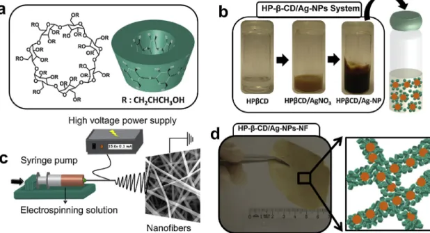
Ag-NPs were synthesized over the reduction of Ag2+to Ag° by the alkaline solutions of CD molecules after overnight mixing (Fig. 1a and b). The formation of Ag-NPs can be visualized by the color change to dark-brown (Fig. 1b) (Kim et al., 2007;Sanghi & Verma, 2009; Vilchis-Nestor et al., 2008). The electrospinning of the colored solution led to a brownishfiber mat (Fig. 1c and d). On the other hand, the color of the fiber mat produced by only HP-β-CD molecules was white (Celebioglu & Uyar, 2012). Thus, this color change can be attributed to the presence of Ag-NPs in the nanofibers.
Fig. 2shows the nanofiber mats generated by the electrospinning of HP-β-CD/Ag-NPs nanocomposite solutions from DMF and aqueous so-lutions at different Ag loadings. The nanofiber mats electrospun from DMF solution have yellowish appearance, while nanofiber mats pro-duced from aqueous solutions are greyish in color. This may be at-tributed to the size of Ag-NPs where bigger nanoparticles were formed
in DMF. Both nanofiber mats could be folded without any structural damage, demonstrating the self-standing andflexible structure of the HP-β-CD/Ag-NPs nanofibers despite their polymer-free structure.
Regardless of the solvent type and Ag content used, all nanofibers
Fig. 1. (a) Chemical and 3D structure of the HP-β-CD. (b) The optical photos of the HP-β-CD solutions during the Ag-NP synthesis. (c) Cartoon illustration of the electrospinning of the CD/Ag-NPs solution. (d) An optical photo of the respective electrospun nanofiber mat and a cartoon scheme of the HP-β-CD/Ag-NPs fiber matrix.
Fig. 2. The optical photos of the HP-β-CD/Ag-NPs nanofiber mats produced at different formulations. The HP-β-CD/Ag-NPs nanofiber mats produced from DMF solutions at two different Ag loadings: (a) 1 and (b) 2 wt.%. The HP-β-CD/ Ag-NPs nanofiber mats electrospun from aqueous solutions at two different Ag loadings: (c) 1 and d) 2 wt.%. Insets show the optical photos of the folded mats.
have bead-free structures as confirmed by SEM analyses (Fig. 3). The nanofibers produced from DMF solutions were smaller in diameter than those generated from aqueous solutions. Whereas, an opposite trend was observed for the Ag-free HP-β-CD fibers: the nanofibers produced from DMF solutions were almost monodisperse and larger than those electrospun from aqueous solutions (Celebioglu & Uyar, 2012). The mean size of the nanofibers produced from DMF solutions increased from ∼184 to ∼280 nm with an increasing Ag loading by two-fold from 1 to 2 wt.%. A similar increase with Ag loading was observed for the nanofibers produced from aqueous solutions: the mean fiber dia-meter increased from∼207 to ∼440 nm with an Ag content rise from 1 to 2 wt.%. The formation of larger nanofibers for the fibers electrospun from aqueous solutions can be attributed to their higher concentrations, i.e., 160 wt.% whereas thefibers electrospun from DMF solutions could be produced at 120 wt.%.
The morphology, size and distribution of the Ag-NPs were explored by electron microscopy.Fig. 4shows the TEM and STEM images of the HP-β-CD/Ag-NPs nanofibers produced at different formulations. The presence of homogeneously dispersed, uniform Ag-NPs throughout the fiber matrix was clearly visible in both TEM and STEM images. With increasing Ag loading, Ag-NPs appeared denser for both solvent sys-tems. Smaller Ag-NPs were observed for the fibers produced from aqueous solutions than those electrospun from DMF solutions. The mean size of the Ag-NPs in thefiber matrix produced from DMF solu-tions was measured as∼3.5 nm for 1 wt.% Ag loading and increased to ∼4.8 nm for 2 wt.% Ag loading (Fig. 4g(i-ii)). Likewise, for the nano-fibers electrospun from aqueous solutions, the mean particle size in-creased from∼1.9 to ∼2.3 nm with an Ag content rise from 1 to 2 wt. %, respectively (Fig. 4g(iii-iv)). The formation of larger Ag-NPs in DMF solutions can be attributed to the reducing role of DMF. In this regard, Pastoriza-Santos et al. reported the formation and stabilization of silver
nanoparticles through the reduction by DMF (Pastoriza-Santos & Liz-Marzán, 1999). The HRTEM images of the Ag-NPs clearly demonstrate the presence of crystal fringes on the nanoparticles (Fig. 4). For both solvent systems, the HRTEM images of the nanoparticles reveal the dominantly exposed plane of {111} of a face-centered cubic (fcc) Ag with the corresponding interplane spacing of 0.235 nm (Huang, Yang, Zhang, & Xiao, 2016).
Table 1summarizes the characteristics of the Ag-NPs embedded HP-β-CD nanofibers produced at different formulations. The electrospin-ning from DMF solutions requires lower HP-β-CD content (∼120 wt.%), whereas the electrospinning from aqueous solutions requires a HP-β-CD concentration of 160 wt.% to form bead-free nanofibers due to the higher viscosity of the respective mixture in DMF, i.e., higher inter-molecular interactions (Celebioglu & Uyar, 2012). The mean size of the nanofibers and Ag-NPs showed variations depending on the solvent type and Ag content used: both increased with increasing Ag content for the nanofibers electrospun from DMF and aqueous solutions.
The presence of the Ag-NPs in the nanofiber matrix was also con-firmed by solid-state UV–vis spectra (Fig. 5). Owing to the characteristic surface plasmonic resonance (SPR) band of metallic Ag-NPs, UV–vis spectra display a broad absorption peak between 300 and 500 nm for the Ag-NPs sizing in the range of 2–5 nm, which is consistent with lit-erature reports (Bhui et al., 2009). As clearly seen inFig. 5a, a strong broad peak was visible in the respective range because of the in situ formed Ag-NPs. The wavelength of the absorption peak is directly re-lated to the particle size: larger nanoparticles induce a bathochromic shift in the spectrum (Maciollek & Ritter, 2014). Therefore, the ab-sorption maximum of the HP-β-CD/Ag-NPs nanofibers from DMF so-lutions shifted to longer wavelengths owing to the formation of larger Ag-NPs. Similar absorption spectra were obtained from the UV–vis spectra of the HP-β-CD/Ag-NPs solutions (Fig. 5b).
Fig. 3. Scanning electron micrographs of the HP-β-CD/Ag-NPs nanofibers. The nanofibers produced from DMF solutions at two different Ag loadings: (a) 1 and (b) 2 wt.%. The nano fi-bers produced from aqueous solutions at two different Ag loadings: (c) 1 and d) 2 wt.%. Insets show the size distributions of the re-spective nanofibers.
FTIR analysis was carried out to explore interactions between HP-β-CD and Ag-NPs in the HP-β-CD/Ag-NPs nanofibers.Fig. 6shows the FTIR spectra of the HP-β-CD/Ag-NPs nanofibers where the OH stretching band of HP-β-CD molecules shifted to lower frequency owing to the interactions between OH groups of CDs and Ag-NPs. The pure HP-β-CD nanofibers revealed a broad peak centered at 3427 cm−1due
to the stretching vibration of OH groups. Upon addition of the Ag-NPs, it shifted to 3419 cm-1for 1 wt.% Ag loading and 3413 cm-1for 2 wt.% Ag loading for the nanofibers produced from DMF solutions. On the other hand, the peak shifted to 3417 and 3411 cm−1for the HP-β-CD/ Ag-NPs nanofibers produced from aqueous solutions with 1 and 2 wt.% Ag loadings, respectively. These shifts suggest the presence of
Fig. 4. TEM and STEM images of the HP-β-CD/Ag-NPs nanofibers produced by different formulations. The nanofibers produced from DMF solutions at two different Ag loadings. (a-i, a-ii) 1 and (b-i, b-ii) 2 wt.%. The nanofibers produced from aqueous solutions at two different Ag loadings. (c-i, c-ii) 1 and (d-i, d-ii) 2 wt.%. TEM and HRTEM images of the Ag-NPs (2 wt.%) embedded in the HP-β-CD nanofibers, which were electrospun from DMF (e-i, e-ii) and aqueous solutions (f-i, f-ii). The size distribution and average particle sizes (APS) of the Ag-NPs. (g-i) HP-β-CD/Ag-NPs-1%-NF (DMF), (g-ii) 2%-NF (DMF), (g-iii) HP-β-CD/Ag-NPs-1%-NF (water) and (g-iv) HP-β-CD/Ag-NPs-2%-NF (water).
Table 1
The averagefiber diameter of the electrospun nanofibers (NF) and the mean size of the Ag-NPs that were produced at different Ag loadings from DMF and aqueous solutions. The values are expressed as mean ± SD.
Sample Solvent HP-β-CD concentration (%, w/v) NP concentration
(%, w/w)
Meanfiber diameter (nm) Average nanoparticle size (nm)
HP-β-CD/AgNPs-1%-NF DMF 120 1 184 ± 45 3.5 ± 1.0
HP-β-CD/AgNPs-2%-NF DMF 120 2 280 ± 85 4.8 ± 1.4
HP-β-CD/AgNPs-1%-NF Water 160 1 207 ± 125 1.9 ± 0.5
interactions between CD and Ag-NPs, which enables the stabilization of the Ag-NPs during the particle growth. Similar shifts were reported for NPs loaded CD nanofibers owing to the interactions between Au-NPs and CD molecules (Celebioglu & Uyar, 2013). Furthermore, a small peak appeared at 1385 cm-1for the HP-β-CD/Ag-NPs nanofibers due to the stretching band of NO3related to AgNO3precursor (Fig. 6a and b). The presence of Ag-NPs in the HP-β-CD/Ag-NPs nanofibers was also confirmed by surface-enhanced Raman scattering (SERS) analysis (Fig. 7). Ag-NPs have been commonly exploited as a SERS active sub-strate (Yang et al., 2015). For SERS measurements, the homogenous distribution of Ag-NPs in the solution or material is highly critical: there are several irresistible factors that can lead to the weakening or dis-appearing of the repulsive forces. However, the use of CD molecules can stabilize them and prevent their aggregation. The nanofibers with 1 wt. % Ag were deformed during the measurements due to excessive heating, and therefore, the SERS measurements were performed on the nanofibers containing 0.1 wt.% Ag-NPs. The Raman bands of the HP-β-CD molecules were weak for pure HP-β-CD nanofibers. Whereas, the presence of 0.1 wt.% Ag-NPs led to drastic changes in the Raman spectrum, where all peaks were clear and sharp. The Raman bands of
the HP-β-CD appeared in following ranges: 800–1500 cm−1 , 2800-3000 cm−1 (CH stretching of hydroxypropyl (HP) group), and 3100–3600 cm−1(OH stretching), and agree well with literature re-ports (Zoppetti, Puppini, Ospitali, & Fini, 2007). Regardless of the solvent-type used in the production of the HP-β-CD/Ag-NPs nanofibers, the intensities of Raman bands of the CD molecules were drastically enhanced because of the presence of homogenous distributed Ag-NPs in thefiber matrices (Fig. 7, insets). It is known that the localized surface plasmon resonance of the nanoparticles can cause strong and highly localizedfields for SERS (Wu, Li, Lin, & Chen, 2017). In this context, the distribution of the Ag-NPs was explored by TEM analysis.Fig. 7(insets) shows the TEM images of the respective nanofiber samples containing 0.1 wt.% Ag. Even though the nanoparticle content was very low, TEM images showed the dark dots related to the formed Ag-NPs without any aggregation: they were homogenously distributed throughout the na-nofiber matrix as observed for the HP-β-CD/Ag-NPs nana-nofibers con-taining 1 or 2 wt.% Ag.
The structural properties of the nanofibers were explored by XRD analysis. Unlike nativeβ-CD (Fig. 8a), HP-β-CD is an amorphous com-pound and gives a broad peak centered at 19° (Fig. 8b). Apart from this
Fig. 5. (a) The solid-state UV–vis spectra of the HP-β-CD/Ag-NPs nanofibers and (b) the liquid-state UV–vis spectra of the HP-β-CD/Ag-NPs solutions.
Fig. 6. FTIR spectra of the pure HP-β-CD nanofibers and HP-β-CD/Ag-NPs nanofibers produced at different formulations. (a-i, a-ii) The nanofibers produced from DMF solutions. (b-i, b-ii) The nanofibers generated from aqueous solutions.
broad peak, many small peaks related to Ag-NPs appeared between 25 and 80°. The diffractions peaks at 38.4°, 44.4°, 64.6° and 77.6° can be attributed to {111}, {200}, {220} and {311} crystalline planes of a fcc
Ag (Celebioglu et al., 2014). Additional diffraction peaks appeared at 2θ of 28°, 32.4°, 46.4° and 57.9° due to the presence of oxidized Ag (Ag2O). The chemical states of Ag in the nanofibers were explored by XPS analysis.Fig. 8c shows the deconvoluted Ag 3d XPS spectra of the samples. Two broad peaks were observed at 368.5 and 374.5 eV related to 3d3/2and 3d5/2orbits and separated by 6 eV (Fig. 8c). These values are consistent with literature reports (Liu et al., 2015; Tzani, Koutsoukos, Koukouzelis, & Detsi, 2017). However, negative shifts were observed at the binding energy for the HP-β-CD/Ag-NPs nanofibers produced from aqueous solutions than those electrospun from DMF solutions due to the higher ratio of Ag2O in the nanofiber sample pro-duced in water (Fig. 8c). Thesefindings were confirmed by XRD ana-lysis with the appearance of peaks related to oxidized Ag (seeFig. 8b). The antibacterial properties of the HP-β-CD/Ag-NPs nanofibers were explored against Gram-negative (E. coli) and Gram-positive (S. aureus) bacteria (Fig. 9). The nanofibers produced in the absence and presence of the Ag-NPs were tested for comparison. The minimize possible errors, the samples were taken at different locations from the same fibrous mat, which was placed on E. coli and S. aureus grown agar plates and visualized after 24 h incubation. Afterwards, the diameter of the in-hibition zones was measured and averaged. No antibacterial activity was observed for pure HP-β-CD nanofibers. On the other hand, the in-corporation of the Ag-NPs into the nanofibers caused antibacterial ac-tivity, and the fibers showed significant inhibition against bacterial growth (Fig. 9). Since pure CD nanofibers do not have any antibacterial activity, the antibacterial activity of the HP-β-CD/Ag-NPs composite nanofibers is directly associated to the presence of the formed Ag-NPs. However, the increase of Ag content by two-fold led to∼10% en-hancement in the antibacterial activity, demonstrating that even 1 wt.% of Ag-NPs is high enough to possess antibacterial activity. The re-spective diameters of inhibition zones for the nanofiber samples con-taining 1 wt.% Ag were found as 1.07 ± 0.038 and 1.16 cm for E. coli and S. aureus, respectively. Similar results were obtained with Ag-NPs embedded electrospun poly(L-lactide)fibers and antibacterial activity, i.e., 98.5% and 94.2% against S. aureus and E. coli, was observed, de-monstrating high antibacterial activity of Ag-NPs (Xu et al., 2006).
4. Conclusion
The nanocomposite nanofibers of HP-β-CD with the in situ formed Ag-NPs were successfully produced by solution electrospinning. CDs are
Fig. 7. Surface-enhanced Raman spectra (SERS) of the HP-β-CD/Ag-NPs and pure HP-β-CD nanofibers produced from (a) aqueous and (b) DMF solutions at the Ag loading of 0.1 wt.%. Insets show the TEM images of the respective HP-β-CD/Ag-NPs nanofibers having 0.1 wt.% Ag loading.
Fig. 8. (a) Wide-angle XRD patterns of the pureβ-CD powder (a) and pure HP-β-CD and HP-β-CD/Ag-NPs nanofibers (b) produced at different formulations. (c) The deconvoluted Ag 3d spectra of the HP-β-CD/Ag-NPs nanofibers.
cyclic oligosaccharides obtained from starch and therefore, en-vironmentally benign molecules, which can be used as the reducing and stabilizing agent during the synthesis of noble-metal nanoparticles. CD could catalyze the reduction of Ag(II) to metallic Ag(0) without the need of any reducing agent. Further, the HP-β-CD/Ag-NPs nanofibers could be produced without the need of a polymeric carrier. Thefiber size could be tailored depending on the solvent type used. Thefibers with smaller diameters were observed in the presence of DMF as the solvent. Regardless of solvent-type, the meanfiber diameter increased with an increasing Ag loading. The formed Ag-NPs were in the size range of 2–5 nm depending on the AgNO3content and solvent system used. The formation of larger Ag-NPs was observed from the DMF-based system. The TEM and STEM analyses of the HP-β-CD/Ag-NPs nanofibers clearly revealed the presence of the embedded Ag-NPs in the fiber matrix. UV–vis experiments revealed the characteristic SPR band of Ag-NPs in the wavelength range of 300–500 nm. Likewise, SERS mea-surement confirmed the presence of Ag-NPs over the enhancement of the Raman bands of the HP-β-CD fiber due to the highly plasmonic properties of the Ag-NPs. The WAXS analysis revealed the poly-crystalline structure of the formed Ag-NPs over {111}, {200}, {220} and {311} crystalline planes. The HP-β-CD/Ag-NPs nanofibers showed an antibacterial activity against the growth of E. coli and S. aureus over 24 h treatment. The concept presented here is not limited to the pro-duction of electrospun CDfibers loaded with the in situ formed Ag-NPs but can be extended to generate functional CDfibers embedded with other types of noble metal nanocrystals. Moreover, since CDs are functional molecular cages that can be adapted for drug delivery and adsorption-based applications, the resultant materials can be exploited in multipurpose applications in addition to their antibacterial applica-tion.
References
Abdelgawad, A. M., Hudson, S. M., & Rojas, O. J. (2014). Antimicrobial wound dressing nanofiber mats from multicomponent (chitosan/silver-NPs/polyvinyl alcohol) sys-tems. Carbohydrate Polymers, 100, 166–178.
Bannerman, D. D., Paape, M. J., Lee, J.-W., Zhao, X., Hope, J. C., & Rainard, P. (2004). Escherichia coli and Staphylococcus aureus elicit differential innate immune re-sponses following intramammary infection. Clinical and Diagnostic Laboratory
Immunology, 11(3), 463–472.
Bhui, D. K., Bar, H., Sarkar, P., Sahoo, G. P., De, S. P., & Misra, A. (2009). Synthesis and UV–vis spectroscopic study of silver nanoparticles in aqueous SDS solution. Journal of Molecular Liquids, 145(1), 33–37.
Celebioglu, A., & Uyar, T. (2012). Electrospinning of nanofibers from non-Polymeric systems: Polymer-free nanofibers from cyclodextrin derivatives. Nanoscale, 4(2), 621–631.
Celebioglu, A., & Uyar, T. (2013). Green and one-step synthesis of gold nanoparticles incorporated into electrospun cyclodextrin nanofibers. RSC Advances, 3(26), 10197–10201.
Celebioglu, A., Aytac, Z., Umu, O. C. O., Dana, A., Tekinay, T., & Uyar, T. (2014). One-step synthesis of size-tunable Ag nanoparticles incorporated in electrospun PVA/cy-clodextrin nanofibers. Carbohydrate Polymers, 99, 808–816.
Chanter, N., Hall, G. A., Bland, A. P., Hayle, A. J., & Parsons, K. R. (1986). Dysentery in calves caused by an atypical strain of Escherichia coli (S102-9). Veterinary Microbiology, 12(3), 241–253.
Crini, G. (2014). Review: A history of cyclodextrins. Chemical Reviews, 114(21), 10940–10975.
Cui, H., Wu, J., Li, C., & Lin, L. (2017). Improving anti-listeria activity of cheese packa-ging via nanofiber containing nisin-loaded nanoparticles. LWT – Food Science and Technology, 81, 233–242.
Cui, H., Bai, M., & Lin, L. (2018). Plasma-treated poly(ethylene oxide) nanofibers con-taining tea tree oil/beta-cyclodextrin inclusion complex for antibacterial packaging. Carbohydrate Polymers, 179, 360–369.
Cui, H., Bai, M., Rashed, M. M. A., & Lin, L. (2018). The antibacterial activity of clove oil/ chitosan nanoparticles embedded gelatin nanofibers against Escherichia coli O157:H7 biofilms on cucumber. International Journal of Food Microbiology, 266, 69–78.
de Souza, V. C., Barros, C. H. N., Tasic, L., Gimenez, I. F., & Teixeira Camargo, Z. (2018). Synthesis of cyclodextrin polymers containing glutamic acid and their use for the synthesis of Ag nanoparticles. Carbohydrate Polymers, 202, 11–19.
Destaye, A. G., Lin, C.-K., & Lee, C.-K. (2013). Glutaraldehyde vapor Cross-linked nano-fibrous PVA mat with in situ formed silver nanoparticles. ACS Applied Materials & Interfaces, 5(11), 4745–4752.
Devi, L. B., & Mandal, A. B. (2013). Self-assembly of Ag nanoparticles using hydro-xypropyl cyclodextrin: Synthesis, characterisation and application for the catalytic reduction of p-nitrophenol. RSC Advances, 3(15), 5238–5253.
Du, W.-L., Niu, S.-S., Xu, Y.-L., Xu, Z.-R., & Fan, C.-L. (2009). Antibacterial activity of chitosan tripolyphosphate nanoparticles loaded with various metal ions. Carbohydrate Polymers, 75(3), 385–389.
González-Sánchez, M. I., Perni, S., Tommasi, G., Morris, N. G., Hawkins, K., López-Cabarcos, E., ... Prokopovich, P. (2015). Silver nanoparticle based antibacterial me-thacrylate hydrogels potential for bone graft applications. Materials Science & Engineering. C, Materials for Biological Applications, 50, 332–340.
Hong, K. H., Park, J. L., Sul, I. H., Youk, J. H., & Kang, T. J. (2006). Preparation of antimicrobial poly(vinyl alcohol) nanofibers containing silver nanoparticles. Journal of Polymer Science Part B: Polymer Physics, 44(17), 2468–2474.
Huang, L., Yang, H., Zhang, Y., & Xiao, W. (2016). Study on synthesis and antibacterial properties of Ag NPs/GO nanocomposites. Journal of Nanomaterials, 2016, 9.
Fig. 9. The representative photos of the antibacterial activity of HP-β-CD/Ag-NPs nanofibrous mats electrospun from aqueous solutions containing different Ag-NPs contents, which were tested against E. coli (top panel) and S. aureus (bottom panel).
Huang, T., Meng, F., & Qi, L. (2009). Facile synthesis and one-dimensional assembly of cyclodextrin-capped gold nanoparticles and their applications in catalysis and sur-face-enhanced raman scattering. The Journal of Physical Chemistry C, 113(31), 13636–13642.
Kaper, J. B., Nataro, J. P., & Mobley, H. L. T. (2004). Pathogenic Escherichia coli. Nature Reviews Microbiology, 2, 123.
Kim, J. S., Kuk, E., Yu, K. N., Kim, J.-H., Park, S. J., Lee, H. J., ... Cho, M.-H. (2007). Antimicrobial effects of silver nanoparticles. Nanomedicine: Nanotechnology, Biology and Medicine, 3(1), 95–101.
Levine, M. M. (1987). Escherichia coli that cause diarrhea: Enterotoxigenic, en-teropathogenic, enteroinvasive, enterohemorrhagic, and enteroadherent. Journal of Infectious Diseases, 155(3), 377–381.
Li, C., Fu, R., Yu, C., Li, Z., Guan, H., Hu, D., ... Lu, L. (2013). Silver nanoparticle/chitosan oligosaccharide/poly(vinyl alcohol) nanofibers as wound dressings: A preclinical study. International Journal of Nanomedicine, 8, 4131–4145.
Li, H., Chen, Q., Zhao, J., & Urmila, K. (2015). Enhancing the antimicrobial activity of natural extraction using the synthetic ultrasmall metal nanoparticles. Scientific Reports, 5, 11033.
Lin, L., Dai, Y., & Cui, H. (2017). Antibacterial poly(ethylene oxide) electrospun nano-fibers containing cinnamon essential oil/beta-cyclodextrin proteoliposomes. Carbohydrate Polymers, 178, 131–140.
Lin, L., Zhu, Y., Li, C., Liu, L., Surendhiran, D., & Cui, H. (2018). Antibacterial activity of PEO nanofibers incorporating polysaccharide from dandelion and its derivative. Carbohydrate Polymers, 198, 225–232.
Lin, L., Zhu, Y., Thangaraj, B., Abdel-Samie, M. A. S., & Cui, H. (2018). Improving the stability of thyme essential oil solid liposome by usingβ-cyclodextrin as a cryopro-tectant. Carbohydrate Polymers, 188, 243–251.
Liu, R., Xian, Z., Zhang, S., Chen, C., Yang, Z., Li, H., & Cao, H. (2015). Electrochemical-reduction-assisted assembly of ternary Ag nanoparticles/polyoxometalate/graphene nanohybrids and their activity in the electrocatalysis of oxygen reduction. RSC Advances, 5(91), 74447–74456.
Lustosa, A. K. M. F., de Jesus Oliveira, A. C., Quelemes, P. V., Plácido, A., da Silva, F. V., Oliveira, I. S., ... de Almeida Leite, J. R. S. (2017). In situ synthesis of silver nano-particles in a hydrogel of carboxymethyl cellulose with phthalated-cashew gum as a promising antibacterial and healing agent. International Journal of Molecular Sciences, 18(11), 2399.
Maciollek, A., & Ritter, H. (2014). One pot synthesis of silver nanoparticles using a cy-clodextrin containing polymer as reductant and stabilizer. Beilstein Journal of Nanotechnology, 5(1), 380–385.
Makvandi, P., Jamaledin, R., Jabbari, M., Nikfarjam, N., & Borzacchiello, A. (2018). Antibacterial quaternary ammonium compounds in dental materials: A systematic review. Dental Materials, 34(6), 851–867.
Martin-Trasanco, R., Cao, R., Esparza-Ponce, H. E., Montero-Cabrera, M. E., & Arratia-Pérez, R. (2017). Reduction of Au(III) by aβ-cyclodextrin polymer in acid medium. A stated unattainable reaction. Carbohydrate Polymers, 175, 530–537.
Pande, S., Ghosh, S. K., Praharaj, S., Panigrahi, S., Basu, S., Jana, S., ... Pal, T. (2007). Synthesis of normal and inverted gold−silver core−shell architectures in β-cyclo-dextrin and their applications in SERS. The Journal of Physical Chemistry C, 111(29), 10806–10813.
Pastoriza-Santos, I., & Liz-Marzán, L. M. (1999). Formation and stabilization of silver nanoparticles through reduction by N,N-dimethylformamide. Langmuir, 15(4), 948–951.
Patel, A. C., Li, S., Wang, C., Zhang, W., & Wei, Y. (2007). Electrospinning of porous silica nanofibers containing silver nanoparticles for catalytic applications. Chemistry of
Materials, 19(6), 1231–1238.
Prochowicz, D., Kornowicz, A., & Lewiński, J. (2017). Interactions of native cyclodextrins with metal ions and inorganic nanoparticles: Fertile landscape for chemistry and materials science. Chemical Reviews, 117(22), 13461–13501.
Qasim, M., Udomluck, N., Chang, J., Park, H., & Kim, K. (2018). Antimicrobial activity of silver nanoparticles encapsulated in poly-N-isopropylacrylamide-based polymeric nanoparticles. International Journal of Nanomedicine, 13, 235–249.
Rujitanaroj, P.-o., Pimpha, N., & Supaphol, P. (2008). Wound-dressing materials with antibacterial activity from electrospun gelatinfiber mats containing silver nano-particles. Polymer, 49(21), 4723–4732.
Ruszczak, Z., & Friess, W. (2003). Collagen as a carrier for on-site delivery of antibacterial drugs. Advanced Drug Delivery Reviews, 55(12), 1679–1698.
Sanghi, R., & Verma, P. (2009). Biomimetic synthesis and characterisation of protein capped silver nanoparticles. Bioresource Technology, 100(1), 501–504. Sharma, N., & Baldi, A. (2016). Exploring versatile applications of cyclodextrins: An
overview. Drug Delivery, 23(3), 729–747.
Shi, Q., Vitchuli, N., Nowak, J., Noar, J., Caldwell, J. M., Breidt, F., & Zhang, X. (2011). One-step synthesis of silver nanoparticle-filled nylon 6 nanofibers and their anti-bacterial properties. Journal of Materials Chemistry, 21(28), 10330–10335. Son, W. K., Youk, J. H., Lee, T. S., & Park, W. H. (2004). Preparation of antimicrobial
ultrafine cellulose acetate fibers with silver nanoparticles. Macromolecular Rapid Communications, 25(18), 1632–1637.
Tzani, A., Koutsoukos, S., Koukouzelis, D., & Detsi, A. (2017). Synthesis and character-ization of silver nanoparticles using biodegradable protic ionic liquids. Journal of Molecular Liquids, 243, 212–218.
Vilchis-Nestor, A. R., Sánchez-Mendieta, V., Camacho-López, M. A., Gómez-Espinosa, R. M., Camacho-López, M. A., & Arenas-Alatorre, J. A. (2008). Solventless synthesis and optical properties of Au and Ag nanoparticles using Camellia sinensis extract. Materials Letters, 62(17), 3103–3105.
Wang, L., Hu, C., & Shao, L. (2017). The antimicrobial activity of nanoparticles: Present situation and prospects for the future. International Journal of Nanomedicine, 12, 1227–1249.
Williamson, D. H., Maroudas, N. G., & Wilkie, D. (1971). Induction of the cytoplasmic petite mutation in Saccharomyces cerevisiae by the antibacterial antibiotics ery-thromycin and chloramphenicol. Molecular and General Genetics MGG, 111(3), 209–223.
Wu, L.-A., Li, W.-E., Lin, D.-Z., & Chen, Y.-F. (2017). Three-dimensional SERS substrates formed with plasmonic core-satellite nanostructures. Scientific Reports, 7(1), 13066. Xu, X., Yang, Q., Wang, Y., Yu, H., Chen, X., & Jing, X. (2006). Biodegradable electrospun
poly(l-lactide)fibers containing antibacterial silver nanoparticles. European Polymer Journal, 42(9), 2081–2087.
Yang, L., Chen, Y., Li, H., Luo, L., Zhao, Y., Zhang, H., ... Tian, Y. (2015). Application of silver nanoparticles decorated withβ-cyclodextrin in determination of 6-mercapto-purine by surface-enhanced Raman spectroscopy. Analytical Methods, 7(16), 6520–6527.
Zhao, L., Wang, H., Huo, K., Cui, L., Zhang, W., Ni, H., ... Chu, P. K. (2011). Antibacterial nano-structured titania coating incorporated with silver nanoparticles. Biomaterials, 32(24), 5706–5716.
Zhuang, X., Cheng, B., Kang, W., & Xu, X. (2010). Electrospun chitosan/gelatin nanofibers containing silver nanoparticles. Carbohydrate Polymers, 82(2), 524–527.
Zoppetti, G., Puppini, N., Ospitali, F., & Fini, A. (2007). Solid state characterization of progesterone in a freeze dried 1:2 progesterone/HPBCD mixture. Journal of Pharmaceutical Sciences, 96(7), 1729–1736.