Archives of Insect Biochemistry and Physiology © 2005 Wiley-Liss, Inc.
DOI: 10.1002/arch.20094
Characterization and Biochemical Analyses of Venom
From the Ectoparasitic Wasp Nasonia vitripennis
(Walker) (Hymenoptera: Pteromalidae)
David B. Rivers,
1* Fevzi Uckan,
2and Ekrem Ergin
2During parasitism, the ectoparasitic wasp Nasonia vitripennis (Walker) (Hymenoptera: Pteromalidae) induces a developmental arrest in host pupae that is sustained until the fly is either consumed by developing larvae or the onset of death. Bioassays using fluids collected from the female reproductive system (calyx, alkaline gland, acid gland, and venom reservoir) indicated that the venom gland and venom reservoir are the sources of the arrestant and inducer(s) of death. Infrared spectroscopic analyses revealed that crude venom is acidic and composed of amines, peptides, and proteins, which apparently are not glycosylated. Reversed phase high performance liquid chromatography (HPLC) and sodium dodecyl polyacrylamide gel electro-phoresis (SDS-PAGE) confirmed the proteinaceous nature of venom and that it is composed mostly of mid to high molecular weight proteins in the range of 13 to 200.5 kilodaltons (kDa). Ammonium sulfate precipitation and centrifugal size exclusion membranes were used to isolate venom proteins. SDS-PAGE protein profiles of the isolated venom fractions displaying biologi-cal activity suggest that multiple proteins contribute to arresting host development and eliciting death. Additionally, HPLC fractionation coupled with use of several internal standards implied that two of the low molecular weight proteins were apamin and histamine. However, in vitro assays using BTI-TN-5B1-4 cells contradict the presence of these agents. Arch. Insect Biochem. Physiol. 61:24–41, 2006. © 2005 Wiley-Liss, Inc.
KEYWORDS: wasp venom; infrared spectroscopy; HPLC; SDS-PAGE; size exclusion
1Department of Biology, Loyola College, Baltimore, Maryland
2Department of Biology, Faculty of Science-Literature, Balikesir University, Balikesir, Turkey
Contract grant sponsor: USDA-NRICGP; Contract grant number: 2001-1005; Contract grant sponsor: Loyola College, Baltimore, Maryland.
Abbreviations used: HPLC = high performance liquid chromatography; I.R. = infrared spectroscopy; kDa = kilodalton; LC99 = lethal concentration to kill 100% of population; LD = light-dark; MWCO = molecular weight cutoff; SDS-PAGE = sodium dodecyl sulfate polyacrylamide gel electrophoresis; TFA = trifluoroacetic acid; VRE = venom reservoir equivalent.
*Correspondence to: Dr. David Rivers, Department of Biology, Loyola College in Maryland, 4501 North Charles Street, Baltimore, MD 21210. E-mail: [email protected]
Received 24 February 2005; Accepted 21 July 2005
INTRODUCTION
Venoms produced by parasitic Hymenoptera possess unique regulatory agents that functionally aid in subduing an insect host. These venom com-ponents are known to facilitate host invasion through induction of paralysis, suppression of host immune responses, stimulation or augmentation of the activity of other parasitoid factors (e.g., en-dosymbiotic viruses), and/or initiation of
develop-mental arrest (Beard, 1963; Coudron, 1991; Piek et al., 1982; Rivers and Denlinger, 1994a; Rivers et al., 2002a; Strand and Pech, 1995; Whitfield and Asgari, 2003). In certain endoparasitic species, other maternally derived secretions (e.g., virus-like particles, polydnaviruses, or ovarian proteins) are also needed for successful parasitism (Beckage, 1998; Webb and Luckhart, 1994; Vinson, 1990). Regardless of the source of regulatory material, only a rudimentary understanding of the mode of
ac-tion or the molecular target sites is available. Con-sequently, precise pathways triggered by parasitic wasp venoms remain unclear.
The availability of specific venom proteins re-sponsible for disruption of host development and physiology as well as inducers of death would greatly enhance efforts aimed at understanding the mechanisms of action of regulatory proteins found within venoms. Unfortunately, only a limited num-ber of studies have attempted to characterize the composition and biochemical properties of para-sitic wasp venoms (Quicke, 1997). Quistad et al (1994) identified several paralytic toxins in the venom from the ectoparasitoid Habrobracon (=
Bracon) hebetor, with at least two of the isolated
proteins demonstrating high insecticidal activity toward 6 species of lepidopteran larvae. Two mid-range molecular weight proteins (33 and 52 kDa) have been isolated and sequenced from the endo-parasitoid Chelonus near circumaculatus (Jones et al., 1992), and though the 52-kDa protein shares a high degree of homology with an insect chitinase (Krishnan et al., 1994), no functional role in para-sitism has been established. By contrast, a 66-kDa protein was isolated from the ectoparasitic wasp
Euplectrus comstockii. When injected into host
lvae, it was capable of inducing developmental ar-rest that resembled natural parasitism (Coudron and Brandt, 1996). Molecular and biochemical analyses of two wasp species (Pimpla hypochondriaca and P. turionella) from the genus Pimpla suggest that these endoparasitoid venoms possess a large and diverse number of enzymes, including laccase, serine protease, reprolysin-like metalloprotease, phospholipases, and phenoloxidases (Parkinson et al., 2001, 2002a,b, 2003; Uçkan et al., 2004). Uçkan et al. (2004) also observed the presence of noradrenaline, apamin, and melittin in venom from P. turionella, consistent with the paralytic ac-tion of the venom in multiple life stages of lepi-dopteran hosts (Kansu and Uur, 1984). Similarly, an aspartylglucosaminidase-like protein has been identified in the venom of Asobara tabida (Moreau et al., 2004), with a speculative role in host pa-ralysis through induction of aspartate-dependent excitatory pathways. Other identified venom
pro-teins are thought to have an immunosuppressive role (Asgari et al., 2003a,b; Parkinson et al., 2001, 2003) or even induce host castration (Digilio et al., 2000).
Nasonia vitripennis (Walker) (Hymenoptera:
Pteromalidae), like other species of parasitoids, has been the subject of several studies focused on de-ciphering how the wasp functions in the parasitic relationship (reviewed by Rivers et al., 1999a). This intensively studied wasp has a well-developed venom system, which produces a proteinaceous venom in the acid gland and stores the venom in active form within a single reservoir (Ratcliffe and King, 1967, 1969). Functionally, venom inhibits host cellular immune responses (Rivers et al, 2002a), depresses respiratory metabolism within 6–8 h (Rivers and Denlinger, 1994b), stimulates increases in lipid levels within hemolymph and fat body (Rivers and Denlinger, 1995; Rivers et al., 1998), and ultimately induces death. In muscoid flies, N. vitripennis venom can retard adult fly mo-bility within 1–2 h when injected artificially, and the flies will succumb to death in less than 24 h (Beard, 1964; Rivers et al., 1993). The venom has been shown to be highly active toward several life stages (wandering larvae, pupae, pharate adults, adults) of flies from at least 4 families (Muscidae, Drosophilidae, Calliphoridae, and Sarcophagidae) (Rivers et al., 1993). Additionally, in vitro assays using cultured insect cells have revealed venom stimulation of G-protein dependent signal trans-duction pathways that promote mobilization of intracellular calcium from mitochondria and en-doplasmic reticulum, elevations in cAMP, and cell death via an oncotic lytic mechanism (Rivers et al., 2002b, 2005).
The diversity of biological functions ascribed to this wasp venom argues that multiple venom pro-teins or factors are involved in subduing the fly host, a feature that would be consistent with most parasitoid venoms that have been at least partially purified (Digilio et al., 2000; Doury et al., 1997; Jones et al., 1992; Parkinson et al., 2002a). How-ever, despite the accumulation of evidence that has shed light on how venom from N. vitripennis op-erates in vivo and in vitro, scarce information
ex-ists on the chemical composition of the venom, or on how many different proteins may be involved in manipulating the host condition. This lack of knowledge has contributed to the precise target tis-sues in the host remaining obscure, as have key aspects of the cellular pathways associated with host biochemical changes, developmental disrup-tion, and death. A necessary next step to address these issues is to characterize and identify venom components responsible for the observed host al-terations. This study was aimed at biochemical analyses of venom components through high per-formance liquid chromatography, infrared spectros-copy, and protein gel electrophoresis (SDS-PAGE). We also attempted to isolate active venom proteins by using ammonium sulfate precipitation and size exclusion centrifugal membranes. Isolated venom fractions were then further analyzed electrophoreti-cally and through in vivo and in vitro biological assays.
MATERIALS AND METHODS
Insect Rearing
A laboratory colony of N. vitripennis was main-tained on pupae and pharate adults of the flesh fly, Sarcophaga bullata, as described previously (Riv-ers and Denlinger, 1994a). Adults and larvae were reared under a 15:9 h light-dark cycle at 25°C. Twenty to thirty females (3–7 days after emergence from host puparia) were placed in a plastic con-tainer (15 × 100 mm) with 30–50 nondiapausing pupae (4 days after pupariation at 25°C) of S.
bullata and a 50% (v/v) honey solution. After 24
h, the adult wasps were removed and parasitized pupae maintained at 25°C, LD 15:9 h. Under these conditions, N. vitripennis develops from egg to adult (eclosion) in 12 days.
A colony of S. bullata was reared according to Denlinger (1972). Larvae and adults were fed beef liver throughout development at 25°C with a light-dark cycle of LD 15:9 h. To synchronize fly devel-opment for assessing host age, third instar larvae that had begun to wander from food (but prior to crop emptying) were collected and placed in a
vented glass jar (1 liter) with 1–2 ml tap water. Larvae were held under these conditions for 3 days at 25°C with frequent (3–5 times/day) water changes. This “wet” treatment temporarily inhib-its the release of ecdysteroids until the larvae are placed in dry conditions, thereby synchronizing pupariation (Ohtaki, 1966).
Cell Culture
BTI-TN-5B1-4 cells (embryos from T. ni) (Davies et al., 1993) (also called High Five™) were pur-chased from Invitrogen (San Diego, CA) and grown in TC-100 (Sigma Chemical Co., St. Louis, MO) containing 10% fetal bovine serum (FBS) (Sigma) at 27°C.
Isolation of Crude Wasp Venom and Other
Reproductive Tissues
Unless otherwise indicated, crude venom from
N. vitripennis was isolated from host-fed females
in phosphate-isolation buffer [10 mM sodium phosphate (pH 8.0), 0.9% (w/v) NaCl, 15% (w/ v) sucrose, 1 mM ethylenediamineteraacetic acid, and 1 mM phenylmethylsulfonyl fluoride] (Rivers et al., 1993) and stored frozen at –70°C. Venom activity was confirmed in vitro and in vivo with BTI-TN-5B1-4 cells and young pharate adults (5 days after pupariation at 25°C) of S. bullata, re-spectively, as described previously (Rivers et al., 1993).
Fluids from calyx tissue, alkaline glands, venom glands (acid gland), and venom reservoirs were col-lected essentially as described above for crude venom isolation. Fine forceps were used to pull the ovipositor and sheath from host-fed adult fe-males covered in phosphate-isolation buffer. This technique allowed removal of ovaries, oviducts, and accessory glands along with the ovipositor. Specific glands or tissues were dissected free from the re-maining tissue using iris scissors and forceps, the identity of each tissue was confirmed using the cri-teria of Ratcliffe and King (1967), and then the tissue was processed as described above. Extract from each tissue was tested for biological activity
as described below for young pharate adult injec-tions and cell venom assays.
Protein Determination
Total protein in crude venom was determined colorimetrically at 562 nm using a micro-BCA Pro-tein Assay kit (no. 23235, Pierce, Rockford, IL). Bo-vine serum albumin (Sigma) served as the standard.
Effect of pH on Venom Activity
To determine the optimal pH for maintenance of venom activity (lethality), the pH of crude venom was adjusted using buffers of different buff-ering capacities. Crude venom was isolated as de-scribed above, with the exception that venom reservoirs were homogenized in buffers of differ-ent pH [citrate-phosphate (pH 5–6.8), sodium phosphate (pH 7.0–8.0), and Tris-HCl (pH 8.2– 9) (Stoll and Blanchard, 1990)], but otherwise the same composition as phosphate isolation buffer. The ability of venom in each buffer to elicit death toward cultured cells and adult flies was assessed as described.
Psoralen Treatment
To determine whether endosymbiotic viruses or other microorganisms may be harbored in wasp venom, and thus responsible for venom biologi-cal activity, crude venom was incubated with psoralen, a DNA cross-linking agent. The venom-psoralen mixture was then placed in a UV-cross-link oven for 55 min (StrateUV-cross-link). Under these conditions, psoralen has been shown to cross-link viral DNA, thereby preventing viral replication and inhibiting all activity (Strand and Noda, 1991).
Infrared Spectroscopy
Infrared spectroscopic analysis was performed essentially as described previously (Uçkan et al., 2004). In brief, a 50-µl sample of lyopholized crude venom, reconstituted in phosphate isolation buffer (final protein concentration = 1.42 µg/µl),
was maintained at 30°C until complete evapora-tion. The dried material was homogeneously ground with potassium bromide before infrared spectroscopic analyses were performed at room temperature using a Perkin-Elmer Spectrum BX-II infrared spectrometer (Perkin-Elmer, Beaconsfield Buks, England).
High Performance Liquid Chromatography
High performance liquid chromatography (HPLC) was used to separate and identify venom components. Reverse-phase liquid chromatography was performed by loading 20 µl of a crude venom solution (150 lyopholized venom reservoirs recon-stituted in 150 µl isolation buffer) onto an AceIII C18 reverse phase column (12.5 cm by 4.0 mm i.d.;
MAC-MOD Analytical, Chadds, Ford, PA) as de-scribed by Uçkan et al. (2004). The mobile phases were (A) 0.1% trifluoroacetic acid (TFA) in aceto-nitrile:water (80:20) and (B) 0.1% TFA in water. Crude venom was fractioned by linear gradient (5– 80%) using mobile phase A at 40 min. The flow-rate was maintained at 1.0 ml/min, and the elution monitored at 280 nm.
Venom Injections
Young pharate adults (5 days after pupariation at 25°C) of S. bullata were injected with venom lat-eral to the dorsal midline of the thorax, just behind the neck. To prepare pharate adults for injection, the operculum of each puparium was removed to expose the head-neck region, and then pricked with an insect pin to bleed-off approximately 1–2 µl of hemolymph. The latter was performed to tempo-rarily alleviate the high hemocoelic pressure result-ing from longitudinal contraction of the body and shrinkage of the cuticle during puparium forma-tion (Zdarek et al., 1979). Injecforma-tions were accom-plished by means of finely drawn glass capillaries (Rivers et al., 2004). Mortality was assessed at 24-h intervals for 30 days (Rivers et al., 1993). Induc-tion of developmental arrest was determined using the criteria of Rivers and Denlinger (1994a).
eclosion at 25°C) were injected (1 µl/fly) with crude venom lateral to the dorsal midline of the abdo-men using finely pulled capillaries. Adults injected with phosphate isolation buffer served as controls.
Venom Assays
BTI-TN-5B1-4 cells were counted with a hema-cytometer and seeded (2 × 10³
cells/well) into 96-well plates (Falcon) with 100 µl TC-100 containing 10% FBS. Cells were grown at 27°C for 2–3 days. Confluent monolayers were washed with PBS (pH 7.4) by removing spent culture media, adding 100 µl PBS, and then gently rocking the plate for 10–20 sec before discarding the saline. After the wash, 100 µl TC-100 with 10% FBS was added, and wasp venom (0.0003–0.01 VRE/µl) was pipetted into each well. Cell viability was assessed with trypan blue dye exclusion staining (final concentration was 0.04%) as previously described (Rivers et al., 1993). HPLC analyses suggested that both apamin and histamine were present in crude venom. To confirm the presence of these agents, venom assays were performed with pure apamin (Sigma) and anti-his-tamine antibodies (Sigma), either alone or in com-bination with a LC99 dose of crude venom from N. vitripennis (Rivers et al., 1993). Similarly,
phenol-oxidases have been identified as major venom com-ponents of some wasp species (Dani et al., 2003; Parkinson et al., 2001), as well as components of parasitoid larval secretions, including N. vitripennis (Gerling and Legner, 1968; Thompson, 1986; Whit-ing, 1967). Consequently, parallel experiments were conducted with anti-phenoloxidase antibodies (Re-search Diagnostics, Flanders, NJ) either alone or si-multaneously with crude venom. Cell viability was assessed with trypan blue dye exclusion staining (fi-nal concentration was 0.04%) as previously de-scribed (Rivers et al., 1993).
Protein Precipitation
The proteinaceous nature of crude venom was examined by mixing a crude venom solution (1 VRE/µl) prepared either from isolated venom glands or venom reservoirs with 3 volumes of
ice-cold acetone, a reagent that promotes protein pre-cipitation. The solution was mixed by gentle vor-texing for 30 sec, and then placed at –20°C for 24 h to facilitate protein precipitation. The mix-ture was then centrifuged at 10,000 rpm for 30 min at 4°C. The protein precipitate that formed was washed twice in phosphate isolation buffer (pH 8.0) by centrifugation at 7,000 rpm for 10 min (4°C), followed by re-suspending the pellet in 100 µl (the original volume of crude venom) phosphate isolation buffer. Venom activity was assessed in vivo and in vitro as described above.
The above method coupled with protein elec-trophoresis confirmed that the active components in crude venom that trigger developmental arrest and death are proteins. Therefore, in an attempt to isolate these active proteins, ammonium sulfate precipitation was performed. Ammonium sulfate was added to crude venom solutions (1 VRE/µl) as described by Englard and Seifter (1990) stepwise until 100% saturation. The solutions were gently stirred for 30 min at 4°C, and then centrifuged at 12,000g for 10 min at room temperature. The pre-cipitate was washed twice (centrifugation at 7,000 rpm for 5 min at room temperature) with phos-phate isolation buffer, and the final pellet re-sus-pended in 200 µl phosphate isolation buffer. To facilitate de-salting, these solutions were then loaded onto a 10,000 molecular cut-off filter (Amicon, Bedford, MA) and centrifuged at 3,000 rpm for 30 min at room temperature. The retentate was removed and adjusted to 50 µl with phosphate isolation buffer. The ammonium sulfate superna-tants from above were loaded into dialysis mem-branes (10,000 MWCO, Pierce, Rockford, IL) and dialyzed with several exchanges of 10 mM PBS (pH 8.0) overnight at 4°C. Following dialysis, the samples were concentrated using 10,000 MWCO centrifuge filters (Amicon) as detailed above. Venom fractions were analyzed by biological as-says and SDS-PAGE.
Size Exclusion Separation of Crude Venom
Size exclusion membranes (centrifuge filters) were used to estimate the molecular size of venom
components responsible for host developmental ar-rest and death. Crude venom samples (30–50 ml, 1.51 µg protein/µl) in phosphate isolation buffer were loaded onto low-protein binding (cellulose acetate) centrifuge membrane filters of different molecular weight cut-off exclusion limits [10,000 to 100,000 MWCO (Spectrum, Gardena, CA or Viviscience, Hannover, Germany)]. Filters were cen-trifuged at 12,000g (4°C) for 30–45 min, or until the volume had been reduced to approximately 10 µl. Retentates were transferred to a microcentrifuge tube (0.5 ml) and the volume adjusted to 25 µl with phosphate isolation buffer. Retentates and fil-trates were then tested for biological activity in vivo and in vitro as described above, and also subjected to SDS-PAGE (below).
Protein Electrophoresis
Sodium dodecyl sulfate polyacrylamide gel elec-trophoresis (SDS-PAGE) was performed by the modified Laemmli method described by Garfin (1990) using Tris-HCl pre-cast gels [10% and lin-ear (4–15%)] (BioRad, Hercules, CA) in a Mini Protean III apparatus (BioRad). After electrophore-sis, gels were placed in fixative [40% methanol (w/ v), 10% trichloroacetic acid (w/v)] for 1 h at room temperature on a Labnet agitator (Orbit P4), fol-lowed by Coomassie blue staining (Garfin, 1990). Gel images were captured using a Versadoc image analysis system (BioRad) connected to a Macintosh G-4 computer (Apple) equipped with Quantity One 1-D analysis software (V. 4.4, BioRad). Mo-lecular weights of protein bands were estimated with reference to molecular weight markers (Ka-leidoscope standards, BioRad).
Statistical Analyses
Means were compared using one- and two-way analysis of variance (ANOVA) and Student New-man-Keul’s multiple comparisons tests using Stat-View statistical software (v. 5.01, α = 0.05). Percentage data was arcsine transformed prior to analysis.
RESULTS
Source of Biological Activity
Upon encountering a fly host in nature, females of N. vitripennis inject secretions into pupae and pharate adults via the ovipositor that evoke a de-velopmental arrest and eventual host death (Riv-ers and Denlinger, 1994a). The source of the arrestant and inducer of death appears to be the venom gland and venom reservoir as evidenced by injection of material from either tissue into young pharate adults of S. bullata inducing a developmen-tal arrest of the fly reminiscent of natural parasit-ism that eventually culminated in death (Table 1). Similarly, incubation of BTI-TN-5B1-4 cells with fluids from venom glands and reservoirs resulted in cell death (Table 1). Prior to death, the plasma and nuclear membranes swelled, which was fol-lowed by cell lysis, a series of events previously shown for crude wasp venom (Rivers et al., 1999b). By contrast, saline, calyx fluid, and fluids from al-kaline glands displayed no biological activity in vivo or in vitro (Table 1).
Psoralen and Acetone Treatments
Incubation of psoralen with crude venom, fol-lowed by UV cross-linking did not alter wasp venom’s ability to trigger developmental arrest or its toxicity toward young pharate adults of S. bullata (Table 1).
When venom (collected from either venom glands or venom reservoirs) was incubated with ice-cold acetone, a precipitate formed. Following centrifugation and pellet washes with PBS, the re-suspended protein pellet was injected into fly hosts. This fly treatment induced host arrestment and death consistent with natural parasitism (Table 1). In contrast, the supernatant resulting from acetone precipitation displayed no biological activity to-ward pharate adults of S. bullata.
In parallel experiments, the supernatant and re-suspended pellets were incubated in vitro with confluent monolayers of T. ni cells and yielded similar results to those observed in vivo: the
acetone supernatant was not lethal toward BTI-TN-5B1-4 cells but the re-suspended precipitate dis-played high cytotoxicity (Table 1).
Effect of pH on Venom Activity
The stability of the arrestment and lethal activ-ity of crude wasp venom at different pH levels was
examined using solutions with differing buffer ca-pacities. Toxicity of crude venom toward adult flies and cultured insect cells was highest at alkaline pH values at or near pH 8.0 (Fig. 1). As the pH became more alkaline than 8.2 or more acidic than 7.4, the toxicity of venom in vivo declined steadily. In con-trast, in vitro lethality decreased much more slowly with increasing or decreasing buffer pH (Fig. 1).
Fig. 1. Effect of pH on insecticidal
activ-ity of crude venom from N. vitripennis. A
LC99 or LD99, respectively, dose of crude
venom was either incubated with con-fluent monolayers of BTI-TN-5B1-4 cells at 27°C, or artificially injected into adults of the flesh fly, S. bullata. Mortality was assessed 24 h later.
TABLE 1. Sources of Insecticidal Activity From Tissues and Fluids of Nasonia vitripennis*
% Response
In vivo In vitro
Source N Developmental arrest Death N Death
Saline (isolation buffer) 45 0a 0a 9,781 0a
Alkaline gland 45 0a 0a 8,643 0a Venom gland 45 95.9± 2.7b 100b 10,175 100b Venom reservoir 45 98.7± 3.0b 100b 7,421 100b Calyx fluid 45 0a 0a 7,704 0a Acetone precipitate Venom gland 45 78.1± 4.6c 87.4 ± 2.2c 10,305 86.3 ± 1.8c Venom reservoir 45 75.4± 6.8c 83.6 ± 1.8c 8,895 88.6 ± 3.2c Psoralen treatment Venom gland 45 97.8± 4.2b 100b 9,547 100b Venom reservoir 45 96.0± 2.9b 100b 9,036 100b
*Fluids were collected by dissecting tissues from host-fed adult females in isolation buffer [10 mM so-dium phosphate (pH 8.0) containing 15% (w/v) sucrose, 0.9% (w/v) NaCl, and 1 mM EDTA]. In vivo assays were performed by injecting samples with finely drawn glass capillaries into the dorsal surface of adults of the flesh fly, S. bullata. In vitro assays used confluent monolayers of BTI-TN-5B1-4 cells grown in 96-well plates at 27°C.
Total Protein Determination
Total protein in crude venom extracted from venom glands and venom reservoirs was deter-mined colorimetrically using bovine serum albu-min as standards. The mean protein content of venom reservoirs (X ± SEM = 1.48 ± 0.3 µg, n = 8 samples containing 450–500 venom reservoirs in phosphate isolation buffer) was significantly higher (t = 25.7, df = 7, P < 0.001) than that of venom glands (0.78 ± 0.2 µg, n = 8). However, this may simply reflect the difference in tissue size, and hence fluid volume, between glands and reservoirs.
Infrared Spectroscopy
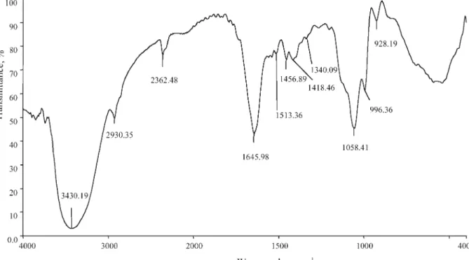
Infrared spectral analyses were performed to char-acterize the peptide and protein composition of wasp venom. The resulting infrared spectrum of crude venom from N. vitripennis is shown in Figure 2 with interpretation of characteristic absorption bands in Table 2. Absorption bands at 3,430, 1,645, and 1,513 cm–1 are consistent with the presence of secondary amine and amide groups, as would be
expected for proteinaceous venoms (Stuart, 1997). The observed band at 2,930 cm–1 is characteristic of alkanes, while the vibration at 1,418 cm–1 reflects the acidic (carboxylic) nature of venom (Leonard, 1972). Absorption bands at 1,058, 996, and 928 cm–1 suggest that some venom components, possi-bly enzymes, contain phosphorous. The noticeable absence of bands at 3,600 (OH peak), 2,900 (C-H stretching), and 1,700 (C=O stretching) cm–1 (Fig.
Fig. 2. Infrared radiation spectroscopic analysis of crude
venom from N. vitripennis. Fifty microliters of crude venom
in isolation buffer (pH 8.0) were used for i.r. analysis.
TABLE 2. Analysis of Characteristic Infrared Absorption Bands Associated With Crude Venom From N. vitripennis*
Frequency (cm–1) Possible assignment
3,430 N–H stretching, secondary amines 2,930 Asymmetric C–H stretching of CH2
2,362 CO2
1,645, 1,513 C = O stretching, amides
1,456 C–N stretching
1,418 C–O–H in plane bending, carboxylic acids
1,340 O–H bending
1,058 P–O–H bending
996, 928 P–O stretching
*Interpretation of infrared absorption bands characteristic of crude venom as well as band assignments are based on 3 replicates of i.r. spectroscopic analysis using 50-µl lyopholized samples of crude venom reconstituted in isolation buffer (Fig. 1).
2) indicates that the contents of venom reservoirs lack a carbohydrate moiety (Fritz and Schenk, 1979; Stuart, 1997).
HPLC Analysis
Crude venom isolated from venom reservoirs was fractioned using a reversed phase C18 column.
Based on the absorption peaks monitored at 280 nm, venom appears to be a complex mixture of peptides and proteins (Fig. 3), which was further supported by SDS-PAGE profiles of crude venom
(Fig. 4). Comparisons made to the retention times of several internal standards suggest that apamin (Fig. 3, t = 9.90 min, peak 4) and histamine (Fig. 3, t = 25.38 min, peak 7) are major components of crude venom. However, by comparison to crude venom alone, confluent monolayers of BTI-TN-5B1-4 cells displayed very little sensitivity to pure apamin, and this toxin did not augment venom activity when co-incubated with T. ni cells (Table 3). Similarly, incubation of crude venom with anti-histamine or anti-phenoloxidase did not lower the toxicity of venom toward cultured cells,
Fig. 3. Fractionation of crude venom (150 venom
reser-voir equivalents of lyophilized venom dissolved in 150 µl
isolation buffer) from N. vitripennis by reversed phase HPLC
(Acelll C18 column, 12.5 cm × 4.0 mm). Eluent A: 0.1%
TFA in acetonitrile: water (80:20); eluent B: 0.1% TFA in water. Fractionation was performed using a linear gradient of 5–80% A at 40 min with a flow rate of 1.0 ml/min and
an injection volume of 20 µl. Absorbance was monitored at 280 nm for (A) crude venom and (B) standards (1) noradrenaline, 0.7 mg/ml; (2) dopamine, 10.3 mg/ml; (3)
serotonin, 11.2 mg/ml; (4) apamin, 1 µg/µl; (5)
phospho-lipase B, 0.5 µg/µl; (6) phosphophospho-lipase A2, 1 µg/µl; (7)
even at antibody levels (1:10) that should have saturated binding sites on any venom proteins (Table 3).
Salt Precipitation
Ammonium sulfate precipitation was used as a means to differentially isolate the venom proteins responsible for developmental arrest and death. Ammonium sulfate was added stepwise to crude venom (in phosphate isolation buffer) until 100% saturation was achieved, and then each fraction or cut was assayed for the ability to induce develop-mental arrest and death in young pharate adults of S. bullata, and also trigger cell death in vitro. Under these conditions, three salt fractions (5, 80, and 100%) displayed biological activity toward S.
bullata in terms of halting fly development and
evoking death, with the 80% cut most closely re-sembling crude venom (Table 4). Likewise, when the fractions was assayed in vitro using T. ni cells, all 3 fractions were cytotoxic, but the 80% cut was most lethal toward the cultured cells (Table 4).
SDS-PAGE analysis of the salt fractions indicated that the 5% cut had a nearly identical protein pro-file as crude venom (Fig. 5, Lanes 2–3). Protein bands with estimated molecular weights ranging from 13 to 146 kilodaltons (kDa) were detected in both crude venom and the 5% fraction (Fig. 5). However, two high molecular weight proteins (188.1 and 200.5 kDa) observed in crude venom were absent from the 5% cut, and the 200.5-kDa band was also not present when crude venom had been prepared in phosphate isolation buffer mi-nus sucrose (Fig. 4).
The protein profile of the 80% ammonium sul-fate fraction revealed partial purification of 4 pro-teins with apparent molecular masses of 125, 116.9, 100.3, and 67.7 kDa (Fig. 5). Two of these bands (67.7 and 100.3 kDa) were observed in the 100% cut, although the biological activity of this fraction was significantly less than that of the 80% cut in terms of inducing developmental arrest (F = 132.5, df = 8, 300, P < 0.001) and triggering death in vivo (F = 99.2, df = 8, 300, P < 0.001) and in vitro (F = 92.4, df = 8, 300, P < 0.001) (Table 4).
Membrane Separations
Fractionation of crude venom proteins was also attempted using centrifuge filters containing
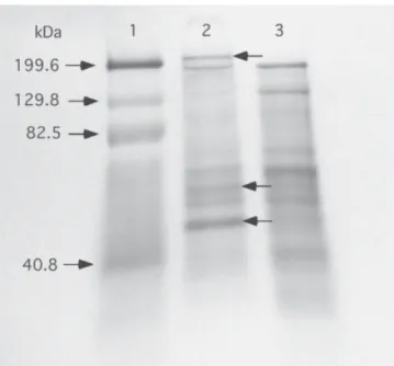
mem-Fig. 4. SDS-PAGE analysis of crude venom from N.
vitripennis using 10% Tris-HCl polyacrylamide gels
(Bio-Rad). Lane 1: Molecular weight standards (10 µl), mysosin
(199.6 kDa), β-galactosidase (129.8 kDa), bovine serum
albumin (82.5 kDa), and carbonic anhydrase (40.8 kDa);
Lane 2: Crude venom (30 µg) in isolation buffer; Lane 3:
Crude venom (30 µg) in isolation buffer lacking sucrose.
Proteins were visualized using 0.1% (w/v) Coomassie bril-liant blue G-250 staining (Garfin, 1990).
TABLE 3. In Vitro Evaluation of Apamin, Histamine, and Phenoloxidase Activity in Crude Venom From N. vitripennis*
Treatment N Cell death (%)
Untreated 9,708 1.2± 0.8a
Saline (isolation buffer) 11,417 2.9± 1.3a
Venom 10,046 100b Apamin (10 µg) 8,643 1.7± 0.9a Venom + apamin 12,479 100b Anti-histamine (1:10) 11,580 2.6± 0.5a Venom + anti-histamine 10,989 100b Anti-phenoloxidase (1:10) 9,056 3.8± 2.1a Venom + anti-phenoloxidase 10,047 100b
*The possible presence of apamin, histamine, and phenoloxidase in crude venom was assessed in vitro by evaluating the insecticidal activity in the presence of apamin, anti-histamine, and anti-phenoloxidase. Assays used confluent monolayers of BTI-TN-5B1-4 cells and mortality was assessed at 24 and 48 h post-incubation using vital staining.
TABLE 4. Insecticidal Activity in Venom Fractions Separated by Ammonium Sulfate Precipitation*
% Response
In vivo In vitro
Fraction N Developmental arrest Death N Death
Saline 30 0a 0a 8,976 0a Crude venom 30 95.1± 6.1b 100b 7,760 100b Salt fraction 5% AS 30 54.2± 5.0c 80.4 ± 3.7c 10,056 91.4 ± 4.5b 15% AS 30 0a 0a 9,004 1.5 ± 0.8c 25% AS 30 0a 0a 11,307 0a 40% AS 30 0a 0a 8,925 1.1 ± 0.4c 65% AS 30 0a 0a 8,458 0.8 ± 0.6c 80% AS 30 93.2± 2.7b 100b 9,350 100b 90% AS 30 0a 0a 10,336 2.3 ± 1.0c 100% AS 30 63.4± 3.8c 56.7 ± 4.8d 11,974 64.8 ± 6.7d *Salt was added stepwise to crude venom from N. vitripennis until 100% saturation, and then the precipi-tate that formed with each fraction was assayed for biological activity. In vivo activity was assessed by injecting 1 µl of test sample into the dorsal surface of adult S. bullata, whereas in vitro activity was mea-sured by incubating BTI-TN-5B1-4 cells with 1 µl of each salt fraction. Mortality was observed 24 h later.
Fig. 5. SDS-PAGE analysis of ammonium sulfate
fraction-ation of crude venom from N. vitripennis using 4–15% Tris-HCl polyacrylamide gels (BioRad). Lane 1: Molecular
weight standards (10 µl), mysosin (199.6 kDa),
β-galactosi-dase (129.8 kDa), bovine serum albumin (82.5 kDa), car-bonic anhydrase (40.8 kDa), soybean trypsin inhibitor (31.5 kDa), and lysozyme aprotinin (17.0 kDa); Lane 2: Crude
venom (30 µg) in isolation buffer; Lane 3: 5% ammonium
sulfate (AS) cut (precipitate); Lane 4: 15% AS cut; Lane 5: 25% AS cut; Lane 6: 40% AS cut; Lane 7: 65% AS cut;
Lane 8: 80% AS cut; Lane 9: 90% AS cut; Lane 10: 100%
AS cut. Proteins were visualized using 0.1% (w/v) Coom-massie brilliant blue G-250 staining (Garfin, 1990).
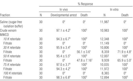
branes with specific size exclusion limits (10,000 to 100,000 MWCO). Retentates of all centrifuge membranes with MWCOs between 10,000–75,000 were highly toxic when injected into adult flies or exposed to confluent monolayers of BTI-TN-5B1-4 cells (Table 5). Only the 100-K MWCO retentate failed to display any lethal activity. By contrast, in vitro and in vivo toxicity were observed with fil-trates from membrane filters with MWCOs rang-ing from 30,000–100,000 (Table 5).
When samples were assayed for the ability to induce developmental arrest in young pharate adults of S. bullata, retentates from all filters ex-cept the 100K MWCO membrane elicited a halt in fly development comparable to crude venom (Table 5). In contrast, only filtrates derived from 75- and 100-K MWCO membranes were capable of stimulating developmental arrest (Table 5). As a control, phosphate isolation buffer alone was loaded onto each type of membrane filter, and retentates and filtrates assayed. In all cases, no bio-logical activity was observed.
SDS-PAGE analyses of the membrane fractions revealed that retentates from all membranes be-tween 10–75-K MWCO had nearly identical
pro-tein profiles as crude venom (Fig. 6, only the 50-and 75-K MWCO membranes are shown as repre-sentations). The protein profile of the 50- and 75-K MWCO filtrate indicated partial purification of at least 3 proteins with apparent molecular masses of 125.7, 114.6, and 67.1 kDa (Fig. 6). This pro-file is nearly identical to that of the 80% ammo-nium sulfate fraction with the exception of a protein band of 100.3 kDa in the 80% cut. The latter protein may be particularly important for venom activity as filtrates from the 50- and 75-K MWCO membranes showed much lower biologi-cal activity than the 80% ammonium sulfate frac-tion (Tables 4 and 5).
DISCUSSION
During natural parasitism, N. vitripennis induces a developmental arrest in fly hosts that lasts until the fly is consumed by parasitoid larvae (Rivers and Denlinger, 1994a). Developmental arrest as well as death result from envenomation, and in the ab-sence of feeding wasp larvae, the halt in fly devel-opment is sustained for an extended period of time (30–60 days at 25°C) (Rivers and Denlinger, 1995).
TABLE 5. Insecticidal Activity in Venom Fractions Separated by MWCO Membranes*
% Response
In vivo In vitro
Fraction N Developmental arrest Death N Death
Saline (sugar-free 30 0a 0a 11,987 0a isolation buffer) Crude venom 30 97.1± 4.2b 100b 10,983 100b MWCO 10 K retentate 30 94.3± 6.7b 100b 12,348 100b Filtrate 30 0a 0a 11,149 0a 30 K retentate 30 95.9± 3.4b 100b 10,806 100a Filtrate 30 0a 56.1± 3.6c 8,359 71.9± 4.8c 50 K retentate 30 95.4± 5.5b 100b 12,007 100b Filtrate 30 0a 47.8± 7.16c 9,939 65.9± 5.0c 75 K retentate 30 97.0± 3.7b 100b 10,035 100b Filtrate 30 94.3± 4.2b 100b 11,972 100b 100 K retentate 30 0a 0a 8,365 0a Filtrate 30 98.3± 6.8b 100b 12,894 100b
*Retentates and filtrates were assayed for insecticidal activity in vivo by injecting 1 µl of test sample into adult S. bullata, or examined in vitro by incubating BTI-TN-5B1-4 cells with 1 µl of each venom fraction. Mortality was observed 24 h later.
Here, we show that the developmental arrestant and the agent(s) that induce host death are present in the venom gland and venom reservoir but not in other tissues associated with the female repro-ductive system. Psoralen treatment of crude venom preparations indicated that biological activity was associated with maternally derived secretions in the gland and reservoir and not attributed to products from symbiotic organisms (endosymbiotic viruses) as shown for some endoparasitic species (Quicke, 1997; Stolz and Whitfield, 1992) or ectoparasitoids harboring microsporidia (Geden, 1996). These ob-servations are consistent with those of Ratcliffe and King (1967), who argued that contents of the venom gland (acid gland) alone are responsible for induction of death in adults of M. domestica, and that the venom reservoir stores venom in ac-tive form (Ratcliffe and King, 1969).
Despite this agreement, previous electrophor-etic analysis of the wasp venom (Ratcliffe and King, 1967) revealed unique protein profiles for
fluids from venom glands versus reservoirs, sug-gesting that venom is modified in the venom res-ervoir. Our study did not find any evidence to support the idea of venom modification after duction in the acid gland. In fact, SDS-PAGE pro-tein profiles were identical for crude venom prepared from venom glands and venom reser-voirs. When venom was extracted from wasp tis-sues in buffer lacking a protein-stabilizing agent such as sucrose (at high concentrations), or enzyme inhibitors (Rivers, unpublished observations) of buffer pH was less than optima, biological activ-ity of venom decreased. Based on SDS-PAGE analyses of crude venom prepared in sucrose-free buffer, the protein composition of venom was likely altered by these treatments. Venom analy-ses performed by Ratcliffe and King (1967) did not report controlling such factors when compar-ing the contents of venom reservoirs and glands. It is also important to note that these early inves-tigations (Ratcliffe and King, 1967, 1969) failed to detect developmental arrest as a feature of en-venomation, largely because non-natural host de-velopmental stages (adult) were used in the biological assays. Previous characterization of the insecticidal properties of venom glands and res-ervoirs (Beard, 1964; Ratcliffe and King, 1967; Tiegs, 1922) must also be interpreted cautiously as the method (i.e., dipping an insect pin in venom prior to pricking the body) of venom in-jection was not necessarily quantifiable or con-sistently reproducible.
Biochemical and electrophoretic analyses of crude venom indicate that venom is predominantly composed of mid to high molecular weight teins that are acidic in nature. This range of pro-tein sizes as well as complexity of the propro-tein profile in terms of total number of proteins is con-sistent with most parasitic wasp venoms that have been examined (Digilio et al., 2000; Leluk et al., 1989; Nakamatsu and Tanaka, 2003; Parkinson et al., 2002a; Uçkan et al., 2004). By contrast, low molecular weight proteins and peptides are typi-cal of venoms of the aculeate Hymenoptera (Leluk et al., 1989; Schmidt, 1982), as are proteins abun-dant in neutral and basic amino acids (Piek and
Fig. 6. SDS-PAGE analysis of MWCO (molecular weight
cut-off) membrane fractionation of crude venom from N.
vitripennis using 4–15% Tris-HCl polyacrylamide gels
(BioRad). Lane 1: Molecular weight standards (10 µl),
mysosin (199.6 kDa), β-galactosidase (129.8 kDa), bovine serum albumin (82.5 kDa), carbonic anhydrase (40.8 kDa), and soybean trypsin inhibitor (31.5 kDa); Lane 2: 75 K (MWCO) retentate; Lane 3: 50 K retentate; Lane 4: 75 K filtrate; Lane 5: 50 K filtrate. Proteins were visual-ized using 0.1% (w/v) Coommassie brilliant blue G-250 staining (Garfin, 1990).
Spanjier, 1986). Infrared spectral analyses suggest that venom proteins do not possess a carbohydrate moiety, a feature shared with venom from P.
turionella (Uçkan et al., 2004) and many social
hy-menopterans (Leluk et al., 1989) but unique from other parasitic wasp venoms that appear to be rich in glycosylated proteins (Leluk et al., 1989; Piek and Spanjier, 1986). The infrared spectrum also revealed the presence of phosphorus-containing structures, typical of enzymes, in the venom. Though the presence of enzymatic activity in N.
vitripennis venom has yet to be confirmed, venoms
from several endoparasitic species and social Hy-menoptera have been shown to contain multiple types of enzymes (Moreau et al., 2004; Parkinson et al., 2001; 2002a,b; 2003; Piek and Spanjier, 1986; Schmidt, 1982; Uçkan et al., 2004). Enzyme activity, however, does not likely account for the ability of venom from N. vitripennis to arrest host development or evoke death as crude venom prepa-rations contained the protease inhibitor PMSF as well as the chelating agent EDTA, and high con-centrations of anti-phenoloxidase antibodies had no influence on venom activity.
Venom stability at alkaline pH is in agreement with wasp toxins identified in venom from the ectoparasitoids H. hebetor (Quistad et al., 1994) and E. comstockii (Coudron and Brandt, 1996). H.
hebetor produces a potent venom with at least two
highly paralytic protein toxins (Brh-I and Brh-V, estimated molecular masses 71–73 kDa) (Quistad et al., 1994) and one smaller, less insecticidal toxin (ca. 20–40 kDa). All three proteins appear to be glycosylated. A 66-kDa venom protein has been isolated from E. comstockii that does not induce paralysis but does arrest host development by in-hibiting larval–larval ecdysis (Coudron and Brandt, 1996). This arrestment protein is thought to be comprised of at least two subunits, suggesting that the native protein is possibly a dimer composed of two 31–33-kDa proteins (Coudron and Brandt, 1996). Size and activity estimates for venom pro-teins from these two parasitoids are in agreement with SDS-PAGE profiles for partially purified venom proteins from N. vitripennis: ammonium sulfate and MWCO membrane fractions of crude
venom yielding biological activity (induction of developmental arrest and death) all contained a protein with an estimated molecular weight of 67– 70 kDa. It seems highly improbable, however, that a single protein in venom from N. vitripennis is responsible for inducing both host arrestment and death. Indeed, though a protein band correspond-ing to approximately 67 kDa was observed in the 100% ammonium sulfate fraction, this salt cut in-duced developmental arrest in significantly fewer pharate adults of S. bullata than the 80% fraction that possessed two additional high molecular weight proteins (114.6 and 125.7 kDa). A similar reduction in toxicity, as well as ability to arrest fly development, was observed with venom fractions (filtrates from the 50- and 75-K MWCO) isolated by centrifugal membranes with correspondingly similar but not identical protein profiles to the 80% salt cut: a 100.3-kDa protein was detected in the salt fraction in addition to 3 proteins present in the membrane fractions. Collectively, these ob-servations argue that multiple proteins are required for full venom activity. This certainly is consistent with the complex nature of parasitic wasp venoms that have been characterized and the milieu of host effects that they evoke (Quistad et al., 1994; Parkinson et al., 2002a,b, 2003).
Some lethal activity was also detected in filtrate collected from the 30-K MWCO membranes. How-ever, this toxicity was significantly less by compari-son to filtrates from membranes with molecular cutoffs higher than 75 K and fly development was not arrested by these samples. Surprisingly, no pro-tein bands were observed by SDS-PAGE that corre-sponded to the filter size exclusion limits. Failure to detect proteins smaller than 30 kDa may reflect protein levels below the detection limits of the Coomassie staining (Garfin, 1990). It is also pos-sible that the actual lethal protein is larger than the size cutoff limit of the membranes used since, like dialysis membranes, the listed size represents a median pore size, with both larger and smaller pores also present on the membranes (Pohl, 1990). Consequently, reduced venom activity may be at-tributed to a small amount of the protein passing through the membrane. SDS-PAGE analyses
sup-port the latter scenario for both the 30- and 50-K MWCO membranes.
HPLC separations of crude venom coupled with the use of internal standards suggested that low molecular peptides or proteins exist in venom in the form of apamin and histamine. It is not sur-prising, then, that neither were detected electro-phoretically since their low molecular masses would allow them to migrate much more rapidly than other venom proteins in 10–12% acrylamide gels (Garfin, 1990). Though these paralytic agents are common in venoms from social Hymenoptera (Piek and Spanjier, 1986) and the endoparasitoid
P. turionella (Uçkan et al., 2004), biological assays
using cultured cells indicate that either apamin or histamine do not affect the cells tested or neither was present in active form within venom from N.
vitripennis. This finding is not unexpected
consid-ering that apamin and histamine are typical of paralytic venoms used primarily for defense (Blum, 1981; Schmidt, 1982). In contrast, venom from N.
vitripennis is non-paralytic (Rivers and Denlinger,
1994a), and is used to manipulate a non-mobile host (pupa) to maximize progeny production (Riv-ers et al., 1998). Clearly, additional studies are needed to further characterize and isolate venom proteins. The stability of venom at alkaline pH sug-gests that anion-exchange chromatography is a vi-able next step to purify individual venom proteins (Quistand et al., 1994).
ACKNOWLEDGMENTS
The authors thank Kenneth Bujold and Timothy Crawley for providing assistance in isolation of crude wasp venom. We also express thanks to Drs. Brain Barr, Neena Din, and Terry Bird (Loyola College) for many conversations concerning protein purifi-cation techniques. This work was supported in part by USDA-NRICGP Seed grant 2001-1005 (D.B.R.) and by a Loyola College Faculty Development Grant (D.B.R.)
LITERATURE CITED
Asgari S, Zhang G, Zareie R, Schmidt O. 2003a. A serine pro-teinase homolog venom protein from an endoparasitoid
wasp inhibits melanization of the host hemolymph. In-sect Biochem Mol Biol 33:1017–1024.
Asgari S, Zareie R, Zhang G, Schmidt O. 2003b. Isolation and characterization of a novel venom protein from an endoparasitoid, Cotesia rubecula (Hym.: Braconidae). Arch Insect Biochem Physiol 53:92–100.
Beard RL. 1963. Insect toxins and venoms. Annu Rev Entomol 8:1–18.
Beard RL. 1964. Pathogenic stinging of house-fly pupae by
Nasonia vitripennis (Walker). J Invertebr Pathol 6:1–4.
Beckage NE. 1998.. Modulation of immune responses to para-sitoids by polydnaviruses. Parasitology 116:S57–S64. Blum MS. 1981. Proteinaceous venoms. In: Blum M, editor.
Chemical defenses of arthropods. New York: Academic Press. p 288–302.
Coudron TA. 1991. Host-regulating factors associated with parasitic Hymenoptera. In: Hedin PA, editor. Naturally oc-curring pest bioregulators. ACS Symp Ser 449:41–65. Coudron TA, Brandt SL. 1996. Characteristics of a
develop-mental arrestant in the venom of ectoparasitoid wasp
Euplectrus comstockii. Toxicon 34:1431–1441.
Dani MP, Richards EH, Isaac RE, Edwards JP. 2003. Antibac-terial and proteolytic activity in venom from the endopara-sitic wasp Pimpla hypochondriaca (Hymenoptera: Ichneu-monidae). J Insect Physiol 49:945–954.
Davies TR, Wickham TJ, McKenna KA. 1993. Comparative recombinant protein production of eight insect cell lines. In Vitro Cell Dev Biol Anim 29:388.
Denlinger DL. 1972.. Induction and termination of pupal dia-pause in Sarcophaga (Diptera: Sarcophagidae). Biol Bull 142:11–24.
Digilio MC, Isidoro N, Tremblay E, Pennacchio F. 2000. Host castration by Aphidius ervi venom proteins. J Insect Physiol 46:1041–1050.
Doury G, Bigot Y, Perguet G. 1997. Physiological and bio-chemical analysis of factors in the female venom gland and larval salivary secretions of the ectoparasitoid wasp Eupelmus orientalis. J Insect Physiol 43:69–81.
Englard S, Seifter S. 1990. Precipitation techniques. In: Deutscher MP, editor. Guide to protein Purification. Lon-don: Academic Press. p 285–300.
Fritz JS, Schenk GH. 1979. Electronic absorption spectra, fluo-rescence and infrared spectroscopy. In: Fritz JS, Schnek GH, editors. Quantitative analytical chemistry. Boston: Allyn and Bacon. p 430–460.
Garfin DE. 1990. One-dimensional gel electrophoresis. In: Deutscher MP, editor. Guide to protein Purification. Lon-don: Academic Press. p 425–440.
Geden CJ. 1996. Nosema disease of the house fly parasitoid
Muscidifurax raptor and its impact on fly IPM programs.
Proceedings of IOBC conference Technology Transfer in Biological Control: From Research to Practice, Montpellier, FR Bull IOBC 19:208.
Gerling D, Legner EF. 1968. Developmental history and re-production of Spalangia cameroni, parasite of synathropic flies. Ann Entomol Soc Am 61:1436–1443.
Jones D, Sawicki G, Wozniak M. 1992. Sequence, structure, and expression of a wasp venom protein with a negatively charged signal peptide and a novel repeating internal struc-ture. J Biol Chem 267:14871–14878.
Kansu IA, Uur A. 1984. Pimpla turionellae (L.) (Hym,-Ichneu-monidae) ile konukçusu bazý lepidopter pupalarý arasýn-daki biyolojik iliÕkiler üzerine araÕtýrmalar. (Investigations on the biological relationships between Pimpla turionella (L.) (Hym.-Ichneumonidae) and some lepidopterous pupae hosts J. Nat. Sci.) Doga Bilim Dergisi 8:160–173.
Krishnan A, Nair PN, Jones D. 1994. Isolation, cloning, and characterization of new chitinase stored in active form in chitin-lined venom reservoir. J Biol Chem 269:20971–20976. Leluk J, Schmidt J, Jones D. 1989. Comparative studies on the protein composition of hymnenopteran venom reser-voirs. Toxicon 27:105–114.
Leonard GJ. 1972. The isolation of a toxic factor from sawfly (Lophyrotoma interrupta klug) larvae. Toxicon 10:597–603. Moreau SJM, Cherqui A, Doury G, Dubois F, Fourdrain Y,
Sabat-ier L, Bulet P, Saarela J, Prevost G, Giordanengo P. 2004. Identification of an apartylglucosaminidase-like protein in the venom of the parasitic wasp Asobara tabida (Hymenop-tera: Braconidae). Insect Biochem Mol Biol 34:485–492. Nakamatsu Y, Tanaka T. 2003. Venom of ectoparasitoid,
Euplectrus sp. Near plathypenae (Hymenoptera: Eulo-phidae) regulates the physiological state of Pseudaletia
separata (Lepidoptera: Nocuitdae) host as a food resource.
J Insect Physiol 49:149–159.
Ohtaki T. 1966. On the delayed pupation of the flesh fly,
Sarcophaga peregrina Robineau-Desvoiday. Jpn J Med Soc
Biol 19:97–104.
Parkinson NM, Smith I, Weaver R, Edwards JP. 2001. A new form of arthropod phenoloxidase is abundant in venom of the parasitoid wasp Pimpla hypochondriaca. Insect Biochem Mol Biol 31:57–63.
Parkinson NM, Conyers C, Smith I. 2002a. A venom protein from the endoparasitoid wasp Pimpla hypochondriaca is similar to snake venom reprolysin-type metalloproteases. J Invertebr Pathol 79:129–131.
Parkinson NM, Richards EH, Conyers C, Smith I, Edwards JP. 2002b. Analysis of venom constituents from the parasi-toid wasp Pimpla hypochondriaca and cloning of cDNA en-coding a venom protein. Insect Biochem Mol Biol 32: 729–735.
Parkinson NM, Conyers CM, Keen JN, MacNicoll AD, Smith I, Weaver RJ. 2003. cDNAs encoding large venom proteins from the parasitoid wasp Pimpla hypochondriaca identi-fied by random sequence analysis. Comp Biochem Physiol 134C:513–520.
Piek T, Spanjier W. 1986.. Chemistry and pharmacology of solitary wasp venoms. In: Piek T, editor. Venoms of the Hymenoptera. London: Academic Press. p 161–307. Piek T, Veenedaal RL, Mantel P. 1982. The pharmacology of
Microbracon venom. Comp Biochem Physiol 72C:303–309.
Pohl T. 1990. Concentration of proteins and removal of sol-utes. In: Deutscher MP, editor. Guide to protein purifica-tion. London: Academic Press. p 68–82.
Quicke DLJ. 1997. Parasitic wasps. London: Chapman and Hall.
Quistad GB, Nguyen Q, Bernasconi P, Leisy DJ. 1994. Purifi-cation and characterization of insecticidal toxins from venom glands of the parasitic wasp, Bracon hebetor. Insect Biochem Mol Biol 24:955–961.
Ratcliffe NA, King PE. 1967. The venom system of Nasonia
vitripennis (Walker) (Hymenoptera: Pteromalidae). Proc R
Ent Soc Lond 42:49–61.
Ratcliffe NA, King PE. 1969. Morphological, ultrastructural, histochemical, and electrophoretic studies on the venom system of Nasonia vitripennis Walker (Hymenoptera: Pteromalidae). J Morph 127:177–204.
Rivers DB, Denlinger DL. 1994a. Developmental fate of the flesh fly, Sarcophaga bullata (Diptera: Sarcophagidae), envenomated by the pupal ectoparasitoid, Nasonia
vitri-pennis (Hymenoptera: Pteromalidae). J Insect Physiol
40:121–127.
Rivers DB, Denlinger DL. 1994b. Developmental fate of the flesh fly, Sarcophaga bullata (Diptera: Sarcophagidae), envenomated by the pupal ectoparasitoid, Nasonia
vitri-pennis (Hymenoptera: Pteromalidae). J Insect Physiol
40:121–127.
Rivers DB, Denlinger DL. 1995. Venom-induced alterations in fly lipid metabolism and its impact on larval develop-ment of the ectoparasitoid Nasonia vitripennis (Walker) (Hymenoptera: Pteromalidae). J Invertebr Pathol 66: 104–110.
Rivers DB, Hink WF, Denlinger DL. 1993. Toxicity of the venom from Nasonia vitripennis (Hymenoptera: Ptero-malidae) toward fly hosts, nontarget insects, different de-velopmental stages, and cultured insect cells. Toxicon 31:755–765.
Rivers DB, Pagnotta MA, Huntington ER. 1998. Reproduc-tive strategies of 3 species of ectoparasitic wasps are modu-lated by the response of the fly host, Sarcophaga bullata (Diptera: Sarcophagidae) to parasitism. Ann Entomol. Soc Am 91:458–465.
Rivers DB, Ruggiero L, Yoder JA. 1999a. Venom from Nasonia
vitripennis: a model for understanding the roles of venom
during parasitism by ectoparasitoids. Trends Entomol 2: 1–17.
Rivers DB, Genco M, Sanchez RA. 1999b. In vitro analysis of venom from the wasp Nasonia vitripennis: susceptibility of different cell lines and venom-induced changes in plasma membrane permeability. In Vitro Cell Dev BiolAnim 35:102–110.
Rivers DB, Rocco MM, Frayha AR. 2002a. Venom from the ectoparasitic wasp Nasonia vitripennis increases Na+ influx and activates phospholipase C and phospholipase A2 de-pendent signal transduction pathways in cultured insect cells. Toxicon 40:9–21.
Rivers DB, Ruggiero L, Hayes M. 2002b. The ectoparasitic wasp
Nasonia vitripennis (Walker) (Hymenoptera: Pteromalidae)
differentially affects cells mediating the immune response of its flesh fly host, Sarcophaga bullata Parker (Diptera: Sarcophagidae). J Insect Physiol 48:1053–1064.
Rivers DB, Zdarek J, Denlinger DL. 2004. Disruption of pupariation and eclosion behavior in the flesh fly,
Sarco-phaga bullata Parker (Diptera: Sarcophagidae), by venom
from the ectoparasitic wasp Nasonia vitripennis (Walker) (Hymenoptera:Pteromalidae). Arch Insect Biochem Physiol 57:78–91.
Rivers DB, Crawley T, Bauser H. 2005. Localization of intrac-ellular calcium release in cells injured by venom from the ectoparasitoid Nasonia vitripennis (Walker) (Hymenoptera: Pteromalidae) and dependence of calcium mobilization on G-protein activation. J Insect Physiol 51: 149–160.
Schmidt JO. 1982. Biochemistry of insect venoms. Annu Rev Entomol 27:339–368.
Stoll VS, Blanchard JS. 1990. Buffers: Principles and practice. In: Deutscher MP, editor. Guide to protein purification. London: Academic Press. p 24–37.
Stoltz DB, Whitfield J. 1992. Viruses and virus-like entities in the parasitic Hymenoptera. J Hymenoptera Res 1:125– 139.
Strand MR, Noda T. 1991. Alterations in the hemocytes of
Pseudoplusia includens after parasitism by Microplitis demol-itor. J Insect Physiol 37:839–850.
Strand MR, Pech LL. 1995. Immunological basis for compat-ibility in parasitoid-host relationships. Annu Rev Entomol 40:31–56.
Stuart B. 1997. Biological applications of infrared spectros-copy. New York: John Wiley and Sons.
Thompson SN. 1986. Nutrition and in vitro culture of insect parasitoids. Annu Rev Entomol 31:197–219.
Tiegs OW. 1922. Researches on insect metamorphosis, Parts I and II. Trans R Soc S Aust 46:319–527.
Uçkan F, Sinan S, SavaÕçý S, Ergin E. 2004. Determination of venom components from the endoparasitoid wasp Pimpla
turionellae L. (Hymenoptera: Ichneumonidae). Ann
Ento-mol Soc Am 97:775–780.
Vinson SB. 1990. How parasitoids deal with the immune sys-tem of their host: Overview. Arch Insect Biochem Physiol 13:3–27.
immuno-suppressive role for related Campoletis sonorensis venom and ovarian proteins in Heliothis virescens. Arch Insect Biochem Physiol 26:147–163.
Whitfield JB, Asgari S. 2003. Virus or not? Phylogenetics of polydnaviruses and their wasp carriers. J Insect Physiol 49:397–405.
Whiting AR. 1967. The biology of the parasitic wasp
Mor-moniella vitripennis (= Nasonia brevicornis) (Walker). Q Rev
Biol 42:333–406.
Zdarek J, Slama K, Fraenkel G. 1979. Changes in internal pressure during puparium formation in flies. J Exp Zool 207:187–196.