..4.O. Yet. 'Ilk. De7.
39 (3): 359-371. 1992
LIVER RESTORATlON POLLOWlNG PARTIAL BEPATBCTOMY AND CIIOLECYSTlLOBECTOMY IN DOG
Nassef.M.T.; KA. Ali; A.S. Saleh _d
M.M. Abd El.Hafeez
Kapeldenle x-nı llepateec:tomyyeCIaoIec:yıtd1obectomy'1 Taklbea Ka•.•dlefta Yealdea 0DaruD.
Özet: Bu falıJ11UZklinik hakımdan sağlıklı erkek "Dediş; toplam 28 olgun köpek üzerinde yürütülmi4tür. Köpekler 4- eşit gruha €!yrılmıştır. Birin-ci "De3.grupdaki köpeklere
%
30 ve%
4-O'lık olTTUJküzere sıras!yla sol lateral loh "Deyine sol lateral ile sol central loh/ara eksi{Yonla kısmi hepatectamy uygulanmıştır. ıkinci "De4-. grupdaki köpeklere de sıras!yla%
30 ve 4-O'lık olTTUJküzere eholerystiloheetomy yapılmıştır.Tüm gruplardaki köpeklerin rejenerasyon yeteneği, rejenerasyon yüzdesi hesahı ile saptanmıştır. Rejenerasyon yetenekleri sıras!}la I. 2 . 3. ve 4-. grup-larda
%
3ı.
I,%
36. O,%
4-2.76 ve%
4-5.4-0 olarak hesaplanmıştır. Cholecystilohe.;tomy uygulanan gruplardaki rejenerasyon yüzdesi, karşıtı olan kısmi hepateetomy uygulamalarına kıyasla yüksek olmasına karşın, hu iki ayrı uygulama sonucunda elde edilen rejenerasyon yüzdeleri arasındaki farkların önemli derecede olmadığı saptanmıştır.SUDlIDary: The present stutfy Was ea"led out on 28 elinieally healthy adult dogs of hoth sexes. They were diuided into four equal groups. Dogs of thefirst and third groups were mhjeeted to 30
%
and%
4-0 partial hepateetomy hy the exeision of lefi lateral lohe and left lateral as well as lefi central lohes . respecti"Dely.Dogs of the second and fourth groups were suhjeeted to 30 % and 4-0%
eholecystilohectomy respeeti"Dely.Regeneration eapahility was estimated in all groups hy ealculation of regeneration percentage. It was 3 i .iO, 36.00, 4-2.76 and 4-5.4-0% in tJıe Ist, 2nd, 3rd and the 4-th groups respeetively. In spite of the regeneralion percentage in ease of eholecystilohectomy groups were higher than those of eoms-ponding partial hepateetamy ones, there was no signifieant difference hetween
the eholecystiloheetomy and partial hepateetomy in regeneration pereentage.
360 M.T. NASEF - M.A. ALt - A.S. SALEH - M.M. ABD EL-HM:EEZ
IntrodUctiOIl
Hepatic resection or partial hepatectomy is indicated in many conditions such as traumatic fracture of hepatic parenchyma (14, 16), hepatic absces3atiou (-6, 19)~,primary or $CCondaıy tumors of hepatic and biliary ducts (9) and in cases bf livothydatid 'tyst (5, 23).
Hepa-tic lobectomy is earried out nowadays to obtain aliver graft for a
non auxilili.ary liveı;itran~plantation (9),
Different teehniques for lobeetomy were described by several authors. The partial hepateetomy was described in dogs (I 7, 18, 22). The teehnique conSisted of isolation of tht struetures in the hepatic bed with individual dosure and division of bile duets and blood ves-sels. Finger-fraeture teehnique was applied for partial hepatic
reeest-ion (7,8). Mass ligatreeest-ion at the base of reseeted lobe including
hepatic vesselsand duets was used (1I) in partial hepateetomy by using a clamp (15). Comparison between the partial hepateetomy by the aid of laser knife and diathermy eut instrement was studied (21). In dogs and rats, when they were subjeeted to partial hepa-teetomy, the remaining liver tissues inereased in bulk. This behaviour ofliver tissue was noUrue regeneratin (4,15,22).
The aim of the present experimental study is to deseribe the teehnique for choleeystilobeetomy and partial hepateetomy in dogs and estimation of the liver restoration following them.
Material and Methods
The present study was performed on 28 elinieally healthy mongrel dogs of both sexes. The animals were weighing from 7 to 21 kg.
Animals were classified into four groups (Fig., 1):
I - 30
%
partial hepateetomy (7 dogs).II - 30
%
eholeeystilobeetomy (7 dogs).III - 40
%
partial hepatectomy (7 dogs).KÖPEKLERDE KARACiGERIN YENIDEN ONARıMı. . . 3(,1
i.. L: Left lateral Iobe. L. C: left central lobe. Q. : Qııadraıe lobe. P. C: Right central lobe. R. L: Right lateral lobe. C, : Caudate process of the
caudate lobe.
P. R: Papiııary proccs of the caudate Iate.
G,n: Gall bladder.
LQ
40% parıial hepatecıomy.rLLiJ ~
30% 1"""'1 I"p"worny.Fig.
ı:
Diagram for hepatic lobes, 30 % and 40 ~;) parıiaI hcpatectomies. Fig. i: Karaci~er Loblarının diyagramı, %30 ve %40 parsiyal hcpatektomi.Animal preparation and preanesthetic medication:
Animals were starved for about i2 hours 'oofore the operation. They were sedated by i
iM
injection of chloropromazine HCl(1 mg Ikg bw.) Clipping and shaving of the ventral abdominal waıı from the xyphoid to the pubis. The operaliye field was prepared for
till aseptic surgery by disinkction of tinctl,lre of iodine 3 %.
Anaethesia ,vas induced by inlravenous injcetion of thiopental sod 5
%
(Biochemie G mb H, ViCniG) until all reficx acıions were abuli.~hed. Dogs WL:rc pO!;İI.İoncd in du:,,;,d r,:cumbency and ehest raised upward to fa'.'i!İtate l~lC exp()"Lll'C or the li\cr.Surgical procedllrcs:
Group i: 30
%
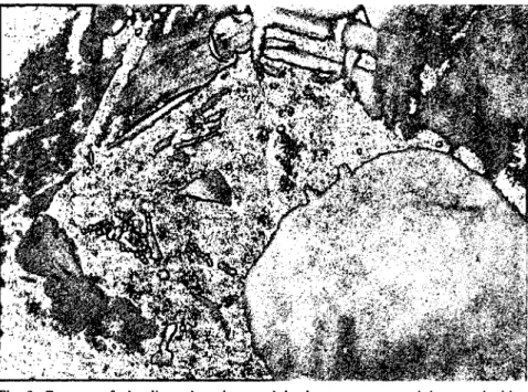
partial hcpa (,ctonıy.An inci',ion was mac1c thro~ıgh the skiıı and abdominal muscle,> on the mİdline from the xyplıoid process dowmvard to the post umblical region. The abdaıninal sheath together wİtlı the peritaneum were cut along the wound. The wound edges were drown wide by applying a self-retaining wound dilator. The pylorus of stomaeh and the duo-denum were grasped downward with a moİst sterile drape (Fig. 2).
362 MoT. NASEF - M.A. ALI - A.S. SALEH - M.M. ABD EL.HAFEEZ
Fig. 2: Exposure of the liver; the pylonıs and duodenum were grasped downward with a moist sıerile drape.
Fig. 2: Karacİğerin açırnı; pİlorus ve duedenurn ıslak sıeril bez ile tuıuluşu.
The triangular eoronary and hepatogastrie ligaments were transeeted
to free the left lateral lobe from its attaehment. The intended lobe
was gently lefted and grasped downward and medially. The pedicle of the lobe was exposed and encircled with a erushing ligatture (silk NO. 2) using Desehamb's needle (Fig. 3). The ligation included the vaseular and biliary pedicle. By the use of diathermy knife eleetrode, the excission of the lobe was made about 0.5 cm distal to the liga-turc (Fig. 4). Any blood oozing from the pedical stump eould be eontrolled individually by a dry guaze swab. Abdominal eavity had
been dried earefully from any remained blood. Abdominal wound
was closed as usual. A eovering suturc was applied to the skin wound.
Group II: 30
%
eholceystilobeetomy.After the same exposure to the livcr as described in group J,
the gall bladder was exposed by pulling the duodenum downward
and over hanging of the central mass of the liver (quadrate lobe medi-aIJy and right central laterally). Gentle pressure was done to evaeuate
the bladder eontents in the duodenum. The wall of the bladder
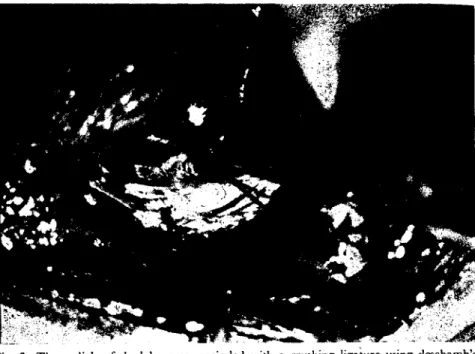
was damped by a Houptner damp (Fig. 5). The gall bladder was
bluntly disseeted from the liver bed by dosely snipping of[ its wall till reaing its upper part of the neek (Fig. 6). Bleeding oceurred during
KÖPEKLERDE KARACiGERiN YENiDEN ONARIMı. ..
Fig. 3: The pediclc of the looc \Vas mcirclcd with a crııshing ligaıurc ıısing drschamb's nerdk.
Fig. 3: Lob bağlantısının deschamb iğnesi kullanarak baskı ligaturu ile çevrimi.
Fig. 4: Excision of the lobe by diathermy knife electrOOe 1/2 cm. distal to the ligature. Fig. 4: Lobun ligaturin 0.5 cm distalinde diatermik elektrOO ile oksizyonu.
3(;L M.T. NA5EF - MA ALİ - A.5. 5ALEH - M.M. ABD EL-HAFEEZ
Fig. 5: Clamping of the fund us of the g:dl bladdar by a hauptner clamp after gentle
eva-("uation.
Fig. 5: Hassas olarak boşaltıldıktan sonra safra kese.;i fıındusunun bir hauptner biri eş-ıiricisi iLCsabitlenme;i.
Fig. 6: TIıe disseetion of the gall bladder from the livcr bed and ligation of the s)'Setic duct. Fig. 6: Safra kesesinin karaciğerden ayrılması ve sistik kanalın ligatürii.
KÖPEKLERDE KARACİGERİN YENİDEN ONARıMı' .. 365
the ~cparation of the gall bladder from the liver bed could be
control-Ied by pre;~urc of hot moist pack or by light coagulation with
diathermy round coautery e1ectrode. The cystic duct was dissected
bluntly from its ~urrounding tİssues. The junction between the
cys-tic and hepatic ducts was doubly ligated with Silk No.
ı.
Cuttingin betwcen the two ligatures and then the gall bladder was excised.
The same technique of 30
%
partial hepatectomy was performed asin group, i. A part of omentum was placed İn-between the quadrate
and right central lobes. Abdominal wall was closed as usual manner.
Group III: 40 % partial hepatectomy.
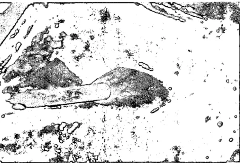
The left lateral lobe was excised as described in group, i. The
lcft central lobe was easily and clearly exposed (Fig. 7). it was
grasped medially to manipuIate its pedicle by applying a double
crushing ligaturc with silk No. 2 using Desham's needle. The ligature
crushed the hepatic parenchyma and closed the hepatic vessels and
ducts.
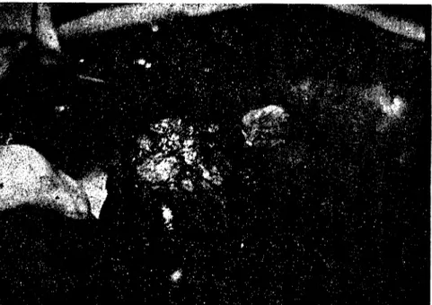
The Ieft central lobe was excised about 0.5 cm distally to the
embedded ligature (Fig. 8). Abdominal wall was closed in the usual
Fig. 7: Exposure of the left central lobc. Fig. 7: Sol merkezi locun açılımı.
366 M.T. NASEF - MA ALİ - kS. SALEH - M.M. ABD EL-HAFEEZ
Fig. 8: The stump of the lcft central lobe alter complete excision. Fig. 8: T",m eksizyondan sonra merkezi lobun kalıntısı.
Group IV: 40
%
cholecystilobectomy.In this gorup the left central and lcft lateral lobes as well as the gall bladder were resected as described before.
Posl-operalive care:
The animals of all groups were subjected to fluid therapy which consists ,of replacement of extraceIIular fluid losses with dextrose 5
%
and normal saline 500 ml of each for 2 cusuccessive days. Parentral
admİnistration of broad spectrum antibiotic ampiciIIin vials wİth
the dose 250 mg jdaily for 5 successive days. Delerminali(jn of regeneration rale:
Total liver weight could be estimated
as
it forms aout 3.5%
of total body weight(22). Remnant Iiver weİghts were obtained by
subtraction of the resected lobes from the total liver weights. Two
months post -operativeIy, anİmals were sacrİfİced. AII liver masses
were removed from attachments and adhesİons. In case of partial
KÖPEKLERDE KARACIGERiN YENIDEN ONARıMı' . . 367
common bile duct at the same lcvel of ligation in case of eholecystio-bectomy. Total liver masses were weighed. Liver gain weights were
calculated by subtraction of the remnant liver weights after
opera-tion from the liver weights at the time of authausia. Regeneration
rates were ealculated (4) from the formula:
Liver gain weight
Regeneration rate = --- --- ---- X 100
Removed -weigh t
Results
Clinical Observatioııs:
Animals recovered from anaesthesia and were ambulant two
hours after completion of the operation without any eomplications.
Convaleseence was unremarkable. They were off food on the second
day depending upon fluid therapy only. By the third day, animals
were in normal gencral healthy eondition, ate their own offered food and water. Skin ~titches were removed 10 days after operation. Only two cases show ed smail stitch abscesses resultin~ from euttin~ the stiteh through the whole thickness of the skin by biting the animal
himself. The abscesses nceded only three successive days deressing.
A dean healin~ was achieved. Aıı animals remained alert, active
without any signs of ilIncss. Animals of all groups survived up to the terminal point of all the experiment.
Liver regenemtion percentage:
The animal body weight, estimated liver wei~ht, the resccted
lobes weight, the remnant liver weight, the liver weight at sacrificing and the liver gain weight were shown in (Table, 1). From these data,
regeneration percentage were (24.66-42.85 %), (22.79-46.63 %),
(30.27-64.28 %) and (25.82-82.14 %) with the means overall of
31.1::1:3.9
%,
36.0 ::1:3.97%,
and 42.76:1:5.90%
in the groups,I, II, III and IV respectively. A non signifieant incrcase was observed in both choleeystilobectomized groups comparing with corresponding
hepatic lobectomized oneS.
Discussion
The results of this study confirmed that the mass ligation techni-que was the simplest and accurate for a complete hepatie lobectomy.
368 M.T. NASEF - MA ALİ - A.S. SALEH - M.M. ABD EL-HAFEEZ
Table i. Mcan valucs and standar error of rcgeneration percentagc in prc and post-hcpatie lobeetomy and eho1ceystilobcetomy in dogs.
Group, i
i
Group, II . Group, III i Gro,'p, IV !Animal weight 9.20:1: 0.86 12.00+ 1.51 i 13.601: 2.00 IO.40:!: 0.92 '
(kg). (7-12) i (8-17) i (9-21) : (8-13) ,
---~~--.-- - I-.---.---i~---i
Est. livcr weight i 322.00:1:30.LO 420.00=53.08: 476.00:!:73.08 364.00:!:32.45 .
__ (g_~_). , (245-420) (280 --595) 1_~~5-7~)
~~~~~~_1
Rcsectcd lobc weight i%~40:l: 9. LO 125.801:15.82 i 190.401:29.23 145.60:1:12.98 'i
(gm). (73-126) (48-178); (126-294) (112--182) .
_._ . _~ __ . --- 1 - _
I
Rcmenant livcr weight 1225.60::1:21.00 288.80:1:41.01 285.60::1:43.84 2IS.40:!:19.47 'i'Igm). (172-294) (196-417) _(I~?~41)_J_~168-~~ __
Liver weight at saerifieing i 256JlO:1:24.
81-
358.00-:'48.'92 362.001:47.47 280.00i: 14.14 i(gm). (190-330) (230-500) (270-530) (240-320)'
Livcr
!:f:h;(~~~.
--i
3°i{~~t/9-
4\~~-=8~~~-,
-76dg~~~6Ö-
61i~f:!:~~).67-1
I
RCg~tion- 1-~o::ı:-3:'ıo- 36.00=~97 -i-42.7[,+5.'90-1-45 .401:ıQ.'15-
1~ __ p_erccntage. . (24.66-42.85) (22.79--46.63)_i._(3_0.p-64.28) , (25_._82_-~8~~_
Group i == 30 % partial hcpatectomy. Group III = 40 % partial hcpateetomy.
Group II = 30 % cholceystiIobcetomy. Group IV = 40 % eholceystilobectomy.
The use of Deschamb's ncedIe for application of an encirding
liga-ture at the most proxima I part of the lobe pedides made insurance
()f accurate complete lobectomy. Placing the ligature and tying the
knot, the thread cut out through the parenchymatous tissue and tied
together with hepatic vessds (10, II). The cut parenchymal tissue
prevent the ligature from slipping. The hepatic and biliary vessels
were induded inside the encirding ligature. In the present study
the Mass ligation technique overcomes the vascular and bilary
ana-tamical variations, whereas these lobal' structures were included in-side the encircling ligature.
The most comman complicatiom of hepatir lobectomy was the
haemorrhagc (I, 2). Neither minor nar major hepatic haemorrhage
occurred in any of the dogs in the present study. Cut of parenchymal
£tump by means of diathermy knife dectrode was more effective in
achieving hacmostasis.
Simply, chromic catgut ligature was placed around the root of
the pedides of the lobe to be resected
(ı
O,ı
I). The ligatuı-c cut through the parenchyma, gathered and constricted the blood vessels and biliary ducts. Cu! of the stump was done by means of surgieal scalpeI. ThisKÖPEKLERDE KARACtGERtN YENİDEN ONARIMT '.' . 369
technique had the advantage of avoiding the difficult and dangerous dissection of the tis~ues and hepatic blood vessels. In the present study, silk was used for pedide ligation of the reseeted lobe. This
nonabso;bab-le material had the advantages of its ability to retain strength in
vivo and its low tissues reaetivity.
Biliary decompression was prefered prior to the disseetion as it
has same advantages(3). The gaIl bladder becomes smaIler and
oe-cupie:.;
a
lcss surgical field and less danger exists of spillage of bile dur~ İng operative disseetion whieh results in biliary peritonitis. The authorhad injected sterilc saline solution subserosaIly where thegaIl blad~
dcr and the liver adhere. This teehnique allawed ~erosa to be attached
to the liver bed a.ıd prevented a row liver surfaee. In the present
study, biliary deeompression was done when gentile pressure to the fundus of the bladder and empting its content into the duadenum. Subserosal injection of saline was not performed in the present proee-dures to avoid the risk of injuring the hepatic parenehyma Or puneture of the gall bladder ncek as it has a very thin walı.
The results of regeneration percentage in the prescnt study were
30. i % and 42.76 % in 30 % and 40 % partial hepatic lobeetomy
after eigh t weeks and 36% and 45.4 % in case of 30 and 40 % eholeeys-stilobeetomy after the same time respeetively. There results were
sup-ported by other researehers (4,12,13,17,18) who conduded that the
regeneration response was influenced by the amount of liver removed
at operation. The present ~;tudyconduded that there was no significant
differences in regeneration percentage between partial hepatectomy
and corresponding choleeystilobectomy.
Complete hepatic regeneration required 8-10 weeks (20) as
they resulted in 99.2% hepatic regeneration percentage after LO weeks
post 42 % partial hepatectomy. Increase and enlargement in
hepa-tocytes whieh was true hepatic restoration or restitution was more
evi-dent in 70 % than 40 % parti al hepatectomy (24, 25). A complete
regeneration İn a shorter time (6 weeks after surgery ) was gained
when 95 % hepatectomy and the reminant papillary process of the
caudate lobe 5 % reconstituted the original liver mass (26).
References
I. Bjorling, D.E., Prasse, K.W. and HollUes, R.A. (1985). Partial hepatectomy in dogs. Compend Cont Educ. Pract.Vet. 7: 257-263.
2. Blass, C.E. and Seinı, G.H. (1985). Surgical techııigue for the liver and biliary tracl. Vet. Clin. North Am. (smail animal practicc). 15: 257-275.
370 M.T. NASEF - M.A. ALt - A.S. SALEH - M.M ABD EL-HAFEEZ
3. Breznock, E.M. (1983). Surgery of hepatie parendıymal and biliary tissues. In: Bojrab MJ .• ed. Current teehniqucs in smail anima! surgery 2 nd Ed. Philadc1phia Lea
& Febİger. 212-225.
4. Chlld, C.G., Barr, D., Holswade, G.R. and Harrison, G.S. (1963). Liver regenera-tion following portaeaval transposition in dDgs. Annals of Surgery 138: 600-608.
5. Coppa, G.F. and Ranson, J.H.C. (1985). Hepatu: reseetion for metastastu: eolon and rectal eanur. Ann Surg.202-203.
6. De Bakey, M.E. and Jordan, G.L. (1977). Hepatic abscesses, both intra and extra hepa-tU:. Surg. Clin. North Am.57 (2): 430-433.
7. De Boer, J.; Downie, H.G. and Archibald, J. (1970). Intra hepatie billio digestive anastomosis in dogs. Surgery 68: 646--{)52.
8. Dingwall, I. S., De Boer, J. and Archibald, J. (1970). A new leehnique for liver reseetion
in the dDg.
9. Drazner, J. (1985). The liver and biliary tract. In Goarley, I.M. and Vassear, P.B. General small animal surgery. Lippieott Company Philadelphia London, Mexİeo Cİty, New York, Sydney.
ıo. E1.Anırousi, S., E1-Guindi, M., E1-Monzaly, M. and Mottelib, A. (1971)
Ex-perimental studies on partial hepaleetomy in dogs. 1- Surgieal proeedures and alteration in serum tramamİnases. U.A.R. J.Vet. Sei.,8, (i) 17-24.
ll. EI Guindi, M.H. and Mottelib, A.A. (1974). The effeet of partial hepaleetomy on some blood eonstituents in dogs. Assiut Vet,. Med.,J. 1, 1-2: 139-145.
12. Francavllla, A., Ove, P., Polimeno, L., Coetzee, M., Makowka, L.; Rose J., Van Thiel, D.H. and Strazel, T.E. (1987). Extraetion and partial purijuation of hepa-tU: stimulatory substance in trats, mu:e and dogs. Cancer Res. ;\Iov. 1 47 (21): 5600-5605. 13. Francavnıa, A. Porter, K., Benicbou, J., Jones, A. and Starzel, T. (1978).
Liver regeneration in dDgs: Morphologu:al and ehemieal changes . .Joumal of Surgieal Rese-arch 25, 409-419.
14. Kirk, R.W. and Bistner, S.I. (1975). Hand Book of Veterinary Procedures and Emergeney
Treatment. 2 nd Ed. W.B. Saunders Company, Philadc1phia, London, Toronto.
15. Lopukhin, Yu. M. (1976). Experimental Surgery. ıst Ed. Mirr Publishers, Moseow.
16. Lucas, C.E. and Walt, A.J. (1970). Critieal deeisions in liver trauma. Areh. Surg. 101: 1207-212.
17. Mackenzie, RoJ., Franciva1, C.M., Wood, C.B." Maoreen, A., O'Keane, M. and Blunıgart, L.H. (1977). The effeet of prologed hepatic isehaemia before 70percent partial hepateetomy in the dDg. Br.J. Surg.64: 66-69.
18. Mackenzie, Roj. Furnival, C.M. and O'Keane, M. (1975). The effeets of hepatu: ischaemü on liver function and the restoration of liver mases after 70 %hepaleetomyx iı,the dog.
Br.J.Surgery, 62: 431-437.
19. Martin, E.C. (1981). Pereutaneous drainage ın the management of hepatu: abseess. Sur.
KÖPEKLERDE KARACİÖERİN YENİDEN ONARIMI. .. 371
20. Mizumoto, R.;Kawarada, Y., Yamiawaki, T., Noguchi, T. and Nishida, S. (I 979). Resectability and Junctional reseroe
~r
the lioer with obstruttioe jaundice in dogs. Am.J. of Surg. 137 (6): 768-778.
21. Orda, R., Wiznitzer, T., Goldwasser, B., Bubis, J., Lcvene, S. and Kaye, R.
(1977). Partial hepateetomies with alzand - held lasser knife: An experimenl'lt'study. Br.J.
Surg. 64: 857--B61.
22. Sigel, B., (1963). Partial hepatectomy in the dog. Areh. Surg. 87 (IL): 788-,.791. 23. Singh, H., (1987). Hepateetamy simpli.fıed with the use oj water sealpel. Imiian J.
Gastro-enterology, 6 (3): 151-152.
24. Starzel, T.E.; Porter, K.A., Hayashide, N., Schechter, P. and Terblanche, J.
(1980). Fur!her studies on hepatie stimulatory substance (SS.)after partial hepalectomy. J.
Surg. Res. 29: 471-474.
25. Starzel, T.E., Terblanche, J., Porter, K. A., Jones, A. F., Usui, S. and Maz-zoni, G. (1979). Growth stimulating Jaetor in the reger.erating tanirıe lioer. Laneet, 1:
12/-131.
26. Szawlowski, A. W., Saint-Aubert, B., Gouttebel, M.C., Asttre, C. and Jouyeux, H. (1982). Experimenlal model oj extended repeated partiallzepatectomy in the dog. Eur. Surg.