Diagn Interv Radiol 2014; 20:172–177 © Turkish Society of Radiology 2014
The importance of craniovertebral and cervicomedullary angles in
cervicogenic headache
Gökçen Çoban, İlker Çöven, Bilal Egemen Çifçi, Erkan Yıldırım, Ayşe Canan Yazıcı, Bahriye Horasanlı
NEURORADIOLOGY
ORIGINAL ARTICLE
PURPOSE
Many studies have indicated that cervicogenic headache may originate from the cervical structures innervated by the up-per cervical spinal nerves. To date, no study has investigated whether narrowing of the craniovertebral angle (CVA) or cer-vicomedullary angle (CMA) affects the three upper cervical spinal nerves. The aim of this study was to investigate the effect of CVA and/or CMA narrowing on the occurrence of cervicogenic headache.
MATERIALS AND METHODS
Two hundred and five patients diagnosed with cervicogen-ic headache were included in the study. The pain scores of patients were determined using a visual analog scale. The nonheadache control group consisted of 40 volunteers. CVA and CMA values were measured on sagittal T2-weighted magnetic resonance imaging (MRI), on two occasions by two radiologists. Angle values and categorized pain scores were compared statistically between the groups.
RESULTS
Intraobserver and interobserver agreement was over 97% for all measurements. Pain scores increased with decreasing CVA and CMA values. Mean angle values were significantly differ-ent among the pain categories (P < 0.001). The pain score was negatively correlated with CMA (Spearman correlation coefficient, rs, -0.676; P < 0.001) and CVA values (rs, -0.725; P < 0.001).
CONCLUSION
CVA or CMA narrowing affects the occurrence of cervico-genic headache. There is an inverse relationship between the angle values and pain scores.
C
ervicogenic headache (CH) is a referred pain from the cervical structures innervated by the three upper cervical spinal nerves. Anesthetic blocks of the cervical structures or related nerves can provide temporary pain relief, suggesting that the pain may be due to a neck disorder (1–4). Possible causes of CH are atlantooccipital joint, at-lantoaxial joint, zygapophyseal joint, intervertebral disc, and upper cer-vical spinal nerve pathologies. However, there is agreement that degen-erative changes in the cervical spine do not necessarily correlate with pain (1, 5). The pain in CH may originate from various anatomic struc-tures in the cervical spine. The diagnostic value of such changes remains controversial, and their relevance in CH is unknown. Many studies have indicated that CH may originate from the cervical structures innervated by the upper cervical spinal nerves and trigeminal nerve branches (6, 7). To date, no study has investigated whether narrowing of the craniover-tebral angle (CVA) or cervicomedullary angle (CMA) affects the trigemi-nal nerve branches and three upper cervical spitrigemi-nal nerves via stretching, thereby causing pain. Herein, we aimed to determine whether CVA and/ or CMA narrowing has any effect on CH occurrence.Materials and methods
A total of 4122 patients were admitted to the Neurology and Neuro-surgery Department of Bakent University with a complaint of headache between January 2011 and May 2012, and 205 of them diagnosed with CH were included in the study. The diagnosis of CH was made according to the International Headache Society (IHS) diagnostic criteria for CH (1):
A. Pain, referred from a source in the neck and perceived in one or more regions of the head and/or face, fulfills criteria C and D. B. Clinical, laboratory, and/or imaging evidence of a disorder or
le-sion within the cervical spine or soft tissues of the neck is known to be, or generally accepted as, a valid cause of headache.
C. There is evidence that the pain can be attributed to the neck disor-der or lesion based on at least one of the following:
1. Demonstration of clinical signs that implicate a source of pain in the neck.
2. Abolition of headache following diagnostic block of a cervical structure or its nerve supply using placebo or other adequate con-trols. Abolition of headache means complete relief of headache, indicated by a score of zero on a visual analog scale (VAS). D. Pain resolves within three months after successful treatment of the
causative disorder or lesion.
Following approval by the Institutional Review Board of our institu-tion, the patients were evaluated for symptoms related to CH. All
pa-From the Departments of Radiology (G.Ç. drgokcencoban@ gmail.com, B.E.Ç., E.Y.), Neurosurgery (İ.Ç.), and Neurology (B.H.), Başkent University School of Medicine, Konya, Turkey; the Department of, Biostatistics (A.C.Y.), Başkent University School of Medicine, Ankara, Turkey.
Received 9 May 2013; revision requested 10 June 2013; revision received 3 August 2013; accepted 10 August 2013.
Published online 4 December 2013. DOI 10.5152/dir.2013.13213
tients were informed about the study, and detailed informed consent was ob-tained from each participant.
Patient exclusion criteria were as follows: hyperlipidemia, hyperten-sion, rheumatoid arthritis, abnormal findings on brain magnetic resonance imaging (MRI) (multiple sclerosis [MS], tumors, infarct, ischemic gliosis, vas-cular malformation, fracture, or infec-tion), or a diagnosis of primary head-ache such as migraine, tension-type or cluster-type headache. The nonhead-ache control group (group 0) consist-ed of 40 volunteers with similar de-mographic characteristics to those of the pain group. A neurologist with 10 years of experience (approximately six years of involvement in the subspe-cialty of headaches, particularly mi-graines and four years of experience as a neurosurgeon) made the CH diag-nosis in all 205 patients (pain group) and verified the lack of headache in the control group. Blockage of the occipital nerve was applied, and the patients who had responded to the medications for the last 2–3 months were included in the study. The pain scores were determined using the 100 mm VAS and categorized into six pain groups, ranging 0–5 (no pain, 0–2 mm; mild pain, 2–17 mm; moderate pain, 17–47 mm; severe pain, 47–77 mm; very severe pain, 77–96 mm; and most severe pain imaginable, 96–100 mm). There was no patient in the sixth pain group, which was thus excluded from the study. Brain MRI was performed in all patients within two days of the clin-ical diagnosis.
Brain MRI was performed in a rou-tine natural supine position using a 1.5 Tesla scanner (Intera, Gyroscan, Philips Medical Systems, Best, The Netherlands). This natural position places the head in slight extension and represents the position in all static clinical MRIs that are obtained for the measurement of CVA and CMA. All images were taken according to a stan-dard protocol using axial T2-weighted turbo spin echo (TR/TE, 4466/100 ms; slice thickness [ST], 5 mm; number of excitations [NEX], 3], coronal and sag-ittal T2-weighted turbo spin echo (TR/ TE, 4800/100 ms; ST, 4 mm; NEX, 3), axial fluid-attenuated inversion recov-ery (TR/TE, 6000/100 ms; ST, 5 mm;
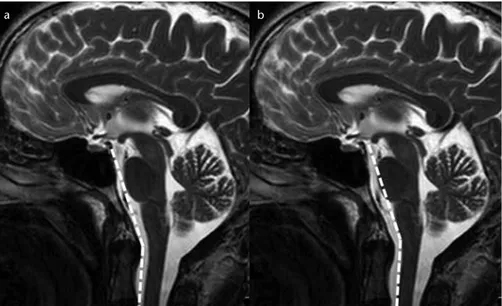
NEX, 3), and axial T1-weighted spin echo sequences (TR/TE, 462/11; ST, 5 mm; NEX, 3) covering the whole brain and upper cervical spine. All brain MRIs of the pain and control groups included the first two upper cervical vertebrae. Midline sagittal images of the T2-weighted series were used to obtain measurements. The CVA was constructed by drawing a line along the clivus and extrapolating it inferior-ly into the upper cervical spinal canal (Fig. 1a). The angle between the two lines on the ventral side of the medul-la oblongata and upper cervical spinal cord constituted the CMA (Fig. 1b).
The CVA and CMA values were mea-sured by two radiologists who were each blinded to the other’s results. CVA and CMA values were measured on two occasions by each radiologist with a four-week interval. The radiol-ogists were also blinded to the clinical findings of the patients.
Statistical analysis
Compliance with the normal distribu-tion of continuous variables was checked using the Shapiro-Wilk test. Levene’s test was used to analyze the homogene-ity of the groups’ variances. The groups’ variances were homogeneous in terms
Figure 1. a, b. T2-weighted mid-sagittal images show the normal craniovertebral angle (a) and normal cervicomedullary angle (b).
a b
Figure 2. a, b. T2-weighted mid-sagittal images show the normal craniovertebral angle (a) and normal cervicomedullary angle (b) in a normal participant with a pain score of 0.
of the pain severity groups, so group means were compared using two-factor analysis of variance, followed by the methods of multiple comparison using Bonferroni’s test. Correlations between variables were evaluated using Spear-man correlation coefficient. Reliability analysis was performed, and intraclass correlation coefficients were calculated for consistency of measurements.
The Statistical Package for the Social Sciences version 17.0 software (SPSS Inc., Chicago, Illinois, USA) was used for the analysis of data sets. A P value less than 0.05 was deemed to indicate statistical significance.
Results
Two hundred and forty-five sub-jects were included in the study (155 females aged 32.1±9.7 years and 90
males aged 31.7±8.2 years; overall mean age, 31.9±8.9 years; range, 14–78 years). The pain group consisted of 205 patients (135 females and 70 males; mean age, 31.8±9.3 years; range, 14–78 years), whereas the nonheadache con-trol group consisted of 40 volunteers (20 females and 20 males; mean age, 31.2±7.3 years; range, 20–55 years) with similar demographic character-istics to those of the pain group. We divided patients into five groups ac-cording to the pain scores. There were 49 patients in pain group 1 (mild pain) (30 females and 19 males; mean age, 33.4±8.8 years; range, 14–50 years), 59 patients in pain group 2 (moderate pain) (30 females and 29 males; mean age, 33.6±10.5 years; range, 18–78 years), 52 patients in pain group 3 (se-vere pain) (38 females and 14 males;
mean age, 31.1±8.2 years; range, 16–47 years), and 45 patients in pain group 4 (very severe pain) (37 females and 8 males; mean age, 30.0±9.8 years; range, 18–60 years). There was no patient in pain group 5 (most severe pain, sixth group of the VAS). The control group was indicated as group 0 (no pain). Ages were comparable among the four pain groups and controls (P = 0.167), and there was also no age difference between the gender groups (P = 0.773). Intraobserver and interobserver agree-ment were found to be over 97% for all measurements (P < 0.001, Table 1).
Pain scores increased with decreas-ing CVA values, and the difference was statistically significant (P < 0.001). There was no difference in CVA values between the control group (group 0) and pain group 1 (P = 0.685). Howev-er, CVA values were different among all other pain groups compared with groups 0 and 1 (P < 0.01).
Pain scores also increased with de-creasing CMA values, and the differ-ence was statistically significant (P < 0.001). There was no difference in CMA values between pain groups 2 and 3 (P = 0.069). However, CMA val-ues were different among the other pain groups and compared with pain groups 2 and 3 (P < 0.05).
The relationship between the CMA and CVA values and pain scores was statistically significant for each group (Tables 2, 3).
The relationship between the pain score and CMA (Spearman correlation coefficient, rs, -0.676; P < 0.001) and the relationship between the pain score and CVA (rs, -0.725; P < 0.001) values were negatively correlated. There was an inverse relationship between the pain scores and mean angle values. In addition, the relationship between the pain score and CMA and CVA values were negatively correlated with gender (Table 4).
In 245 subjects, there was an 83.4% correlation between CVA and CMA values (rs, 0.834; P < 0.001) according to gender.
Ninety five patients showed a re-stricted range of motion on physical examination, and all were in the se-vere pain (50 patients in group 3) and very severe pain (45 patients in group 4) groups.
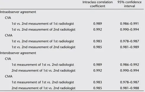
Table 1. Intraobserver and interobserver agreement for CVA and CMA measurements
Intraclass correlation 95% confidence
coefficient interval Intraobserver agreement CVA 1st vs. 2nd measurement of 1st radiologist 0.989 0.986–0.991 1st vs. 2nd measurement of 2nd radiologist 0.992 0.990–0.994 CMA 1st vs. 2nd measurement of 1st radiologist 0.983 0.978–0.987 1st vs. 2nd measurement of 2nd radiologist 0.985 0.981–0.989 Interobserver agreement CVA 1st measurement of 1st vs. 2nd radiologist 0.989 0.986–0.992 2nd measurement of 1st vs. 2nd radiologist 0.992 0.990–0.994 CMA 1st measurement of 1st vs. 2nd radiologist 0.983 0.978–0.987 2nd measurement of 1st vs. 2nd radiologist 0.985 0.981–0.988 CMA, cervicomedullary angle; CVA, craniovertebral angle.
Agreements were found to be over 97% for all measurements (P < 0.001).
Table 2. Craniovertebral angle (°) in the pain groups and significance of differences between the groups
95% confidence P
Pain group (n) Mean±SD interval Group 1 Group 2 Group 3 Group 4 Group 0 (n=40) 153.3±4.9 151.6–155.0 0.685 < 0.001 < 0.001 < 0.001 Group 1 (n=49) 150.9±5.3 149.5–152.7 0.004 < 0.001 < 0.001 Group 2 (n=59) 147.2±6.2 145.8–148.6 < 0.001 < 0.001 Group 3 (n=52) 142.8±5.6 141.0–144.4 < 0.001 Group 4 (n=45) 136.9±5.0 134.5–138.7 SD, standard deviation.
Discussion
Our study showed that CVA and CMA narrowing affects the occurrence of CH. The average CMA and CVA val-ues in CH patients were significantly narrower than those in controls, and there was an inverse relationship be-tween the pain scores and CVA and CMA values (Figs. 2–4).
The differential diagnosis of CH is difficult in practice. Cervical structures facilitate the pain; however, CH is not limited to neck pain. The pain in CH
ra-diates from the back to frontal regions and is also a referred pain from the cer-vical structures innervated by the three upper cervical spinal nerves (3). Consid-ering the difficulty in the clinical diag-nosis, the IHS developed new diagnos-tic criteria for CH in 2004 (1). Tumors, fractures, infections, rheumatoid arthri-tis, posterior cranial fossa lesions, and vertebral artery dissection or aneurysms should be ruled out (7).
Our pain groups (205 patients) were selected according to the following
di-agnostic criteria: patients with hyperlip-idemia, hypertension, rheumatoid ar-thritis, abnormal findings on brain MRI (such as MS, tumors, infarct, ischemic gliosis, vascular malformation, fracture, infection) and those diagnosed with primary headache such as migraine, tension-type or cluster-type headaches were excluded from the study.
CH shows a female predominance (8), which was also observed in our study. Trauma is a predisposing factor (8). In our study, 12 patients (5%) had a history of head trauma; no patient in the control group suffered neck trauma.
Craniovertebral junction (CVJ) align-ment is altered when cervical spine de-generation occurs, and CMA and CVA values also change with advancing age. The pathogenesis of CH is unclear and starts earlier in life (at approximately 32–35 years of age) (9, 10); thus, most cases are not caused by spondylosis. In our study, the mean age of the pain group was 31 years.
The exact pathophysiology of CH is unknown, but dysfunction, weakness, and spasms of the muscles may lead to CH. Some authors have suggested that mechanical cervical spine pathologies and dysfunction in neck muscles may produce painful and limited neck mo-tion (11, 12).
Trigger points in the posterior neck muscles (trapezius, sternocleidomas-toid, and splenius capitis) and peric-ranial tenderness have been associated with CH (1). Many authors have indi-cated muscle imbalance, such as mus-cle tightness and weakness, and others have confirmed decreased strength and endurance of the deep neck flex-ors in CH patients (11, 13).
Schellhas et al. (14) mapped extraspi-nal pain with a radicular distribution as well as distant or referred pain re-lated to different levels of the cervical spine. It was concluded that trigem-inal pain is infrequently related to cervical disc pathology (15). Fukui et al. (16) showed that the C2/3 to C7/ Th1 joints are related to a pain distri-bution pattern that does not involve trigeminal areas. C2/3 and C3 stimu-lation can produce occipital pain but no headache at the forehead (16). All these studies indicate that most neck lesions do not produce CH and cannot explain the CH source.
Table 3. Cervicomedullary angle (°) in the pain groups and significance of differences between the groups
95% confidence P
Pain group (n) Mean±SD interval Group 1 Group 2 Group 3 Group 4 Group 0 (n=40) 159.5±4.1 157.9–161.1 0.017 < 0.001 < 0.001 < 0.001 Group 1 (n=49) 155.7±5.2 154.4–157.4 0.017 < 0.001 < 0.001 Group 2 (n=59) 152.6±5.3 151.3–154.0 0.069 < 0.001 Group 3 (n=52) 149.7±5.7 148.1–151.4 0.026 Group 4 (n=45) 146.0±5.1 143.7–147.8 SD, standard deviation.
Table 4. Spearman correlation coefficient (rs) for pain score-CVA, pain score-CMA, and CVA-CMA correlations
Total Female Male
rs P rs P rs P
Pain score vs. CVA -0.725 < 0.001 -0.717 < 0.001 -0.679 < 0.001 Pain score vs. CMA -0.676 < 0.001 -0.647 < 0.001 -0.678 < 0.001 CVA vs. CMA 0.834 < 0.001 0.836 < 0.001 0.817 < 0.001 CMA, cervicomedullary angle; CVA, craniovertebral angle.
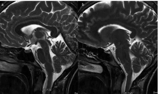
Figure 3. a, b. T2-weighted mid-sagittal images show the narrowed craniovertebral (a) and cervicomedullary (b) angles in a patient with a pain score of 3.
The most common cause of spinal dysfunction in the elderly is spondy-lotic myelopathy. In that group of pa-tients, symptoms include weakness of the arms and legs, upper motor neuron dysfunction, and sensory symptoms; headache is usually not among the symptoms. The clinical observations show that spondylosis is not an import-ant cause of headache (15). Knackstedt et al. (17) indicated no difference in disc degeneration or MRI signal chang-es in the transverse and alar ligaments among patients with CH, migraine, and whiplash-associated headaches. There-fore, CH might not be due to the neck lesions, as indicated in such studies (1, 5, 15), and it would not be appropriate to focus only on the structural changes. The source of CH remains crucial for detecting its main pathology. Like most authors, we suggest that the spi-nal nucleus of the trigemispi-nal nerve can be highly suspected as the pain source in CH (7, 17, 18). The pain originating from the cervical structures innervat-ed by the upper cervical spinal nerves might be sensed in areas innervated by the trigeminal nerve branches. Con-vergence between the cervical afferents and nervus trigeminus may result in CH (7, 18, 19). In our study, we focused on this pathway and investigated wheth-er narrowing of the CVA and CMA af-fects the trigeminal nerve branches and three upper cervical spinal nerves via stretching, thereby causing pain.
The CVJ refers to the occiput, atlas, axis, and supporting ligaments. It also
includes the cervicomedullary junction (medulla, spinal cord, and lower cranial nerves). Signs and symptoms are vari-able in CVJ abnormalities, although most signs and symptoms may lead the patient to consult a neurologist or neurosurgeon. Presently, with the large number of patients undergoing MRI, it is easy to routinely evaluate the CVJ on sagittal MRI of the brain (20).
The CMA was first described by Bundschuh et al. (21) as the angle subtended by the lines drawn parallel to the ventral surfaces of the medulla and upper cervical cord on MRI. Bund-schuh et al. (21) reported that there was a strong correlation between CMA values less than 135° and clinical ev-idence of cervical myelopathy, brain-stem compression, or C2 root pain. In some current studies, the average CMA values were reported to be 155.2°, 163°, and 158.4°, respectively (21–23).
The CVA is formed with a line con-structed along the posterior surface of the axis body and the odontoid pro-cess (Wackenheim clivus baseline or clivus-canal angle) (24). The CVA nor-mally ranges from 150° in flexion to 180° in extension (25). Ventral spinal cord compression may occur when the angle is less than 150° (25).
The mean CMA and CVA values in our control group were 159.5°±4.2° and 153.3°±4.9°, respectively. Howev-er, in the pain groups, the mean CMA (152.9°±6.6°) and CVA (146.6°±7.6°) values were significantly narrower. An inverse and statistically significant
re-lationship was demonstrated between pain scores and CVA and CMA.
Some authors have suggested that the range of motion decreases in CH patients (26, 27). Hall et al. (27) indi-cated that range of motion restriction can be related to headache intensity. This relationship may explain why all 95 patients were in the severe pain (50 patients in group 3) and very severe pain (45 patients in group 4) groups in our study.
One limiting factor in our study was the insufficient number of subjects in the control group. If the subject num-ber in the control group was close to that in the patient group (165 pa-tients), we could have assigned cutoff values for the CVA and CMA for the occurrence of pain. The CMA and CVA values might change following physio-therapy, and medication might not be needed. However, in our study, none of the patients received physiotherapy, and we also did not monitor the CMA and CVA values after the medication; thus, further studies are needed.
In conclusion, our study showed that narrowing of the CVA and CMA affects the occurrence of CH. There was an in-verse relationship between the angle values and the pain scores (Figs. 2–4). Brain MRI is applied in patients pre-senting with headache to exclude or-ganic causes. During the evaluation of the brain MRI in these patients, CVA and CMA measurements obtained from the sagittal images should be mentioned in the report.
Conflict of interest disclosure
The authors declared no conflicts of interest. References
1. Headache Classification Subcommittee of the International Headache Society. The international classification of headache disorders: 2nd edition. Cephalalgia 2004; 24:9–160.
2. Sjaastad O, Fredriksen TA, Pfaffenrath V. The Cervicogenic Headache International Study Group. Cervicogenic headache: di-agnostic criteria. Headache 1998; 38:442– 445. [CrossRef]
3. Sjaastad O, Fredriksen TA. Cervicogenic headache: criteria, classification and ep-idemiology. Clin Exp Rheumatol 2000; 18:3–6.
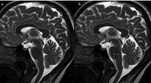
Figure 4. a, b. T2-weighted mid-sagittal images show the prominently narrowed craniovertebral (a) and cervicomedullary (b) angles in a patient with a pain score of 4.
4. Merskey H, Bogduk N. Classification of chronic pain. Description of pain syn-dromes and definitions of pain terms. 2nd ed. Seattle: IASP Press, 1994. 5. Pollmann W, Keidel M, Pfaffenrath V.
Headache and the cervical spine: a criti-cal review. Cephalalgia 1997; 17:801–816.
[CrossRef]
6. Kerr FW. Central relationships of trigem-inal and cervical primary afferents in the spinal cord and medulla. Brain Res 1972; 43:561–572. [CrossRef]
7. Bogduk N. The neck and headaches. Neu-rol Clin 2004; 22:151–171. [CrossRef]
8. Vincent MB. Cervicogenic headache: clin-ical aspects. Clin Exp Rheumatol 2000; 18:7–10.
9. Vincent MB, Luna RA. Cervicogenic head-ache: a comparison with migraine and tension-type headache. Cephalalgia 1999; 19:11–16.
10. Sjaastad O, Bakketeig LS. Migraine with-out aura: comparison with cervicogenic headache. Vaga study of headache ep-idemiology. Acta Neurol Scand 2008; 117:377–383. [CrossRef]
11. Watson DH, Trott PH. Cervical headache: an investigation of natural head posture and upper cervical flexor muscle performance. Cephalalgia 1993; 13:272–284. [CrossRef]
12. Knackstedt H, Bansevicius D, Aaseth K, Grande RB, Lundqvist C, Russell MB. Cer-vicogenic headache in the general pop-ulation: The Akershus study of chronic headache. Cephalalgia 2010; 30:1468– 1476. [CrossRef]
13. Janda V. Muscles and motor control in cervicogenic disorders. In: Grant R, ed. Physical therapy of the cervical and tho-racic spine. Edinburgh: Churchill Living-stone, 1994; 195–215.
14. Schellhas KP, Smith MD, Gundry CR, Pol-lei SR. Cervical discogenic pain. Prospec-tive correlation of magnetic resonance imaging and discography in asymptomat-ic subjects and pain sufferers. Spine 1996; 21:300–311. [CrossRef]
15. Vincent MB. Cervicogenic headache: the neck is a generator: con. Headache 2010; 50:706–709. [CrossRef]
16. Fukui S, Ohseto K, Shiotani M, et al. Re-ferred pain distribution of the cervical zygapophyseal joints and cervical dorsal rami. Pain 1996; 68:79–83. [CrossRef]
17. Knackstedt H, Kråkenes J, Bansevicius D, Russell MB. Magnetic resonance imag-ing of craniovertebral structures: clinical significance in cervicogenic headaches. J Headache Pain 2012; 13:39–44. [CrossRef]
18. Bartsch T, Goadsby PJ. Increased respons-es in trigeminocervical nociceptive neu-rons to cervical input after stimulation of the dura mater. Brain 2003; 126:1801– 1813. [CrossRef]
19. Kerr FW. Central relationships of trigem-inal and cervical primary afferents in the spinal cord and medulla. Brain Res 1972; 43:561–572. [CrossRef]
20. Smoker WR. Craniovertebral junction: normal anatomy, craniometry, and con-genital anomalies. Radiographics 1994; 14:255–277. [CrossRef]
21. Bundschuh C, Modic MT, Kearney F, Morris R, Deal C. Rheumatoid arthritis of the cervical spine: surface-coil MR imag-ing. Am J Roentgenol 1988; 151:181–187.
[CrossRef]
22. Abumi K, Takada T, Shono Y, Kaneda K, Fujiya M. Posterior occipitocervical reconstruction using cervical pedicle screws and plate-rod systems. Spine 1999; 24:1425–1434. [CrossRef]
23. Wang S, Wang C, Passias PG, Li G, Yan M, Zhou H. Interobserver and intraobserver reliability of the cervicomedullary angle in a normal adult population. Eur Spine J 2009; 18:1349–1354. [CrossRef]
24. Wackenheim A. Roentgen diagnosis of the craniovertebral region. New York, NY: Springer Verlag, 1974.
25. VanGilder JC, Menezes AH, Dolan KD. The craniovertebral junction and its ab-normalities. New York, NY: Futura, 1987. 26. Zito G, Jull G, Story I. Clinical tests of
musculoskeletal dysfunction in the diag-nosis of cervicogenic headache. Man Ther 2006; 11:118–129. [CrossRef]
27. Hall TM, Briffa K, Hopper D, Robinson KW. The relationship between cervi-cogenic headache and impairment de-termined by the flexion rotation test. J Manipulative Physiol Ther 2010; 33:666– 671. [CrossRef]