Discussion
Noncompaction cardiomyopathy is a rare genetic disorder with a reported prevalence of 0.05% (2); however, the true measure is believed to be higher. Noncompaction can be an isolated cardiomyopathy or associated with cardiac or extracardiac, particularly neuromuscular disorders. But, the coexistence of NC with genital anomalies is very rare, and we could find only two recently reported cases (3, 4). Both of them were male patients. Our case had genital and skeletal anomalies, in addition to NC. Coexistence of hypergonadotropic hypogonadism with dilated cardiomyopathy was first reported 1973 by Najjar et al. (5), and since that time, only 15-20 similar cases have been reported in the English literature. Since this condition is known to be caused by muta-tions in the LMNA gene, we screened this gene for mutamuta-tions but found nothing. The common features of these cases are dilated cardiomyopa-thy, hypoplastic genitalia, and hypergonadotropic hypogonadism. Although cardiac involvement in our case is distinct from these reports, the extracardiac manifestations are very similar, especially to the case reported by Narahara (6). It is possible that at least some of these cases were noncompaction cardiomyopathy but not dilated cardiomyopathy, because at that time, NC was not yet reported or was not well known.
Conclusion
In conclusion, this is the first case with NC and female genital anomalies. Coexistence of biventricular NC, genital and skeletal anom-alies, and mental retardation leads one to consider the presence of a syndrome, but we have not been able to find a similar combination of symptoms in the literature. Although our case is isolated, the unex-plained death of the patient’s siblings supports the inheritance.
Video 1. Transthoracic parasternal short-axis view, showing mild pericardial effusion and global hypokinesia of the left ventricle Video 2-3. Transthoracic apical four-chambers views, showing hypertrabeculations on the lateral wall and apex of the left ventricle Video 4. Multiple intertrabecular recesses in communication with the ventricular cavity demonstrated by forward and reverse flow of blood on color flow Doppler
Video 5. Cardiac MRI, confirming biventricular hypertrabeculations, suggesting noncompaction cardiomyopathy
References
1. Nakashima K, Kusakawa I, Yamamoto T, Hirabayashi S, Hosoya R, Shimizu W, et al. A left ventricular noncompaction in a patient with long QT syndrome caused by a KCNQ1 mutation: a case report. Heart Vessels 2013; 28: 126-9. [CrossRef]
2. Ritter M, Oechslin E, Sütsch G, Attenhofer C, Schneider J, Jenni R. Isolated noncom-paction of the myocardium in adults. Mayo Clin Proc 1997; 72: 26-31. [CrossRef]
3. Ojala T, Polinati P, Manninen T, Hiippala A, Rajantie J, Karikoski R, et al. New mutation of mitochondrial DNAJC19 causing dilated and noncompaction cardiomyopathy, anemia, ataxia, and male genital anomalies. Pediatr Res 2012; 72: 432-7. [CrossRef]
4. Özcan KS, Osmonov D, Altay S, Güngör B, Eren M. Fertile eunuch syndrome in association with biventricular noncompaction, bicuspid aortic valve, severe aortic stenosis, and talipes equinovarus. Tex Heart Inst J 2013; 40: 204-6. 5. Najjar SS, Kaloustian VM der, Nassif SI. Genital anomaly, mental retardation,
and cardiomyopathy: a new syndrome? J Pediatr 1973; 83: 286-8. [CrossRef]
6. Narahara K, Kamada M, Takahashi Y, Tsuji K, Yokoyama Y, Ninomiya S, et al. Case of ovarian dysgenesis and dilated cardiomyopathy supports exis-tence of Malouf Syndrome. Am J Med Genet 1992; 144: 369-73. [CrossRef]
Address for Correspondence: Dr. Halil Ataş, Marmara Üniversitesi Eğitim ve Araştırma Hastanesi, Pendik, İstanbul-Türkiye
Phone: +90 532 546 25 35 Fax: +90 216 657 09 65 E-mail: [email protected] Available Online Date: 25.12.2014
©Copyright 2015 by Turkish Society of Cardiology - Available online at www.anakarder.com DOI:10.5152/akd.2014.5722
Combined catheter thrombus
fragmentation and percutaneous
thrombectomy in a patient with
massive pulmonary emboli and acute
cerebral infarct
Aylin Özsancak Uğurlu, Özlem Çınar*, İsmail Caymaz**, Halime Çevik**, Burçak Gümüş**
Departments of Pulmonary, *Anesthesiology and Reanimation, **Radiology, Faculty of Medicine, Başkent University; İstanbul-Turkey Figure 2. Transthoracic echocardiography of the patient revealing
prominent trabeculations and intertrabecular recesses in the left ventricle
Figure 3. Cardiac magnetic resonance imaging of the patient confirming the findings of transthoracic echocardiography
Case Reports Anatolian J Cardiol 2015; 15: 69-74
Introduction
Acute massive pulmonary embolism (PE) constitutes approximately one-fifth of all PE events and is a life-threatening condition (1, 2). Although systemic thrombolysis is the recommended first-line treat-ment, in the case of high risk of bleeding, surgical embolectomy and percutaneous interventions are the alternative treatment options (3, 4). Here, we present a patient with massive PE and an acute cerebrovas-cular accident (CVA), treated successfully with a relatively lesser known and practiced treatment option.
Case Report
A 64-year-old female was admitted to the emergency department with increased shortness of breath, chest pain, recurrent syncope, and convulsion. She was normotensive and had dyspnea and tachypnea with oxygen saturation of 85% on room air. Arterial gases confirmed hypoxemic respiratory failure, and an electrocardiogram revealed incomplete right bundle-branch block and nonspecific T wave changes. D-dimer and troponin I levels were 3889 and 0.61 ng/mL, respectively. A subsequent CT pulmonary angiogram (CTPA) showed intraluminal filling defects in both main arteries, extending into the lobar branches (Fig. 1a-d). Brain diffusion MRI demonstrated acute infarct areas, as well as chronic ones (Fig. 2a, b). The echocardiography demonstrated right ventricular dilatation and free-wall akinesis and flattening of the interventricular septum with an estimated pulmonary artery systolic
pressure of 60 mm Hg. However, the patient deteriorated clinically 1 hour after admission, with the need for intubation, Gelofusine, and inotrope infusion. The patient had major contraindications for systemic thrombolytic treatment because of the acute CVA and was also consid-ered a poor surgical candidate for embolectomy. Therefore, she was taken to the interventional radiology catheter laboratory for percutane-ous intervention.
Pulmonary angiography via a 5-F pigtail catheter, advanced through the inferior vena cava to the pulmonary truncus, revealed increased filling defects in both pulmonary arteries extending into the lobar branches compared to CTPA (Video 1). A 6-F guiding catheter was then advanced through both pulmonary arteries, and with assistance of a 0.035-inch hydrophilic guidewire, the guiding catheter was advanced beyond the thrombus, and large amounts of fragmented thrombus were recurrently aspirated. The control angiogram revealed a significant reduction in thrombus burden from both pulmonary arteries, with sig-nificant restoration of blood flow (Video 2). Oxygenation parameters and hemodynamics improved gradually. After the procedure, the patient was re-started on i.v. unfractionated heparin infusion. A reduction in systolic pulmonary arterial pressure (from 55-60 mm Hg to 40-45 mm Hg) with relatively increased right ventricular systolic functions was revealed in the echocardiography 1 day after the procedure. The patient was extubated on the 7th day and discharged on the 16th day.
Discussion
Acute massive PE is a relatively common condition with most hazard-ous life-threatening manifestations of venhazard-ous thromboembolism (1, 3, 4). Systemic thrombolysis, in addition to anticoagulation, is the cur-rently approved treatment of acute massive PE (3, 4). Surgical embolec-tomy is indicated in selected centers as an alternative in massive PE with contraindications to thrombolytics, failed thrombolysis, or shock with a high risk of sudden mortality before thrombolysis can take effect; how-ever, it has rarely been performed even in the large registries (2, 4, 5). Percutaneous embolectomy is the only alternative in massive PE patients with contraindications to or failure of the treatment modalities mentioned above, if appropriate expertise and resources are available (3, 4).
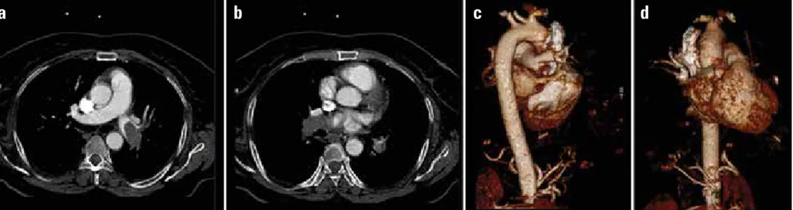
Selective catheterization of the main right and left pulmonary arter-ies is routinely performed for percutaneous interventions for PE, with the frequent necessity of selective or sub-selective catheterization of pulmonary segments (6). Thrombus fragmentation (with or without use of local thrombolytics) breaks apart large emboli by direct mechanical action, with a risk for macroembolization, hemoptysis, or temporary mechanical hemolysis (7). Percutaneous thrombectomy can be per-Figure 1. a-d. CT angiography and 3D images of intraluminal filling defect in the left (a, c) and right (b, d) main pulmonary arteries, extending into lobar branches
a
b
c
d
Figure 2. a, b. Brain diffusion MRI (b1000): demonstrated acute infarct areas in the left occipital (a) and bilateral cerebellar lobes (b)
a
b
Case Reports
formed by direct aspiration of emboli or with rheolytic thrombectomy devices, with later devices (e.g., AngioJet rheolytic devices) being recommended to be avoided because of the higher rate of major com-plications (like bradycardia, heart block, hemoglobinuria, and proce-dure-related deaths) (6, 8-10).
There are no randomized controlled trials comparing the effects of different mechanisms or devices used for percutaneous intervention for PE. According to the most extensive meta-analysis results, the clinical success (defined as the stabilization of hemodynamics, resolu-tion of hypoxia, and survival to hospital discharge) rate was 86.5%, with a major complication rate of only 2.4% (9).
Conclusion
This case shows that percutaneous interventions performed in experienced centers can be the only life-saving treatment option in patients otherwise dying from acute massive PE.
Video 1. Pulmonary angiography: before filling defects in both pul-monary arteries extending into lobar branches
Video 2. Control pulmonary angiography: reduction in thrombus from both pulmonary arteries
References
1. Jaff MR, McMurtry MS, Archer SL, Cushman M, Goldenberg N, Goldhaber SZ, et al. Management of massive and submassive pulmonary embolism, iliofemoral deep vein thrombosis, and chronic thromboembolic pulmonary hypertension: a scientific statement from the American Heart Association. Circulation 2011; 123: 1788-830. [CrossRef]
2. Goldhaber SZ, Visani L, De Rosa M. Acute pulmonary embolism: clinical outcomes in the International Cooperative Pulmonary Embolism Registry (ICOPER). Lancet 1999; 353: 1386-9. [CrossRef]
3. Arseven O, Sevinç C, Alataş F, Ekim N, Erkan L, Fındık S, et al. Pulmoner Tromboembolizm Tanı ve Tedavi Uzlaşı Raporu. Turk Thorac J 2009; 10: 1-46.
4. Kearon C, Akl EA, Comerota AJ, Prandoni P, Bounameaux H, Goldhaber SZ, et al. Antithrombotic therapy for VTE disease: Antithrombotic Therapy and Prevention of Thrombosis, 9th ed: American College of Chest Physicians Evidence-Based Clinical Practice Guidelines. Chest 2012; 141: e419S-94S. 5. Kasper W, Konstantinides S, Geibel A, Olschewski M, Heinrich F, Grosser
KD, et al. Management strategies and determinants of outcome in acute major pulmonary embolism: results of a multicenter registry. J Am Coll Cardiol 1997; 30: 1165-71. [CrossRef]
6. Uflacker R. Percutaneous interventions for pulmonary embolism. In: Mauro MA, Murphy KPJ, et al, eds. Image-Guided Interventions Philadelphia: Saunders, Elsevier; 2008.
7. Nakazawa K, Tajima H, Murata S, Kumita SI, Yamamoto T, Tanaka K. Catheter fragmentation of acute massive pulmonary thromboembolism: distal embolisation and pulmonary arterial pressure elevation. Br J Radiol 2008; 81: 848-54. [CrossRef]
8. Lee L, Kavinsky CJ, Spies C. Massive pulmonary embolism: review of man-agement strategies with a focus on catheter-based techniques. Expert Rev Cardiovasc Ther 2010; 8: 863-73. [CrossRef]
9. Kuo WT, Gould MK, Louie JD, Rosenberg JK, Sze DY, Hofmann LV. Catheter-directed therapy for the treatment of massive pulmonary embolism: sys-tematic review and meta-analysis of modern techniques. J Vasc Interv Radiol 2009; 20: 1431-40. [CrossRef]
10. Kuo WT. Endovascular therapy for acute pulmonary embolism. J Vasc Interv Radiol 2012; 23: 167-79. [CrossRef]
Address for Correspondence: Dr. Aylin Özsancak Uğurlu, Başkent Üniversitesi Tıp Fakültesi, Oymacı Sok. No: 2 Altunizade, İstanbul-Türkiye
Phone: +90 532 742 52 33 Fax: +90 216 651 66 26
E-mail: [email protected] Available Online Date: 25.12.2014
©Copyright 2015 by Turkish Society of Cardiology - Available online at www.anakarder.com DOI:10.5152/akd.2014.5765
Presented at the 2013 ATS International Conference, May 17-22, 2013, Philadelphia, Pennsylvania, as a ‘poster discussion’ in the ‘UNUSUAL CASES IN CRITICAL CARE: INTENSIVIST BEWARE’ session (10.1164/ajrccm confer-ence.2013.187.1_MeetingAbstracts.A5050 A5050).
Case Reports Anatolian J Cardiol 2015; 15: 69-74