Postoperative pain intensity associated with the use of different
nickel-titanium shaping systems during single-appointment
endodontic retreatment: a randomized clinical trial
Tan Firat Eyuboglu, DDS, PhD/Mutlu Özcan, DDS, Dr med dent, PhD
Objective: The objective of this randomized clinical trial was to compare the effect of different NiTi shaping systems on postoperative pain after single-appointment nonsurgical end-odontic retreatment. Method and materials: Between Sep-tember 2016 and December 2016, 99 patients with asymptom-atic root canal-treated teeth requiring nonsurgical endodontic retreatment were randomly divided into three groups (n = 33 per group). After removing previous root canal filling, instrumen-tation was performed using One Shape, Revo-S, and Wave One systems in groups 1, 2, and 3, respectively. Postoperative pain intensity was assessed at 6, 12, 18, 24, 48, and 72 hours, 7 days, and 1 month after the retreatment. Data were analyzed using one-way ANOVA and Mann-Whitney U and Kruskal-Wallis tests
(alpha = .01). Results: Up to 72 hours, postoperative pain was significantly less in group 1 than in groups 2 and 3 (P < .01). From 72 hours to 7 days, postoperative pain was significantly less in groups 1 and 2 (P > .05), compared to group 3 (P < .01). At 1 month, postoperative pain was not significantly different among all three groups (P > .05). Postoperative pain was the highest with WaveOne group. Conclusions: Since One Shape and Revo-S are both based on the rotational approach and WaveOne on reciprocal approach, less incidence of postoper-ative pain intensity with One Shape and Revo-S in single- appointment nonsurgical endodontic retreatment could be associated with the motion type during root canal shaping.
(Quintessence Int 2019;50: 624–634; doi: 10.3290/j.qi.a42693)
Key words: continuous rotation, postoperative pain, randomized controlled clinical trial, reciprocating motion, retreatment, single-appointment endodontic retreatment
Postoperative dental pain is directly associated with host- dependent factors such as history of preoperative pain1 and occlusal trauma.1,2 However, chemical and mechanical injury or bacterial infection during root canal preparation considered as operator-dependent factors may also induce postoperative dental pain.4 During root canal treatment, postoperative pain is mainly caused by apically extruded debris containing bacteria, necrotic tissue, and dentin.5,6 Although it is possible to control certain aspects of the treatment, such as irrigation regime, instrumentation technique, and file characteristics, it is impos-sible to control other aspects such as virulence and bacterial species during the treatment.7
Several in-vitro studies have reported some amount of debris extrusion with both manual and engine-driven instru-mentation techniques.8-10 Furthermore, other studies on api-cally extruded debris have reported a significant difference between various engine-driven nickel-titanium (NiTi) instru-mentation techniques.11-13 Although those studies mentioned an association between apically extruded debris and post-operative pain, due to the factors mentioned above, the clinical relevance of this association may be plausible particularly in nonsurgical endodontic retreatment. Microbial habitat in pre-viously treated teeth is very different and more resistant to che-momechanical treatment than that in untreated teeth,14 which
may alter the clinical association between apically extruded debris and postoperative pain caused by host-dependent fac-tors. Therefore, from a clinical view point, a randomized clinical trial may be more suitable for assessing the effect of different instrumentation techniques and file characteristics on post-operative pain, particularly in the presence of complex micro-bial environments, even though different factors may affect the results. Although clinical studies on postoperative pain are associated with disadvantages such as subjectivity of data and effect of other factors on obtained results,14-17 comparison of in-vitro data on apically extruded debris with in-vivo data of postoperative pain developed during the retreatment of end-odontically failed teeth may improve understanding of post-operative dental pain.
In spite of the fact that previous studies have provided con-tradicting results on single-appointment nonsurgical root canal treatments,18-21 high success rate of these treatments can
be achieved using novel equipment and techniques.20,22 Pre-vention of recontamination, decrease in microleakage, reduc-tion in treatment time, cost and high success rate and patient request of single-appointment treatments have increased the indication of single-appointment nonsurgical retreatments.23-26
Postoperative dental pain may be a poor indicator for long- term success.27 Yet, it may increase the reluctance of clinicians to perform these procedures, necessitating a refinement of the procedure, particularly operator-dependent factors associated with postoperative pain. Therefore, the objective of this clin-ical trial was to investigate the effect of different engine-driven NiTi root canal-shaping systems on postoperative dental pain in patients with asymptomatic teeth after a single-appoint-ment nonsurgical root canal retreatsingle-appoint-ment. The null hypothesis tested was that there would be no significant difference between the root canal-shaping systems on postoperative dental pain.
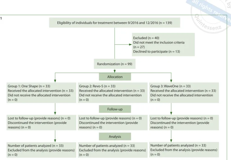
Eligibility of individuals for treatment between 9/2016 and 12/2016 (n = 139)
Excluded (n = 40)
Did not meet the inclusion criteria (n = 27) Declined to participate (n = 13) Randomization (n = 99) Allocation Follow-up Analysis
Lost to follow-up (provide reasons) (n = 0) Discontinued the intervention (provide reasons) (n = 0)
Number of patients analyzed (n = 33) Excluded from the analysis (provide reasons) (n = 0)
Lost to follow-up (provide reasons) (n = 0) Discontinued the intervention (provide reasons) (n = 0)
Lost to follow-up (provide reasons) (n = 0) Discontinued the intervention (provide reasons) (n = 0)
Number of patients analyzed (n = 33) Excluded from the analysis (provide reasons) (n = 0)
Number of patients analyzed (n = 33) Excluded from the analysis (provide reasons) (n = 0)
Fig 1 CONSORT flow chart for eligibility, allocation, follow-up, and analysis of the patients receiving single-appointment nonsurgical retreatment using NiTi root canal-shaping systems.
Group 1: One Shape (n = 33)
Received the allocated intervention (n = 33) Did not receive the allocated intervention (n = 0)
Group 2: Revo-S (n = 33)
Received the allocated intervention (n = 33) Did not receive the allocated intervention (n = 0)
Group 3: WaveOne (n = 33)
Received the allocated intervention (n = 33) Did not receive the allocated intervention (n = 0)
Method and materials
Inclusion and exclusion criteria
This study was designed as a single center, single-blind, pro-spective randomized clinical trial. In addition to approval of the local ethics committee of Istanbul Medipol University, Istanbul, Turkey, the study was registered at clinicaltrials.gov with the ID number NCT03478241. All procedures performed in this study involving human participants were in accordance with the ethical standards of the institutional and/or national re-search committee and with the 1964 Helsinki declaration and its later amendments or comparable ethical standards.
Initially, a power analysis (G*Power 3.1.7) was conducted for sample size calculation. It was estimated that a minimum sam-ple size of 26 individuals per group would be required for an effect size (Cohen’s effect size) of 0.80 (with an alpha error of 0.05 and a power beta of 0.80) in order to achieve 95% confi-dence of a true difference between the groups. In case approx-imately 20% of the patients would not respond, the total adjusted sample size required was calculated to be 96.
Between September 2016 and December 2016, 139 pa-tients with asymptomatic teeth and with no contradictory medical history were assigned for nonsurgical retreatment at the Department of Endodontics, Faculty of Dentistry, Istanbul Medipol University. All teeth presented radiographic evidence of periapical lesion (periapical index > 2) and an initial root ca-nal treatment not shorter than 4 mm.
Exclusion criteria were the following:
■ patients younger than 18 years
■ patients with symptomatic teeth, teeth with vertical root fractures, excessive periodontal disease, teeth that need periodontal surgery prior to coronal restorations due to marginal deficiency, teeth with damaged or resorbed peri-apex, or teeth that were treated with a fiber post
■ patients who received or required surgical endodontic treatment
■ patients diagnosed with systemic diseases
■ patients who used analgesics 12 hours before or antibiotics 1 month before the retreatment
■ patients who could not abide the follow-up time of the study.
Accordingly, in total 27 patients were excluded from the study and 13 patients declined to participate in the study (Fig 1). Teeth that were root canal-treated at least 2 years before with
evident periapical lesions clarifying failure of the previous treatment were included in the study.
Patients who met the inclusion criteria and agreed to par-ticipate in the study were randomly divided into three groups (n = 33 patients per group). Thus, 99 teeth in 99 patients (53 women and 46 men; mean age: 45.7 ± 13.9 years) were nonsur-gically retreated by one endodontics specialist (TFE) who had 14 years’ experience. According to the clinical regulations of the department, all new endodontic devices and instruments are practiced in extracted teeth as part of the calibration proto-col and then practiced in patients.
All enrolled patients volunteered and signed a written informed consent form. A stratified randomization was used in order to distribute different tooth types homogenously into their respected groups. In order to achieve this objective, six envelopes were prepared according to different tooth types, namely maxillary molar, mandibular molar, maxillary premolar, mandibular premolar, maxillary anterior, and mandibular anter-ior teeth. Equal group numbers from all three groups were placed in each envelope to cover the number of teeth to be treated. Hence, 21 numbers (seven from each group) for 21 mandibular molar teeth, 18 numbers (six from each group) for 17 maxillary molar teeth, 15 numbers (five from each group) for 15 mandibular premolar teeth, 27 numbers (nine from each group) for 25 maxillary premolar teeth, nine numbers (three from each group) for eight mandibular anterior teeth, and 15 numbers (five from each group) for 14 maxillary anterior teeth were placed in the respective envelopes. One clinical assistant provided the respective envelopes to the patients and asked them to choose a number from the envelopes.
Removal of previous restorations and root canal
fillings
The retreatments were performed in a single appointment under ×3.5 magnification particularly in the morning in order to make it easier for the patients to abide the follow-up time schedule. Before gaining access to the teeth, coronal restor-ations were removed. Root posts were removed with a surgical clamp (portegue). Previous root canal fillings in the coronal third of the root were removed using no. 1, 2, and 3 Gates Glid-den (GG) burs (Mani) after preparing access cavities. Remaining root canal fillings were removed using no. 15 K-files (Mani). Pre-vious root canal fillings were removed carefully and without shaping the root canal walls as much as possible. No chemical solvent was used to remove gutta-percha or sealer, and extreme care was taken not to proceed beyond the apex. Root
lengths were measured using an apex locater (MM Control, Micro-Méga) before preparing a glide path inside the root canals by using no. 15 K-files. Next, the teeth were divided into three groups for performing the shaping procedure.
Shaping the root canal
■ Group 1: In this group, One Shape (Micro-Méga) was used for cleaning and shaping root canals using the crown-down technique. Each tooth was prepared at a distance of 0.5 mm from the apex. Apical 1 (size 30, 0.06 taper) and 2 files (size 35, 0.06 taper) were used respectively for apical shaping. Api-cal 1 and ApiApi-cal 2 have 0.06 tapers at only apiApi-cal 5 mm from the tip of the files. The rest of the files did not have taper. After each instrumentation, root canals were irrigated using 1 mL 2.5% sodium hypochlorite (NaOCl). Final irrigation was performed using 2.5 mL 5% ethylenediaminetetraacetic acid (EDTA), 2.5 mL 2.5% NaOCl, and 5 mL distilled water. The root canals were filled with Apical 2 gutta-percha (Micro-Méga) using a single-cone technique. MM Seal (Micro-Méga) was used as a filling paste and was introduced inside the root canals using a master cone through brushing motion. Acces-sory gutta-percha cones (SU 25, Revo-S, Micro-Méga) were used if needed using the non-compaction method (Fig 2).
■ Group 2: In this group, Revo-S (Micro-Méga) was used for cleaning and shaping root canals using the crown-down technique. Each tooth was prepared at a distance of 0.5 mm from the apex. SC1, SC2, and SU files were used for shaping, and AS 30, AS 35, and AS 40 files (size 40, 0.06 taper) for apical shaping. Irrigation and final irrigation were per-formed using protocols similar to those perper-formed for patients in group 1. The single-cone technique was used to introduce the MM Seal into the root canal using AS 40 gutta- percha cone in a brushing motion. Accessory gutta- percha cones (SU 25, Revo-S) were used if needed using the non-compaction method (Fig 2).
■ Group 3: In Group 3, WaveOne (Dentsply Maillefer) was used for cleaning and shaping root canals. Each tooth was prepared at a distance of 0.5 mm from the apex. Primary (red; size 25, 0.08) or large (black; size 40, 0.08) files were used according to root canal diameter. Irrigation and final irrigation were performed using protocols similar to those used in patients in groups 1 and 2. The single-cone tech-nique was used to introduce the MM Seal into the root canal by means of a matching gutta-percha cone (Wave-One) in brushing motion. Accessory gutta-percha cones (SU 25, Revo-S) were used if needed employing the non-com-paction method (Fig 2).
2a 2b 2c 2d
2e 2f
Figs 2a to 2f Radiographic evaluation of three groups before and after the retreatment: (a) One Shape group, right maxillary first molar tooth, before treat-ment; (b) One Shape group, right maxillary first molar tooth, after treatment; (c) Revo-S group, left maxillary first molar tooth, before treatment; (d) Revo-S group, left maxillary first molar tooth, after treatment;
(e) WaveOne group, right mandibular first molar tooth,
before treatment; (f) WaveOne group, left mandibular first molar tooth, after treatment.
Coronal restorations were performed using total-etch adhesive system (Single Bond 2, 3M Espe), according to the manufactur-er’s instructions. Root canal orifices were sealed using a flow-able composite resin (Filtek Ultimate Flowflow-able, 3M Espe) as the base material. Remaining coronal restorations were performed using composite resin (Filtek Ultimate, 3M Espe) or a fiber post (Cytec Blanco, HT-Glasfiber, E. Hahnenkratt) cemented with composite resin material (RelyX U200, 3M Espe) followed by composite resin (Filtek Ultimate) restoration before performing fixed restoration depending on the prosthetic plan.
Evaluation of postoperative pain
The participants were given a self-administered questionnaire to assess and record their postoperative pain intensity at 6, 12, 18, 24, 48, and 72 hours, 7 days, and 1 month after the retreatment as described in a previous study.17 Each patient was prescribed naproxen sodium (550 mg) to take only in case of severe pain. Acetaminophen (500 mg) was prescribed if naproxen sodium was contraindicated. In addition, the patients were asked to record the number of days for complete pain resolution. More-over, postoperative pain was recorded using a four-level verbal rating scale, which was validated in previous studies,17,28,29 where 0 indicated no pain, 1 indicated slight pain, 2 indicated moder-ate pain, and 3 indicmoder-ated severe pain. Furthermore, the patients were asked to return to the clinic for the controls and bring the questionnaires at 24, 48, and 72 hours, 7 days, and 1 month after the treatment. One clinician performed all the clinical assess-ments at 24, 48, and 72 hours, 7 days and 1 month. In addition to postoperative pain, unscheduled appointments for emergency treatment or any complication such as postoperative swelling or paresthesia were also recorded in the patient charts.
Statistical analysis
Statistical analysis was performed using Number Cruncher Sta-tistical System 2007 StaSta-tistical Software (NCSS). Data were
expressed as mean, standard deviation, median, frequency, percentage, minimum, and maximum values. One-way analysis of variance (ANOVA) was used for intergroup comparisons of parameters showing normal distribution, and Tukey HSD test was used for post-hoc analysis. Independent sample t test was used to make comparisons between two groups. Kruskal-Wallis test was used for intergroup comparisons of parameters not showing normal distribution, and Mann-Whitney U test for pairwise comparisons. Pearson chi-square test was used for comparing qualitative data. A P value of < .05 was considered statistically significant in all tests.
Results
No drop out was experienced throughout the study. Age, gen-der, and analgesic use were not significantly different among the study groups (P > .05). Patient distribution according to the tooth types is presented in Table 1. The number of patients in each pain category (0 to 3) among the groups at various time points is presented in Table 2.
Statistical analysis indicated significant difference in pain intensity between 6, 12, 18, 24, 48, and 72 hours and 7 days among the groups (P = .001). When pairwise comparisons of 6-, 12-, 18-, and 24-hour pain intensity values were compared, Group 3 (WaveOne) group presented the highest pain intensity values, followed by patients treated using Group 2 (Revo-S) (P = .001) and One Shape (P = .001). Pain intensity values for patients treated using Revo-S (P < .01) were significantly higher than those of patients treated using One Shape (P = .001) (Table 2 and Fig 3).
Pairwise comparisons of 48- and 72-hour pain intensity val-ues showed that patients treated using WaveOne resulted in the highest pain intensity values, followed by patients treated using Revo-S (P = .001) and One Shape (P = .001). Moreover, 48-hour pain intensity values in patients treated using Revo-S were significantly higher than those of the patients treated using One Shape (P = .002). However, no significant difference
Table 1 Distribution of number of teeth in 99 patients assigned for each NiTi root shaping system
Mandibular molar Maxillary molar Mandibular premolar Maxillary premolar Mandibular anterior Maxillary anterior Total One Shape 7 5 5 8 3 5 33 Revo-S 7 5 5 9 2 5 33 WaveOne 7 6 5 8 3 4 33
Table 2 Pain intensity levels associated with different NiTi root canal-shaping systems at different time points
Time Pain score
Group 1: Shape (n = 33) Group 2: Revo-S (n = 33) Group 3: WaveOne (n = 33) P† Pairwise comparison P‡ 6 h 0 [n (%)] 17 (51.5) 5 (15.2) 1 (3.0) .001** 3 > 1, 2; 2 > 1 1 [n (%)] 10 (30.3) 8 (24.2) 4 (12.1) 2 [n (%)] 6 (18.2) 18 (54.5) 7 (21.2) 3 [n (%)] 0 (0.0) 2 (6.1) 21 (63.6) Median (Q1–Q3) 0 (0–1) 2 (1–2) 3 (2–3) Mean ± SD 0.67 ± 0.78 1.52 ± 0.83 2.45 ± 0–83 12 h 0 [n (%)] 20 (60.6) 6 (18.2) 1 (3.0) .001** 3 > 1, 2; 2 > 1 1 [n (%)] 8 (24.2) 10 (30.3) 7 (21.2) 2 [n (%)] 5 (15.2) 16 (48.5) 4 (12.1) 3 [n (%)] 0 (0) 1 (3) 21 (63.6) Median (Q1–Q3) 0 (0–1) 2 (1–2) 3 (1.5–3) Mean ± SD 0.55 ± 0.75 1.36 ± 0.82 2.36 ± 0.93 18 h 0 [n (%)] 21 (63.6) 6 (18.2) 1 (3.0) .001** 3 > 1, 2; 2 > 1 1 [n (%)] 7 (21.2) 12 (36.4) 7 (21.2) 2 [n (%)] 5 (15.2) 14 (42.4) 8 (24.2) 3 [n (%)] 0 (0.0) 1 (3.0) 17 (51.5) Median (Q1–Q3) 0 (0–1) 1 (1–2) 3 (1.5–3) Mean ± SD 0.52 ± 0.76 1.30 ± 0.81 2.24 ± 0.90 24 h 0 [n (%)] 23 (69.7) 7 (21.2) 1 (3.0) .001** 3 > 1, 2; 2 > 1 1 [n (%)] 8 (24.2) 12 (36.4) 9 (27.3) 2 [n (%)] 2 (6.1) 13 (39.4) 5 (15.2) 3 [n (%)] 0 (0.0) 1 (3.0) 18 (54.5) Median (Q1–Q3) 0 (0–1) 1 (1–2) 3 (1–3) Mean ± SD 0.36 ± 0.60 1.24 ± 0.83 2.21 ± 0.96 48 h 0 [n (%)] 27 (81.8) 16 (48.5) 6 (18.2) .001** 3 > 1, 2; 2 > 1 1 [n (%)] 5 (15.2) 8 (24.2) 6 (18.2) 2 [n (%)] 1 (3.0) 8 (24.2) 7 (21.2) 3 [n (%)] 0 (0.0) 1 (3.0) 14 (42.4) Median (Q1–Q3) 0 (0–0) 1 (0–2) 2 (1–3) Mean ± SD 0.21 ± 0.48 0.82 ± 0.92 1.88 ± 1.17 72 h 0 [n (%)] 29 (87.9) 24 (72.7) 13 (39.4) .001** 3 > 1, 2 1 [n (%)] 4 (12.1) 6 (18.2) 4 (12.1) 2 [n (%)] 0 (0.0) 2 (6.1) 6 (18.2) 3 [n (%)] 0 (0.0) 1 (3.0) 10 (30.3) Median (Q1–Q3) 0 (0–0) 0 (0–1) 1 (0–3) Mean ± SD 0.12 ± 0.33 0.39 ± 0.75 1.39 ± 1.30 7 d 0 [n (%)] 32 (97.0) 31 (93.0) 20 (60.6) .001** 3 > 1, 2 1 [n (%)] 1 (3.0) 2 (6.1) 10 (30.3) 2 [n (%)] 0 (0.0) 0 (0.0) 3 (9.1) Median (Q1–Q3) 0 (0–0) 0 (0–0) 0 (0–1) Mean ± SD 0.03 ± 0.17 0.06 ± 0.24 0.48 ± 0.67 1 mo 0 [n (%)] 33 (100.0) 33 (100.0) 33 (100.0) **P < .01. †Kruskal-Wallis test.
was observed in 72-hour pain intensity values between patients treated using Revo-S and One Shape (P = .101) (Table 2 and Fig 3).
Pairwise comparison of 7-day pain intensity values indi-cated that patients treated using WaveOne experienced the highest pain intensity values compared to the patients treated with Revo-S (P = .01) and One Shape (P = .01) files. However, no significant difference was observed in pain intensity values between patients treated using Revo-S and One Shape (P = .558) (Table 2 and Fig 3).
None of the patients reported any postoperative pain at 1-month follow-up. Two patients from the One Shape group, three patients from the Revo-S group, and six patients from the WaveOne group used analgesics (naproxen sodium) with no statistically significant difference among the groups (P > .05) (Table 2 and Fig 3).
Discussion
This clinical trial investigated the effect of different engine-driven NiTi root canal-shaping systems on postoperative dental pain in patients with asymptomatic teeth after a single-appoint-ment nonsurgical root canal retreatsingle-appoint-ment. Patients in all the groups experienced the highest postoperative pain at 6 hours after the retreatment but pain decreased gradually with time, and no pain was reported at 1 month after the retreatment. Thus, the null hypothesis could be rejected. These results are consis-tent with those of previous studies evaluating the incidence and severity of postoperative pain at different time points.28-31
All treatments were completed in a single appointment due to a previous research concluding favorable postoperative pain
intensity results in nonsurgical endodontic retreatments for this approach compared to two-appointment endodontic retreat-ments.32 Persistent pain after root canal treatment is a common occurrence, with a frequency of 5.4%.33 It can be classified as either or both odontogenic and nonodontogenic in etiology.34,35 While odontogenic origin might be the root canal-treated tooth or the adjacent tooth, nonodontogenic origin was shown to be temporomandibular disorder pain or dentoalveolar pain disor-der.35 In order to eliminate the risk of persistent pain, which would affect the results of this study, follow-ups were contin-ued up to 1 month to ascertain the complete relief of pain.
Several factors such as age, gender, pulpal and periradicular status, tooth type, preoperative pain, and technical aspects affect postoperative dental pain.30 Of these factors, only techni-cal aspects, including instrumentation technique, file charac-teristics, and irrigation and obturation protocols could be con-trolled by the clinician. These technical aspects, also referred to as operator-dependent factors, are the main causes of nonbio-logic (chemical and mechanical) or biononbio-logic (bacterial) injuries during root canal preparation.4,36 Therefore, in the present study, in order to limit the effect of variables in the procedure and prevent unwanted interaction with the apical tissues, no chemical solvents were used during the removal of the previ-ous root canal filling.
Subjectivity of measurement due to the perception of pain in patients is one of the main problems in pain intensity evalu-ation studies.14 Therefore, questionnaire design, patient fol-low-up, and inclusion and exclusion criteria play a significant role in such studies.16 Although in some studies tooth type selection was kept to teeth with a single root canal,17,37 other studies have included multiple tooth types.4,37 Since multiple
P ost oper a tiv e P ain I n tensit y 6h 12h 18h 24h 48h 72h 1 week 1 month
One Shape Revo-S WaveOne
Mean+SE 3.0 2.5 2.0 1.5 1.0 0.5 0.0 -0.5
Fig 3 Postoperative pain intensity level among patients in different groups as a function of time.
host- and operator-dependent factors affect postoperative pain, homogenous randomization of different tooth types and inclusion of increased numbers of patients should be consid-ered when investigating the relevance and performance of dif-ferent NiTi root canal-shaping techniques on pain induction. In the present study, the patients were homogenously random-ized according to the tooth types as much as possible in order to decrease this effect on postoperative pain. Although in a previous study 23 patients were assigned,38 the power analysis employed in the present study required inclusion of 33 patients for each group. Furthermore, all the treatments were per-formed by a single operator to decrease procedural bias.
Previous studies reported a strong association between apically extruded debris due to different instrumentation tech-niques and postoperative pain,40,41 while other studies have suggested some association between apically extruded debris and postoperative pain.17,37 However, the complexity of differ-ent factors affecting the postoperative pain have raised doubts about this association. Since host-dependent factors have an increased effect on teeth requiring nonsurgical retreatment, it can be suggested that apically extruded debris does not play an important role in the development of postoperative pain, as implicated in previous studies.37-41 As the teeth that do not respond to endodontic treatment may contain persistent bac-terial species compared with the species present during the initial endodontic treatment,42 host-dependent factors may mask or affect the relationship between apically extruded debris and postoperative pain. Moreover, persistent bacterial species in teeth not responding to endodontic treatment may increase the effect of other operator-dependent factors com-pared with that of apically extruded debris on the develop-ment of postoperative pain from a clinical viewpoint. Numer-ous studies have highlighted the insufficient cleaning and shaping of apical complexity as important factors in failure of endodontic treatments and persistence of periapical lesions,14,43-45 indicating the central role of apical shaping in both treatment success and decreased postoperative pain in these teeth. Therefore, it is important to better understand the relationship between postoperative pain and apically extruded debris, particularly in apically compromised teeth requiring nonsurgical retreatment. Although two meta-analyses have included in-vitro debris extrusion studies while evaluating operator-dependent factors in postoperative pain due to their strong correlation,46,47 the effect of other operator- and host- dependent factors on postoperative pain are yet to be verified.
In the present study, patients treated using a reciprocal sys-tem (WaveOne) reported significantly higher postoperative
pain intensity values than patients treated using the continu-ous rotational files (One Shape and Revo-S) until 7 days after the treatment, similar to previous studies,37,48,49 concluding that reciprocal files induced significantly more severe postoperative pain than those of continuous rotational files. Therefore, the null hypothesis cannot be accepted when 7-day data are con-sidered. Moreover, these results are consistent with the results of studies that compared apically extruded debris with recipro-cal and continuous rotational files, concluding greater apirecipro-cal extrusion of debris in reciprocal motion than continuous rotary motion.11,12,50,51 However, the present results contradict other studies that reported no significant difference in postoperative pain in patients treated using reciprocal and continuous rota-tional files.38,37,52-54 The discrepancy among the studies could be explained by different instrument types and study designs where only one type of teeth were used.
A significant difference was observed in postoperative pain intensity between patients treated using the continuous rota-tional single-file system (One Shape) and continuous rotarota-tional multiple-file system (Revo-S) until 72 hours’ follow-up, which was inconsistent with the meta-analysis where the instrument design and type of instrument were found more influential on the symptomatic apical periodontitis after root canal treatment than the number of files.53 Moreover, some studies assessing apically extruded debris showed no significant difference between single and multiple continuous rotational file sys-tems.12,50 Although several files were used for patients treated using One Shape, due to procedural necessities in nonsurgical retreatment, the intensity of postoperative pain was signifi-cantly higher in patients treated using Revo-S than those treated using One Shape. In an in-vitro study, although not sig-nificant, reciprocating files was reported to cause the highest amount of debris and irrigant extrusion, followed by single file and multiple file rotary systems.51 In another in-vitro study where mesiobuccal roots of extracted mandibular first molars were used, One Shape presented better centricity and less transportation abilities compared to Revo-S files. Moreover, apical diameter achieved during the instrumentation was dif-ferent between the file systems (size 35, 0.06 taper for One Shape and size 40, 0.06 taper for Revo-S), indicating that apical extrusion increases with an increase in apical diameter.7 Although the same explanation can be given for patients treated using WaveOne, where only one file with apical prepar-ation diameter of size 40, 0.08 taper or size 25, 0.08 taper was used for root canal shaping, the reciprocal motion seemed to effect postoperative pain more than the prepared apical diam-eter or number of files used during root canal shaping in single-
appointment nonsurgical retreatment.53 In a recent meta- analysis, higher intensity of postoperative pain was reported after single-visit root canal treatments with reciprocating files than continuous rotation files, which is consistent with the results of the present study.55 Reciprocating files cause more debris and irrigant extrusion compared to both single and mul-tiple rotary file systems.50 In another randomized clinical trial, the effect on postoperative pain of root canal preparation by a continuous rotation file system (ProTaper Next, Dentsply Sirona) and a reciprocating file system (WaveOne) was evalu-ated on 42 patients who were in need of root canal treatment, where all treatments were carried out at two appointments. The results indicated significantly higher postoperative pain intensity in the WaveOne group compared to the ProTaper Next group in both appointments.28 However, the highest postoper-ative pain intensity was recorded 6 hours after both appoint-ments. Based on the results of this study, both statements are consistent with the present results. Future clinical studies reporting on pain intensity should consider the possible effects of the variation in NiTi root canal-shaping systems.
Conclusion
Clinician-dependent factors affecting postoperative pain in a single-appointment nonsurgical endodontic retreatment could
be restrained to using shaping instruments based on a rota-tional approach with apical-shaping ability rather than those based on a reciprocal approach. The number of files used for root canal shaping and the prepared apical diameter of the root canal did not affect the postoperative pain results as much as the motion type during root canal shaping. The WaveOne NiTi shaping system was associated with the highest postoper-ative pain intensity values. Both One Shape and Revo-S systems have similar apical-shaping features and are based on the rota-tional approach, whereas WaveOne is based on the reciprocal approach, which may explain the lower postoperative pain val-ues associated with One Shape and Revo-S in single-appoint-ment nonsurgical endodontic retreatsingle-appoint-ments.
Declaration
The authors declare no conflict of interest regarding any of the materials used in this study.
Acknowledgment
The authors would like to thank to Ms E. Bor for her assistance with the statistical analysis.
References
1. Glennon JP, Ng Y-L, Setchell DJ, Gulabivala K. Prevalence of and factors affecting post preparation pain in patients undergoing two-visit root canal treatment. Int Endod J 2004;37:29–37.
2. Yu CY. Role of occlusion in endodontic management: report of two cases Aust Endod J 2004;30:110–115.
3. Caviedes-Bucheli J, Azuero-Holguin MM, Correa-Ortiz JA, et al. Effect of experimen-tally induced occlusal trauma on substance P expression in human dental pulp and peri-odontal ligament. J Endod 2011;37:627–630. 4. Siqueira JF Jr, Rôças IN, Favieri A, et al. Incidence of postoperative pain after intraca-nal procedures based on an antimicrobial strategy. J Endod 2002;28:457–460. 5. Siqueira JF Jr. Microbial causes of end-odontic flare-ups. Int Endod J 2003;36:453–463. 6. Kustarci A, Akpinar KE, Er K. Apical extrusion of intracanal debris and irrigant following use of various instrumentation techniques. Oral Surg Oral Med Oral Pathol Oral Radiol Endod 2008;105:257–262.
7. Tanalp J, Gungor T. Apical extrusion of debris: a literature review of an inherent occurrence during root canal treatment. Int Endod J 2014;47:211–221.
8. Reddy SA, Hicks ML. Apical extrusion of debris using two hand and two rotary instru-mentation techniques. J Endod 1998;24: 180–183.
9. De-Deus G, Brandão MC, Barino B, Di Giorgi K, Fidel RA, Luna AS. Assessment of apically extruded debris produced by the single-file ProTaper F2 technique under reciprocating movement. Oral Surg Oral Med Oral Pathol Oral Radiol Endod 2010;110: 390–394.
10. Capar ID, Arslan H, Akcay M, Ertas H. An in vitro comparison of apically extruded debris and instrumentation times with Pro-Taper universal, ProPro-Taper next, twisted file adaptive, and HyFlex instruments. J Endod 2014;40:1638–1641.
11. Bürklein S, Schäfer E. Apically extruded debris with reciprocating single-file and full-sequence rotary instrumentation systems. J Endod 2012;38:850–852.
12. Bürklein S, Benten S, Schäfer E. Quanti-tative evaluation of apically extruded debris with different single-file systems: Reciproc, F360 and OneShape versus Mtwo. Int Endod J 2014;47:405–409.
13. Yılmaz K, Özyürek T. Apically extruded debris after retreatment procedure with Reciproc, ProTaper Next, and Twisted File adaptive instruments. J Endod 2017;43: 648–651.
14. Nair PN. On the causes of persistent apical periodontitis: a review. Int Endod J 2006;39:249–281.
15. Risso PA, Cunha AJ, Araujo MC, Luiz RR. Postobturation pain and associated factors in adolescent patients undergoing one and two-visit root canal treatment. J Dent 2008;36:928–934.
16. Arias A, Azabal M, Hidalgo JJ, de la Macorra JC. Relationship between postend-odontic pain, tooth diagnostic factors, and apical patency. J Endod 2009;35:189–192.
17. Topçuoğlu HS, Topçuoğlu G. Post-operative pain after the removal of root canal filling material using different techniques in teeth with failed root canal therapy: a randomized clinical trial. Acta Odontol Scand 2017;75:249–254. 18. Friedman S. Considerations and con-cepts of case selection in the management of post-treatment endodontic disease treat-ment failures. Endod Topics 2002;1:54–78. 19. Tronstad L, Sunde PT. The evolving new understanding of endodontic infec-tions. Endod Topics 2003;6:55–77. 20. Trope M, Delano EO, Orstavik D. Endodontic treatment of teeth with apical periodontitis: single vs. multi-visit treatment. J Endod 1999;25:345–350.
21. Wang C, Xu P, Ren L, Dong G, Ye L. Comparison of postobturation pain experi-ence following one-visit and two-visit root canal treatment on teeth with vital pulps: a randomized controlled trial. Int Endod J 2010;43:692–697.
22. Ashraf H, Milani AS, Shakeri Asadi S. Evaluation of the success rate of nonsurgical single visit retreatment. Iran Endod J 2007; 2:69–72.
23. Weiger R, Rosendahl R, Lost C. Influence of calcium hydroxide intracanal dressing on the prognosis of teeth with endodontically induced periapical lesions. Int Endod J 2000; 33:219–226.
24. Ørstavik D, Pitt FTR. Apical periodontitis: microbial infection and host responses. In: Ørstavik D, Pitt FTR (eds). Essential Endodon-tology: Prevention and treatment of apical periodontitis. Oxford: Blackwell Science, 1998:1-8.
25. Wrong R. Conventional endodontic failure and retreatment. Dent Clin North Am 2004;48:265–289.
26. Eyuboglu TF, Olcay K, Özcan M. A clinical study on single-visit root canal retreatments on consecutive 173 patients: frequency of periapical complications and clinical success rate. Clin Oral Investig 2017;21:1761–1768. 27. Taintor JF, Langeland K, Valle GF, Krasny RM. Pain: a poor parameter of evaluation in dentistry. Oral Surg Oral Med Oral Pathol Oral Radiol Endod 1981;52:299–303. 28. Nekoofar MH, Sheykhrezae MS, Meraji N, et al. Comparison of the effect of root canal preparation by using WaveOne and ProTaper on postoperative pain: a random-ized clinical trial. J Endod 2015;41:575–578. 29. Kara Tuncer A, Gerek M. Effect of work-ing length measurement by electronic apex locator or digital radiography on postopera-tive pain: a randomized clinical trial. J Endod 2014;40:38–41.
30. DiRenzo A, Gresla T, Johnson BR, Rog-ers M, Tucker D, BeGole EA. Postoperative pain after 1- and 2-visit root canal therapy. Oral Surg Oral Med Oral Pathol Oral Radiol Endod 2002;93:605–610.
31. Pak JG, White SN. Pain prevalence and severity before, during, and after root canal treatment: a systematic review. J Endod 2011;37:429–438.
32. Erdem Hepsenoglu Y, Eyuboglu TF, Öz-can M. Postoperative pain intensity after sin-gle- versus two-visit nonsurgical endodontic retreatment: a randomized clinical trial. J En-dod 2018;44:1339–1346.
33. Nixdorf DR, Moana-Filho EJ, Law AS, McGuire LA, Hodges JS, John MT. Frequency of persistent tooth pain after root canal ther-apy: a systematic review and meta-analysis. J Endod 2010;36:224–230.
34. Klasser GD, Kugelmann AM, Villines D, Johnson BR. The prevalence of persistent pain after nonsurgical root canal treatment. Quintessence Int 2011;42:259–269. 35. Nixdorf DR, Law AS, John MT, Sobieh RM, Kohli R, Nguyen RH. National Dental PBRN Collaborative Group. Differential diag-noses for persistent pain after root canal treatment: a study in the national dental practice-based research network. J Endod 2015;41:457–463.
36. Ng YL, Mann V, Gulabivala K. A pro-spective study of the factors affecting out-comes of nonsurgical root canal treatment: Part 1: periapical health. Int Endod J 2011;44:583–609.
37. Çiçek E, Koçak MM, Koçak S, Sağlam BC, Türker SA. Postoperative pain intensity after using different instrumentation techniques: a randomized clinical study. J Appl Oral Sci 2017;25:20–26.
38. Comparin D, Moreira EJL, Souza EM, De-Deus G, Arias A, Silva EJNL. Postoperative pain after endodontic retreatment using ro-tary or reciprocating instruments: a random-ized clinical trial. J Endod 2017;43:1084–1088. 39. Yoldas O, Topuz A, Isçi AS, Oztunc H. Postoperative pain after endodontic retreat-ment: single- versus two-visit treatment. Oral Surg Oral Med Oral Pathol Oral Radiol Endod 2004;98:483–448.
40. Nair PN, Henry S, Cano V, Vera J. Micro-bial status of apical root canal system of human mandibular first molars with primary apical periodontitis after “one-visit” end-odontic treatment. Oral Surg Oral Med Oral Pathol Oral Radiol Endod 2005;99:231–252. 41. Caviedes-Bucheli J, Moreno JO, Carreno CP, et al. The effect of single-file reciprocating systems on Substance P and Calcitonin gene-related peptide expression in human periodontal ligament. Int Endod J 2013;46:419–426.
42. Sundqvist G, Figdor D, Persson S, Sjögren U. Microbiologic analysis of teeth with failed endodontic treatment and the outcome of conservative re-treatment. Oral Surg Oral Med Oral Pathol Oral Radiol Endod 1998;85:86–93.
43. Wang J, Jiang Y, Chen W, Zhu C, Liang J. Bacterial flora and extraradicular biofilm associated with the apical segment of teeth with post-treatment apical periodontitis. J Endod 2012;38:954–959.
44. Nair PN. Pathogenesis of apical peri-odontitis and the causes of endodontic fail-ures. Crit Rev Oral Biol Med 2004;15:348–381. 45. Rocas IN, Alves FR, Santos AL, Rosado AS, Siqueira JF Jr. Apical root canal micro-biota as determined by reverse-capture checkerboard analysis of cryogenically ground root samples from teeth with apical periodontitis. J Endod 2010;36:1617–1621. 46. Caviedes-Bucheli J, Castellanos F, Vasquez N, Ulate E, Munoz HR. The influence of two reciprocating single-file and two rota-ry-file systems on the apical extrusion of debris and its biological relationship with symptomatic apical periodontitis. A system-atic review and meta-analysis. Int Endod J 2016;49:255–270.
47. Hou XM, Su Z, Hou BX. Post endodontic pain following single-visit root canal prepa-ration with rotary vs reciprocating instru-ments: a meta-analysis of randomized clinical trials. BMC Oral Health 2017;17:86.
48. Gambarini G, Testarelli L, De Luca M, et al. The influence of three different instru-mentation techniques on the incidence of postoperative pain after endodontic treat-ment. Ann Stomatol (Roma) 2013;4:152–155. 49. Nayak G, Singh I, Shetty S, Dahiya S. Evaluation of apical extrusion of debris and irrigant using two new reciprocating and one continuous rotation single file systems. J Dent (Tehran) 2014;11:302–309.
50. Kucukyilmaz E, Savas S, Saygili G, Uysal B. Evaluation of apically extruded debris and irrigant produced by different nickel-tita-nium instrument systems in primary teeth. J Contemp Dent Pract 2015;16:864–868. 51. Küçükyilmaz E, Savas S, Saygili G, Uysal B. Assessment of apically extruded debris and irrigant produced by different nickel- titanium instrument systems. Braz Oral Res 2015;29:1–6.
52. Kherlakian D, Cunha RS, Ehrhardt IC, Zuolo ML, Kishen A, da Silveira Bueno CE. Comparison of the incidence of postopera-tive pain after using 2 reciprocating systems and a continuous rotary system: a prospec-tive randomized clinical trial. J Endod 2016;42:171–176.
53. Caviedes-Bucheli J, Castellanos F, Vasquez N, Ulate E, Munoz HR. The influence of two reciprocating single-file and two rotary-file systems on the apical extrusion of debris and its biological relationship with symptomatic apical periodontitis. A system-atic review and meta-analysis. Int Endod J 2016;49:255–270.
54. Mollashahi NF, Saberi EA, Havaei SR, Sabeti M. Comparison of postoperative pain after root canal preparation with two recip-rocating and rotary single-file systems: a randomized clinical trial. Iran Endod J 2017;12:15–19.
55. Sun C, Sun J, Tan M, Hu B, Gao X, Song J. Pain after root canal treatment with differ-ent instrumdiffer-ents: a systematic review and meta-analysis. Oral Dis 2018;24:908–919.
Tan Firat Eyuboglu
Mutlu Özcan
Tan Firat Eyuboglu Assistant Professor, Istanbul Medipol Univer-sity, School of Dentistry, Department of Endodontics, Istanbul, Turkey
Mutlu Özcan Professor, University of Zurich, Dental Materials Unit, Center for Dental and Oral Medicine, Clinic for Fixed and Removable Prosthodontics and Dental Materials Science, Zürich, Switzerland
Correspondence: Assistant Professor Tan Fırat Eyüboğlu, Istanbul Medipol University, School of Dentistry, Department of Endodontics, Atatürk Bulvarı No:27, Unkapanı, Fatih 34083, İstanbul, Turkey. Email: [email protected]