p53 MUTATIONS AS A SOURCE OF ABERRANT β-CATENIN ACCUMULATION IN CANCER CELLS
A THESIS SUBMITTED TO
THE DEPARTMENT OF MOLECULAR BIOLOGY AND GENETICS AND
THE INSTITUTE OF ENGINEERING AND SCIENCE OF BÝLKENT UNIVERSITY
IN PARTIAL FULFILLMENT OF THE REQUIREMENTS FOR THE DEGREE OF DOCTOR OF PHILOSOPHY
By
TOLGA ÇAGATAY September, 2002
To My Family;
To My Family;
ÇA
ii
I certify that I have read this thesis and that in my opinion it is fully adequate, in scope and in quality, as a thesis for the degree of Doctor of Philosophy.
________________________________ Prof. Dr. Mehmet Öztürk
I certify that I have read this thesis and that in my opinion it is fully adequate, in scope and in quality, as a thesis for the degree of Doctor of Philosophy.
________________________________ Prof. Dr. Wayne Criss
I certify that I have read this thesis and that in my opinion it is fully adequate, in scope and in quality, as a thesis for the degree of Doctor of Philosophy.
________________________________ Prof. Dr. Ahmet Koman
I certify that I have read this thesis and that in my opinion it is fully adequate, in scope and in quality, as a thesis for the degree of Doctor of Philosophy.
________________________________ Assist. Prof. Ugur Yavuzer
I certify that I have read this thesis and that in my opinion it is fully adequate, in scope and in quality, as a thesis for the degree of Doctor of Philosophy.
________________________________ Assist. Prof. Isik. G. Yulug
Approved for the Institute of Engineering and Science
_______________________________
Prof. Dr. Mehmet Baray
iii
ABSTRACT
p53 MUTATIONS AS A SOURCE OF ABERRANT β-CATENIN ACCUMULATION IN CANCER CELLS
TOLGA ÇAGATAY
Ph.D. in Molecular Biology and Genetics Supervisor: Prof. Dr. Mehmet Öztürk
September 2002, 157 Pages
β-catenin is involved in both cell-cell interactions and wnt pathway-dependent cell fate determination through its interactions with E-cadherin and TCF/LEF transcription factors, respectively. Cytoplasmic/nuclear levels of β-catenin are important in regulated transcriptional activation of TCF/LEF target genes. Normally, these levels are kept low by proteosomal degradation of â-catenin through Axin1- and APC-dependent phosphorylation by CKI and GSK-3β. Deregulation of β-catenin degradation results in its aberrant accumulation, often leading to cancer. Accordingly, aberrant accumulation of β-catenin is onberved at high frequency in many cancers. This accumulation correlates with either mutational activation of CTNNB1 (β-catenin) or mutational inactivation of APC and Axin1 genes in some tumors. However, there are many tumors that display β-catenin accumulation in the absence of a mutation in these genes. Thus, there must be additional sources for aberrant β-catenin accumulation in cancer cells. Here, we provide experimental evidence that wild-type β-catenin accumulates in hepatocellular carcinoma (HCC) cells in association with mutational inactivation of p53 gene. We also show that worldwide p53 and β-catenin mutation rates are inversely correlated in HCC. These data suggest that inactivation of p53 is an important cause of aberrant accumulation of β−catenin in cancer cells.
iv
ÖZET
KANSER HÜCRELERiNDEK SIRADISI β-KATENiN BiRiKiMiNiNE p53 MUTASYOUNUNUN KATKISI
TOLGA ÇAGATAY
Doktora Tezi, Moleküler Biyoloji ve Genetik Bölümü Tez Yöneticisi: Prof. Dr. Mehmet Öztürk
Eylül 2002, 157 sayfa
β-katenin, E-kaderin ve TCF/LEF ifade faktörleri ile etkileserek hücre-hücre etkilesim ve wnt-bagimli hücre kaderi belirlenmesinde rol alir. β -kateninin sitoplazmik/çekirdek seviyeleri TCF/LEF hedefi olan genlerin kontrollu ifadesi için önemlidir. Nomalde bu seviyeler β-kateninin Aksin-1’e ve APC’ye bagli olarak CK-I ve GSK-3β tarafindan fosforlanmasini takiben poteozomal olarak parçalanmasi ile düsük seviyelerde tutulur. β-katenin parçalanmasindaki bozukluklar, sIklIkla kansere neden olnan siradisi β -katenin birikimi ile sonuçlanir. Ayni dogrultuda birçok kanserde siradisi β-katenin birikimi yüksek bir sIklIkla gözlenmektedir. Bazi tümörlerde bu birikim ya CTNNB1 (β-katenin)’in mutasyonal olarak aktivasyonu ya da APC veya Aksin1’in mutasyonal olarak inaktive edilmesi ile iliskilidir.Birçok tümörde ise bahsi geçen genlerde herhangi bir mutasyon olmadigi halde β -katenin birikimi gözlenmektedir. Bundan dolayi, kanser hücrelerindeki siradisi β-katenin bitirikimine katkisi olan baska ek nedenler olmalidir. Bu çalismada normal β-katenin tasiyan hepatoselüler kanser hücrelerindeki siradisi β-katenin biriminin p53 genin mutasyonel olarak inaktive edilmesi ile iliskili oldugu deneysel olarak gösteriyoruz. Ayrica, dünya çapindaki p53 ve β -katenin mutayonlarinin ters oratili gidisatini göstermekteyiz. Bu veriler p53 inaktivasyonunun kanser hücrelerinde gözlenen siradisi β-katenin birikimine önemli bir katkisi olduðunu düsündürmektedir.
v
ACKNOWLEDGMENT
“The best part of a scientific research is that it has a joyful, game-like nature in itself. We were saying "we're gonna have fun", we had actually and we have been continuously to have it “ - J. Dausset, 1983-
Thanks first of all and most of all to my advisor, Prof. Mehmet Öztürk who’s been unfailingly brilliant to work with. Every time we’ve talked I’ve come away with new ideas and enthusiasm and whenever I’ve lost the plot completely he’s steered me gently back in the right direction. I wish to express my thanks to him for making it possible to have an excellent working environment in Turkey.
My special thanks go to Dr. Uður Yavuzer for her support and always being there when I needed, and being one of few people who made my time here memorable.
Thanks to Dr. Iþýk G. Yuluð for her guidance during my callow young- science days, and being a good listener in my murmurous -mood.
I appreciate moral support by research assistants of Department of Molecular Biology and Genetics in Bilkent University, especially to Arzu , Cemo, Esra, Hani, K.Tuba and NÖztürk for their endurance toward any trouble that I caused in course of close interactions. Thanks to my laboratory partners Sayans for their reciprocal warm feelings.
Very special thanks to T.K Fountain and Tuba Dincer Erdemir for their presence, their patience and their constant love. I greatly appreciate their support and optimism during both the good and difficult times encountered throughout my studies in Bilkent.
Last, but certainly not least, my ultimate thanks to my family for always giving their unconditioned interest and support. They gave me the chance to explore my own limits.
vi TABLE OF CONTENTS SIGNATURE PAGE ii ABSTRACT iii ÖZET iv ACKNOWLEDGMENTS v TABLE OF CONTENTS vi
LIST OF FIGURES xii
LIST OF TABLES xv
ABBREVIATIONS xvi
CHAPTER 1. INTRODUCTION 1
1.1 Liver Cancers 1
1.2 General Mechanism of Hepatocellular Carcinogenesis 2 1.3 Pathogenesis of Hepatocellular Carcinoma 4
1.3.1 Significance of Viral Hepatitis to Hepatocellular Carcinoma 4 1.3.2 Alcohol and Cirrhosis 6
1.3.3 Hepatotoxic Chemicals 6
1.3.4 Hemochromatosis and Iron 7 1.4 Molecular pathogenesis of Hepatocellular Carcinoma 7
vii
1.4.1 Oncogenes in HCC 8
1.4.2 Tumor Suppressor Genes in HCC 10
1.4.3 Genetic mechanism of Hepatocellular Carcinoma 14
1.5 The p53 Tumor Suppressor 16
1.5.1 Activation of p53 16
1.5.2 Biochemical and physiological Functions of p53 18
1.5.3 p53 and Oncogenic Stress 20
1.5.4 The p53 Tumor Suppressor in HCC 20
1.6 The Canonical WNT/Wg (Wingless) Signaling 22
1.7 Canonical WNT signaling in Cancer 23
1.7.1 Ligand and Receptor 23
1.7.2 Dishevelled (Dsh) 24
1.7.3 Glycogen Synthase Kinase 3â (GSK-3â) 25
1.7.4 Axin 1 25
1.7.5 The Adenomatous Polyposis Coli (APC) 26
1.7.6 Beta Catenin (â-catenin) 26
1.7.7 T cell factor/lymphocyte enhancer binding factor (TCF/LEF) 32 family of transcriptional factors
1.8 The p53 –WNT cross talk 33
1.9 WNT/â-catenin Signaling in Liver Cancer 34
CHAPTER 2. OBJECTIVES AND RATIONALE 37
CHAPTER 3. MATERIALS AND METHODS 39
viii 3.1.1 Reagents 39 3.1.2 Bacterial Strain 39 3.1.3 Enzymes 39 3.1.4 Nucleic Acids 40 3.1.5 Oligonucleotides 40
3.1.6 Electrophoresis and Photography 40
3.1.7 Tissue Culture Reagents and Cell Lines 41
3.1.8 Tumor Specimens 41
3.1.9 Radioisotopes 41
3.1.10 Antibodies and Chemiluminesence 41
3.2 SOLUTIONS AND MEDIA 42
3.2.1 General Solutions 42
3.2.2 Microbiological Media and Antibiotics 43
3.2.3 Tissue Culture Solutions 44
3.2.4 Single Strand-Conformational Polymorphism (SSCP) Solutions 45 3.2.5 SDS (sodium deodecyl sulfate)-PAGE (Polyacrylamide gel 45
electrophoresis) Solutions
3.2.6 Immunoblotting Solutions 46
3.2.7 RNA Study Solutions 47
3.2.7 Immunofluorescence solutions 47
3.3 METHODS 48
3.3.1 General Methods 48
ix
3.3.1.2 Growth and Storage of Bacterial Strains 49 3.3.1.3 Preparation of Genomic DNA from Cultured Cells 49 3.3.1.4 Quantification and Qualification of Nucleic Acids 50 3.3.1.5 Restriction Enzyme Digestion of DNA 50 3.3.1.6 Gel Electrophoresis of Nucleic Acids 51
3.3.2 Computer Analysis of DNA Sequences 52
3.3.3 Tissue Culture Techniques 52
3.3.3.1 Cell Lines 52
3.3.3.2 Thawing Cell Lines 53
3.3.3.3 Growth Conditions 53
3.3.3.4 Cryopreservation of Cell Lines 54
3.3.3.5 Transient Transfection of Eukaryotic Cells 54 Using BES Method
3.3.3.6 Generation of Stable Cell Lines 55
3.3.4 Amplification of DNA by Polymerase Chain Reaction (PCR) 56 3.3.5 Extraction of Total RNA from Tissue Culture Cells and First 58
Strand cDNA Synthesis
3.3.5.1 Primer Design for Expression Analysis by 59 Semi-Quantitative PCR
3.3.5.2 Fidelity and DNA Contamination Control in First 59 Strand cDNAs
3.3.6 Expression Analysis of a Gene by Semi-Quantitative PCR 60
3.3.7 Mutation Screening Methods 60
3.3.7.1 SSCP 60
x
3.3.8 Protein Extraction from Tissue Culture Cells 62
3.3.9 Western Blotting 64
3.3.10 Immunofluorescence Staining 65
CHAPTER 4. RESULTS 67
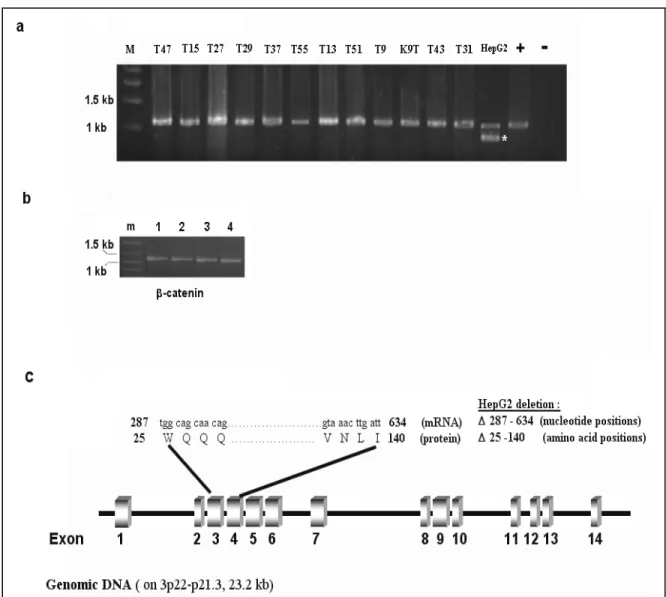
4.1 Mutational Screening of CTNNB1 (â-catenin) gene 67 4.1.1 Interstitial Deletion of â-catenin Gene Are Rare in Primary 69
HCC Tumors and Cell Lines
4.1.2 Missense â-catenin Mutations are Rare in Primary HCC 70 Tumors and Cell Lines
4.2 Mutational Analysis of Axin1 Gene in HCC Cell Lines 77
4.2.1 Analysis of Axin1 transcript by RT-PCR 78
4.2.2 Analysis of point Mutations and Small Deletion in 79
Axin1 Transcript
4.3 Summary of Mutational Screening Analysis 82
4.3.1 Mutational Screening of the CTNNB1 and Axin1 Gene in 82 14 HCC Cell Lines
4.3.2 Mutational Screening of the CTNNB1 in 33 Primary HCC 83 Tumor Samples from Southern Africa and China
4.4 â-catenin Protein Levels in HCC Cell Lines 85
4.4.1 Aberrant Accumulation of Wild-Type â-catenin in Mutant 85 p53 Cell Lines
4.4.2 Accumulated â-catenin Level Evident in Both Cytosolic and 88 Non-cytosolic Protein Fractions
xi
4.5 Analysis of Ectopic Expression of Mutant p53 Effect on Wild-Type 90 â-catenin Protein
4.5.1 Generation of Isogenic Cell Lines Ectopically Expressing 90 Wild-Type and Mutant p53
4.5.2 Ectopic Expression of Mutant p53 Causes the Accumulation 93 of Wild-Type â-catenin Protein
4.6 Subcellular Localization of Accumulated â-catenin Protein 95 in HepG2-derived and SK-Hep1-derived Stable Clones
4.7 Up-regulation of Wild-Type â-catenin Transcripts in SK-Hep1 and 98 HepG2 Cells Following Extopic Expression of Mutant p53
4.8 Expressional Analysis of Known Target Genes of Activate 101 â-catenin in 12 Selected HCC Cell Lines
4.9 Inverse Correlation in the Worldwide Distribution of â-catenin 104 and p53 Gene Mutations in HCC
CHAPTER 5. DISCUSSION 107
REFERENCES 114
APPENDIX 141
xii
LIST OF FIGURES
Figure 1 Proposed model of multistep hepatocarcinoge nesis 11
Figure 2 p53 functional motifs 19
Figure 3 Canonical WNT pathway 24
Figure 4 â-catenin is a key molecule in E-cadherin-mediated cell-cell 28 adhesion and in the WNT signaling pathway
Figure 5 cDNA and protein structure of human â-Catenin 29 Gene (CTNNB1)
Figure 6 â-catenin degradation pathways 35
Figure 7 A summary of genetic alteration of â-catenin gene in HCC 68 Figure 8 Analysis of extracted genomic DNA samples 69 Figure 9 Large deletion analysis in â-catenin gene by PCR of 70
genomic DNA
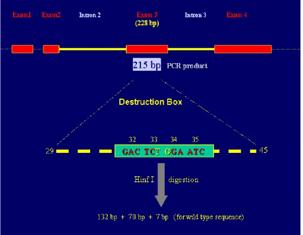
Figure 10 Schematic representation of Hinf I analysis of 72 exon 3 of â-catenin
Figure 11 Hinf I digestion profile of primary HCC tumor samples 73 Figure 12 SCCP autoradiogram of exon 3 of â-catenin gene 74 Figure 13 â-catenin mutation status of hepatocellular carcinoma cell lines 76
xiii
Figure 14 Genomic and protein structure of Axin 1 78
Figure 15 Interstitial deletion analysis of 14 HCC cell lines 79 Figure 16 Sequence analysis of Axin1 cDNA of PLC/PRF/5 and Snu423 80 Figure 17 The Arg(454)His substitution (CGC CAC) in exon 4 of Axin1 81
in the Hep40 cell line
Figure 18 Increased β-catenin protein levels in HCC cell lines displaying 86 a p53 mutation
Figure 19 Anlaysis of â-catenin in NP-40-soluble (cytosolic) and –insoluble 89 (non-cytosolic) protein fractions
Figure 20 Analysis of â-catenin in cytosolic and non-cytosolic fractions 89 extracted by Sucrose-Lysis buffer system
Figure 21 Giemsa staining and colony formation efficiency of SK-Hep1 92 stable tranfection
Figure 22 Selection of SK-hep1 clones expressing the mutant p53-V143A 92 Figure 23 Ectopically expressed mutant p53 proteins cause an increase 94
in the levels of wild-type â-catenin HepG2 stable clones
Figure 24 Ectopically expressed mutant p53 proteins cause an increase 95 in the levels of wild-typed â-catenin SK-Hep1 stable clones
Figure 25 Fluorescent immunocytochemical staining of â-catenin in 96 parental HepG2 cells and HepG2 derived clone 249
Figure 26 Fluorescent immunocytochemical staining of â-catenin in 97 tranfection negative control clone SK-Hep1-pCDNA3
and SK-Hep1 derived clone 143-8
Figure 27 Cycle optimization of Siah1 and â-catenin genes 98 Figure 28 Analysis of Siah1 transcript by semi-quantitative PCR 99
xiv
Figure 29 Analysis of â-catenin transcript by semi-quantitative 100 PCR in HepG2 clones
Figure 30 Analysis of â-catenin transcript by semi-quantitative 101 PCR in SK-Hep1 clones
Figure 31 Cycle optimization of GAPDH, c-myc and cyclinD1 genes 102 Figure 32 Semi-quantitative PCR results of c-myc and cyclin D1 gene 103 Figure 33 Comparative analysis reveals an inverse relationship 106
Between worldwide rates of p53 and â-catenin mutations in hepatocellular carcinoma
xv
LIST OF TABLES
Table 1 Risk factors that contribute to the development of HCC 3 Table 2 Gene mutations in hepatocellular carcinomas 9 Table 3 Beta-catenin mutations in human cancers 31
Table 4 List of primers and PCR conditions 57
Table 5 Frequently observed point mutations in codons 32, 33, 34 71 and 35 in HCCs
Table 6 Results of mutational screening of CTNNB1 and Axin1 genes 83 in HCC 14 cell lines.
Table 7 Lack of â-catenin mutation in hepatocellular carcinomas from 84 Africa and China
Table 8 Comparative analysis of p53 and â-catenin status of HCC 88 cell lines
Table 9 Geographical distribution of reported â-catenin and p53 105 mutation rates in hepatocellular carcinoma
xvi
ABBREVIATIONS
ATP adenine triphosphate â-catenin beta-catenin
bp base pair
cDNA complementary DNA CKI casein kinase I CKII casein kinase II
cm centimeter
dATP adenosine deoxyribonucleoside triphosphate dCTP cytosine deoxyribonucleoside triphosphate dGTP guanosine deoxyribonucleoside triphosphate DMEM Dulbecco's Modified Eagle Medium
DMSO dimethylsulphoxide DNA deoxyribonucleic acid DNase deoxyribonuclease
dNTP deoxynucleotide triphosphate
dTTP thymine deoxyribonucleoside triphosphate EDTA ethylenediaminetetra-acetic acid
xvii FCS foetal calf serum
FITC fluorescein isothiocyanate GSK-3â glycogen synthase kinase 3 beta
xg gravity
g gram
h hour
H33258 the fluorochrome dye H33258
kb kilobase kV kilovolt LB Luria-Bertani medium M molar mM millimolar ml milliliter min minute
mRNA messenger RNA
ms millisecond
OD600 optical density at 600 nm PBS phosphate buffered saline rpm revolution per minute RNA ribonucleic acid RNase ribonuclease
SDS sodium dodecyl sulphate
sec second
TAE tris/acetic acid/EDTA buffer TGF-â transfORming growth factor beta
xviii
Tris 2-amino-2-(hydroxymethyl)- 1,3 propandiol
U unit
V volt
v/v volume for volume w/v weight for volume w/w weight for weight
CHAPTER 1.
INTRODUCTION
1.1 Liver Cancers
The liver is one of the largest organs in the body, filling the upper right side of the abdomen and protected by the rib cage. The liver has many functions. It plays an important role in changing food into energy and also filters and stores blood. Primary liver cancer is different from cancer that has spread from other places in the body to the liver (liver metastases). Roughly, there are two different types of primary liver cancer (Durr and Caselmann, 2000):
• The widespread kinds are called hepatoma, or hepatocellular carcinoma, and arise from the main cells of the liver (the hepatocytes). This type is usually confined to the liver, although occasionally it spreads to other organs.
• The other type of primary liver cancer is called cholangiocellular carcinoma or bile duct cancer, because it starts in the cells lining the bile ducts
Childhood liver cancer, also called hepatoma, is rare. Hepatoblastoma is more common in young children before age 3. Children infected with hepatitis B or C (viral infections of the liver) is more likely to develop hepatocellular cancer than other children. Immunization to prevent hepatitis B may decrease the chance of developing hepatocellular cancer (Aguayo and Patt, 2001).
Hepatocellular carcinoma (HCC) is the most frequent primary liver cancer. A hepatocellular carcinoma cell resembles, to some extent, the morphological features of a hepatocyte. Although it is classified in several types (trabecular, clear cell, etc.), these morphological distinctions have no reflection in biological behavior (Engstrom et al., 1981); the exception being fibrollamelar carcinoma that appears in younger individuals, often arises in non-cirrhotic livers, and has a more benign course. People who have hepatitis B or C (viral infections of the liver) or a disease of the liver called cirrhosis are more likely to develop adult primary liver cancer than other people (Caselmann, 1996; Caselmann and Alt, 1996; Craig, 1997).
1.2 General Mechanism of Hepatocellular Carcinogenesis
Hepatocellular carcinoma (HCC) belongs to the group of epithelial tumors; it is the most common liver tumor and is one of the most frequent tumor forms worldwide (Parkin, 2001). There is a striking difference in tumor incidence with reported incidence rates based on tumor registries all over the world ranging between 0.2/100,000 in Guyana to 54292/100,000 in China (Ince and Wands, 1999; Parkin, 2001).
Based on the accumulated experimental data with chemically induced carcinogenesis in various models, the progress of HCC, as within the other organs, takes place in three stages: initiation, promotion, and progression. A mutational change caused by a carcinogen can lead to the initiation of malignant transformation in an irreversible manner. Promotion can be depicted as the clonal expansion of the abnormal cells and formation of foci. Growth of tumor from the dysplastic lesions refers to progression. The whole cascade is generally supported by an imbalance between oncogene activation and tumor suppressor gene inactivation. Also, during tumor progression gross molecular changes, such as chromosomal aberrations can occur.
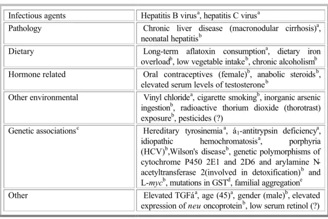
Table 1 Risk factors that contribute to the development of HCC
Infectious agents Hepatitis B virusa, hepatitis C virusa
Pathology Chronic liver disease (macronodular cirrhosis)a,
neonatal hepatitisb
Dietary Long-term aflatoxin consumptiona, dietary iron
overloadb, low vegetable intakeb, chronic alcoholismb Hormone related Oral contraceptives (female)b, anabolic steroidsb,
elevated serum levels of testosteroneb
Other environmental Vinyl chloridea, cigarette smokingb, inorganic arsenic ingestionb, radioactive thorium dioxide (thorotrast) exposureb, pesticides (?)
Genetic associationsc Hereditary tyrosinemiaa, á1-antitrypsin deficiencya,
idiopathic hemochromatosisa, porphyria
(HCV)b,Wilson's diseaseb, genetic polymorphisms of cytochrome P450 2E1 and 2D6 and arylamine N-acetyltransferase 2(involved in detoxification)b and L-mycb, mutations in GSTd, familial aggregatione
Other Elevated TGFáa, age (45)a, gender (male)b, elevated
expression of neu oncoproteinb, low serum retinol (?) aStrong association with the development of HCC. bWeak or inconsistent association with the
development of HCC. cCirrhosis, and not the underlying genetic mutation(s), appears to be the major
risk factor associated with the development of HCC. dA dose-response relationship between AFB
exposure and HCC has been documented in HBV carriers with a GST null genotype. eFamilial
aggregation of HCC may result from environmental agents such as HBV or HCV infections, although a genetic basis for HCC susceptibility in such families has not been excluded.
(Adapted from (Feitelson et al., 2002) ).
Under normal physiological conditions, adult hepatocytes are non-dividing cells. Indeed, only a minor fraction of hepatocyes undergo cell division in response to cell loss due to ageing or apoptosis (programmed cell death) (Ozturk and Cetin-Atalay, In press). HCC is most frequently associated with chronic hepatitis B virus (HBV) and hepatitis C virus (HCV) infections, with chronic exposure to the mycotoxin, aflatoxin B1 (AFB1), and also is a complication of alcoholic cirrhosis. Considering the other risk factors given in Table 1, there is a slight increase in HCC incidence in recent decades (Abel and Becker, 1987; Taylor-Robinson et al., 1997; Deuffic et al., 1998; El-Serag and Mason, 1999).
1.3 Pathogenesis of Hepatocellular Carcinoma
Within the multifactorial pathogenesis of HCC, environmental, infectious, nutritional, metabolic, and endocrine factors contribute directly or indirectly to hepatocarcinogenesis. There is considerable variability in contribution of these factors depending on environmental and socioeconomic influences.
1.3.1 Significance of Viral Hepatitis in Hepatocellular Carcinoma
The epidemiologic association of chronic HBV or HCV infection with HCC has been well established. The availability of cloned HBV and HCV genomes made it possible to detect hepatitis viruses in hepatocellular carcinomas, and their involvement in hepatocarcinogenesis. Oncogenic mechanism of HBV and HCV infection may be simply defined as releasing the growth control of hepatocytes by coding for a factor like the X protein of HBV (HbX) that activates otherwise dormant genes or activates proto-oncogenes or silences anti-proto-oncogenes; by inserting its DNA sequences that can activate and influence the transcription of cellular genes; by causing chronic inflammation with cell death and hepatocyte regeneration and with fibrosis; and by activation of the immune system liberating cytokines at the wrong time at the wrong place.
Regarding the contribution of HBV to hepatocarcinogenesis, the role of integration of HBV DNA into host chromosomes and the subsequent chromosomal instabilities is an interesting issue. Integration of HBV DNA into HepG2 cells or into transgenic mouse chromosomes has resulted in chromosomal instability that may lead to loss of heterozygosity (LOH) in many loci during chronic infection (Hino et al., 1991; Livezey and Simon, 1997). It has also been shown that HBV chronic carriers display a higher incidence of chromosomal instabilities thant he corresponding the uninfected population (Simon et al., 1991; Laurent-Puig et al., 2001).
Transcriptional activation of a wide range of viral, as well as cellular genes such as c-fos ,c-myc, insulin-like growth factor 2 (IGF2), insulin-like growth factor I receptor (IGFR1) and β-interferon , were shown to be induced by HBV encoded X antigen (HBxAg) (Twu and Schloemer, 1987; Colgrove et al., 1989; D'Arville et al., 1991; Caselmann, 1996; Kim et al., 1996). In chronic HBV infection, it has been shown that HBxAg binds and functionally inactivates the tumor suppressor p53 (Ueda et al., 1995; Huo et al., 2001) and the negative growth regulator p55sen (Ueda et al., 1995; Feitelson, 1999), both of which are involved in senescence related pathways. Inactivation of the retinoblastoma (Rb) tumor suppressor by hyperphosphorylation resulting in the activation of E2F1 and the trigger of the cell cycle has been reported in HbxAg-positive HCC cells (Sirma et al., 1999). It has also been shown that HBxAg can down regulate the expression of translational factor sui1, and cyclin-dependent kinase inhibitor p21WAF1/CIP1/SDI1(Feitelson et al., 1999; Sirma et al., 1999). As with HbxAg, carboxyterminal truncated middle hepatitis B surface protein (MHBSt) can activate various viral and cellular gene promoters (Caselmann et al., 1990; Kekule et al., 1990). Recent data suggests that HbxAg contributes to HCC development also by mechanisms other than transactivation. HBxAg binds to the X-associated protein 1 and possibly disturbs its function in nucleotide excision repair mechanism (Becker et al., 1998). It has also been shown that HBxAg-stimulated cell growth is associated with constitutive activation of the ras/raf/MAPK and NKκ-B signal transduction pathways (Lucito and Schneider, 1992; Benn and Schneider, 1994; Shirota et al., 2001).
Studies have recently begun to clarify the molecular mechanisms of HCV-induced carcinogenesis. Studies with HCV proteins showed that viral proteins interact with various cellular proteins, including 14-3-3 protein, apolipoprotein AII, tumor necrosis factor (TNF) receptor, lymphotoxin-ß receptor, DEAD domain of RNA helicase, nuclear ribonucleoprotein, double-stranded RNA protein kinase (PKR), p53 and SNARE-like protein (Ghosh et al., 1999; Shimotohno, 2000; Ray and Ray, 2001).
In chronic HCV infection, inactivation of p53 can be achieved either by HVC core protein which transcriptionally represses the p53 promoter (Ray et al., 1997; Pontisso et
al., 1998), or by nonstructural protein 3 (NS3) and NS5A which bind and most likely inactivates p53 (Ishido and Hotta, 1998; Majumder et al., 2001). Recently, stable expression of HCV core protein in HepG2 cells was shown to result in constitutive activation of the mitogen activated protein kinase/extracellular signal-regulated kinase (MAPK/ERK) pathway (Hayashi et al., 2000).
1.3.2 Alcohol and Cirrhosis
Beside for the HBV and mostly HCV-associated cirrhosis, non-viral induced cirrhosis is also coupled with elevated risk of HCC (Furuya et al., 1988; Floreani et al., 1999). Following liver cell necrosis, inflammation, regeneration, and fibrosis, quiescent hepatocytes start to proliferate. During this physiological process, irregular regeneration of hepatocytes through clonal expansion may lead to loss of control over growth and development of HCC (Ueno et al., 2001) .
It has been shown that alcoholic cirrhosis is associated with both HCC and cholangiocarcinoma. In the absence of cirrhosis, however, alcohol has not been shown to be carcinogenic in animal studies. It appears that, in large part, the relationship of alcoholic cirrhosis to liver cancer may be due to concomitant infection with HCV, although the possibility that alcohol exerts a promoting effect in the absence of HCV remains open (Farber, 1996).
1.3.3 Hepatotoxic Chemicals
Aflatoxin B1 (AFB1), benzo(a)pyrene and vinly chloride were shown to have a well-defined genotoxic effect in hepatocarcinogenesis. Consumption of food contaminated with aflatoxins, toxic metabolites of some species of Aspergillus fungi, has been allied with both human and animal HCCs. It has been shown to induce a specific mutation in codon 249 of the p53 gene leading to G:C to T:A transversion (Bressac et al., 1991; Hsu et al.,
1991). Similarly, genotoxic effect of benzo(a)pyrene and vinly chloride on p53 has been supported by human and animal data (Puisieux et al., 1991; Barbin et al., 1997).
1.3.4 Hemochromatosis and Iron
Iron overload has been associated with a high risk of HCC in patients with untreated hemochromatosis and dietary iron intake in patients without hereditary iron overload (Hann et al., 1989; Fargion et al., 1992). One possible mechanism that involves iron overload as a cofactor for hepatocarcinogenesis is the iron deposition which occurs in hepatocytes, Kupffer cells and bile ducts. It results in increased production of oxygen-free radicals, peroxidation of membrane lipids, and other reactive oxygen species (ROS)-mediated damages (Kicic et al., 2001; Rocken and Carl-McGrath, 2001).
1.4 Molecular Pathogenesis of Hepatocellular Carcinoma
Cancer is not a single disease; it can be defined as great variety of malignant and benign tumors that arise from the same critical process of uncontrolled cell proliferation. Cancer proceeds through accumulation of mutations in genes that govern the cell proliferation and death, and it is believed that hepatocarcinogenesis also shares this common molecular pathogenesis as well as common biological features.
The absence of an evidently inherited predisposition to liver cancer has obstructed the identification of specific critical genes in hepatocarcinogenesis. However, HCCs present various genomic alterations, including DNA rearrangements, loss of heterozygosity (LOH), chromosomal amplification, loss of imprinting and mutations (Buendia, 2000; Pineau and Buendia, 2000).
Table 2 lists the genes that have been implicated in the pathogenesis of HCC, and may be divided into four functional groups: (1) genes regulating DNA damage response,
(2) genes involved in cell cycle control, (3) genes involved in growth inhibition and apoptosis, and (4) genes responsible for cell-cell interaction and signal transduction (Ozturk, 1999).
1.4.1 Oncogenes in HCC
Beta-catenin (β-catenin) gene mutations that lead to oncogenic activation have been recently described as one of the most frequent mutations in both HCCs and hepatoblastomas. Reported incidence of β-catenin mutation in HCC varies between 8% and 24% depending on the geographical origin of the tumors (de La Coste et al., 1998; Miyoshi et al., 1998a; Huang et al., 1999; Kondo et al., 1999; Terris et al., 1999; Hsu et al., 2000; Satoh et al., 2000; Devereux et al., 2001; Fujie et al., 2001; Laurent-Puig et al., 2001; Wong et al., 2001). However, childhood hepatoblastomas represent much higher rate of β-catenin mutations that varies between 48% and 89% (Blaker et al., 1999; Koch et al., 1999; Jeng et al., 2000; Wei et al., 2000; Park et al., 2001; Takayasu et al., 2001; Udatsu et al., 2001).
The amplification and overexpression of the cyclin D1 gene that results in the rapid growth and aggressiveness of tumor was observed in 11-13 % of HCCs (Zhang et al., 1993; Nishida et al., 1994; Albrecht et al., 1995; Kim et al., 2000; Joo et al., 2001). Cyclin A is an S- and G2-M-phase regulatory protein, and its abnormal expression has been implicated in 39% of HCCs (Chao et al., 1998).
The constitutive activation of c-myc expression is key to the genesis of many cancers (Dang et al., 1999; Fearon and Dang, 1999). The overexpression of c-myc mRNA and gene product without amplification may be a characteristic of HCC. The reported frequencies of c-myc gene overexpression diverge between 0% and 87%. (Hansen et al., 1993; Saegusa et al., 1993; Wu et al., 1996).
Table 2 Gene alterations in hepatocellular carcinomas
Mutated gene Alteration (%)
*
Additional referencesc-myc 0–87
N-myc 0
p53 28
p73 0–13 (Mihara et al., 1999; Peng et al., 2000)
p51 0 (Hamada et al., 2000)
IGF II 23-27 (Cariani et al., 1988; Uchida et al., 1997; Eriksson
et al., 2001)
M6P/IGF2R 0–33
TGFRβ2 0
Smad2 0–2
Smad4 0–6
Smad6 0 (Kawate et al., 2001)
Smad7 0 (Kawate et al., 2001)
RB1 15 p15INK4B 0 p16INK4A 0–55 p21CIP1 5 Cyclin D 11–13 Cyclin A 19 E-Cadherin p.u. β-Catenin (CTNNB1) 8–24
AXIN1 7-9 (Satoh et al., 2000; Taniguchi et al., 2002)
MET protooncogene 11 (Park et al., 1999b)
hMSH1 p.u. hMSH2 <30 (Yano et al., 1999) K-ras 0–17 N-ras 0–16 H-ras 0–10 BRCA2 5
BCL10 57 (Jihua et al., 2000; Cheng et al., 2001)
PTEN/MMAC1 3 (Yao et al., 1999; Fujiwara et al., 2000)
TRα1 65 (Lin et al., 1999)
TRβ1 76 (Lin et al., 1999)
M6P/IGF2R: Mannose 6-phosphate/insulin-like growth factor 2 receptor gene, RB1: Retinoblastoma gene,hMSH1: Human Mut L homolog-1, hMSH2 Human Mut S homolog-2, BRCA2: Breast cancer susceptibility gene 2, p.u. = Prevalence unknown, *: mutation, amplification and de nova methylation. (adapted form Ozturk, 1999)
Aberrant proto-oncogene expression of the “ras” family of genes has been implicated in hepatic cell proliferation, transformation and carcinogenesis using rat models (Sills et al., 1999; Boivin-Angele et al., 2000), but there is little evidence of activation in human tumors. The mutation rate in K-ras and N-ras genes have been reported to be between 0% and 17%, and N-ras mutation seems to be less frequent (Tsuda et al., 1989; Challen et al., 1992; Bjersing et al., 1996; Luo et al., 1998; Boivin-Angele et al., 2000; Boix-Ferrero et al., 2000; Weihrauch et al., 2001).
The c-met proto-oncogene encodes the tyrosine kinase receptor for hepatocyte growth factor/scatter factor (HGF/SF), a potent mitogen and motogen for epithelial cells. There is only one report about the c-met mutations in HCC by Park et. al. who described three missense mutations in 10 childhood HCCs, whereas there is no mutation identified in adult HCCs, cholangiocellular carcinomas, or hepatoblastomas (Park et al., 1999b). However, c-met over expression has been reported in some HCC tumors (Ljubimova et al., 1997; Ueki et al., 1997; Okano et al., 1999; Tavian et al., 2000).
The insulin-like growth factor II (IGF II) gene expression is monoallelic in all the normal tissues other than the liver because of the imprinting of the maternal allele. However, interestingly, most of the cancers arising in these tissues are reported to show biallelic expression (relaxation) of the IGF II gene (Suzuki et al., 1994; Quinn et al., 1996; Steller et al., 1996). Allelic imbalance (re-imprinting) of IGF II gene expression is often seen in HCC as well as hepatoblastomas (Li et al., 1995; Sohda et al., 1996; Takeda et al., 1996; Kim and Lee, 1997; Li et al., 1997; Uchida et al., 1997; Li et al., 1998; Sohda et al., 1998; Eriksson et al., 2001).
1.4.2 Tumor Suppressor Genes in HCC
p53 gene is the best characterized and known tumor suppressor in terms of its implications in human carcinogenesis (Levine et al., 1991). In human HCC, LOH at chromosome 17p13 has been observed in 26-60 % of tumors in different studies, and the
global prevalence of p53 can be estimated to be around 28% (reviewed in (Ozturk, 1999; Buendia, 2000).
Reduced p21(WAF1/CIP1) expression, which is mainly associated with p53 gene mutation in HCCs, contributes to hepatocarcinogenesis, but a p53-independent pathway also plays a role in the regulation of p21/WAF1 expression (Hui et al., 1998; Qin et al., 1998; Bhardwaj et al., 1999). It has been reported that reduced p21(WAF1/CIP1) expression with wild-type p53 participate in hepatocarcinogenesis with only 5% occurrence in studied HCCs (Furutani et al., 1997; Hui et al., 1997).
Tumor suppressor function of the mannose 6-phosphate/insulin-like growth factor II receptor (M6P/IGFIIR) was first proposed by De Souza et al. by the identification of 6q26-27 chromosomal deletion in HCC samples and its role in activation of transforming growth factor-beta 1 (TGF-β1) and promoting the degradation of IGFII (De Souza et al., 1995). Mutations in the M6P/IGFIIR have been detected in 0 to 33% of HCCs (Piao et al., 1997; Wada et al., 1999; Saeki et al., 2000; Kishimoto et al., 2001; Oka et al., 2002).
Inactivation of the retinoblastoma (Rb) gene is considered to play a fundamental role in the genesis and progression of several human cancers. In human HCCs, it has been shown that inactivation of Rb gene occurs either via strongly down-regulated pRb protein in 30-50% of tumors or via LOH at chromosome 13q where the Rb locus is found (25-48%); (Murakami et al., 1991a; Fujimoto et al., 1994; Hsia et al., 1994; Zhang et al., 1994; Hada et al., 1996). Although no mutation has been reported in Rb gene itself so far, a recent publication from Higashitsuji et al (2000) proposes a novel pathway that leads to inactivation of Rb via a new oncogene called gankyrin (an ankyrin-repeat protein homologoues to the p28 subunit of 26S proteosome) (Higashitsuji et al., 2000).
Regional de nova methylation of CpG islands in transcriptional regulatory region represents an alternative mechanism for loss of function of a number of tumor suppressor genes in human cancer. Notably, hypermethylation of normally under-methylated CpG islands in transcriptional regulatory regions of CDKN2A, a gene located on 9q21, has been
observed, and shown to be the main mechanism for inactivation of p16INK4 in variety in
HCC. CDKN2A encodes p16INK4, an inhibitor of cyclin-dependent kinase -4 and -6, and p14ARF which target p53 via inhibition of mdm-2. (Lin et al., 1998; Liew et al., 1999a; Wong et al., 1999; Iwata et al., 2000; Jin et al., 2000; Kanai et al., 2000; Azechi et al., 2001; Weihrauch et al., 2001; Shen et al., 2002). LOH at 9q occurs with 20% and homologous deletion of 9q21 has been detected only in four HCC cases (Liew et al., 1999b). In HCC, germline and somatic mutations of CDKN2A seems to be quite rare (<10% of studied cases) (Chaubert et al., 1997; Liew et al., 1999a).
The adenomatous polyposis coli (APC) gene mutations were suggested to occur in HCCs (Koch et al., 1999; Wei et al., 2000). APC mutations result in up-regulated wild-type β-catenin levels and constitutive Wnt signaling. The finding of rare LOH at 5q where the APC gene is located and the absence of truncation mutations in the HCC samples analyzed indicate that APC can not be considered as a major contributor to hepatocarcinogenesis (Ding et al., 1993; Devereux et al., 1999; Su et al., 2001).
Another tumor suppressor gene referred as Human Mut S homolog-1 (hMSH1) is located on chromosome 5q, but its role and contribution to HCC, if any, remains to be identified. Mutations in another mismatch repair gene know as Human Mut S homolog-2 (hMSH2) has been reported at about 30% of HCCs examined (Yano et al., 1999), but this data has not been confirmed yet.
LOH of 16q may also be involved in the HCC transformation via loss of the Axin gene which normally promotes ubiqitine-dependent and -regulated degradation of β-catenin (Laurent-Puig et al., 2001). Recently, input of Axin 1 mutations to HCC development was reported to be around 7-9 % (Satoh et al., 2000; Taniguchi et al., 2002) and that of Axin2 mutations was reported as ~3% in studied HCCs but, not in HBs (Taniguchi et al., 2002). LOH of 16q also correlates with metastatis in HCC and therefore can be a link to another candidate tumor suppressor gene, namely E-cadherin which is located at 16q22 (Niketeghad et al., 2001). Its role as a receptor in adherence junctions is essential both for the maintenance of tissue structure and regulation of free cytoplasmic
β-catenin levels. In addition to LOH (Slagle et al., 1993), hypermethylation of E-cadherin promoter has been proposed as an alternative mechanism for WNT activation in HCC (Kanai et al., 1997).
PTEN/MMAC1 tumor suppressor gene has recently been shown to block growth-stimulatory and survival signals mediated by PI3 kinase and converging on the activation of protein kinase B/Akt (Downward, 1998; Stambolic et al., 1998). Somatic mutations of PTEN gene were found in 4% of HCCs (Kawamura et al., 1999; Yao et al., 1999; Fujiwara et al., 2000), and 27% of HCCs display LOH in the same gene (Kawamura et al., 1999; Yeh et al., 2000).
Low frequency of mutation in the Smad2, Smad4, and TGF-β RII genes has been reported by two independent studies implicating a disruption of TGF-β pathway in less than 10 % of HCCs (Kawate et al., 1999; Yakicier et al., 1999). It has been also shown that mutations of the Smad6 and Smad7 genes are not the main cause of the TGF-beta resistance in human HCC (Kawate et al., 2001).
The recently described bcl10 gene has been suggested to be a major target gene for inactivation in a variety of human cancers and the wild type acts as a tumor suppressor, while mutant type behaves like an oncogene. (Bose et al., 1999; Dyer, 1999; Gill et al., 1999; Lambers et al., 1999; Lee et al., 1999b; Stone et al., 1999). It has been shown that BCL10 promotes apoptosis, pro-caspase-9 maturation and activation of NF-kappaB (NF-κB) via NF-κB-inducing kinase (NIK) and IkB kinase (IKK) (Koseki et al., 1999; Willis et al., 1999; Ruland et al., 2001). Recently, the Bcl 10 gene was reported to have high mutation frequency in liver cancer (57 %); (Jihua et al., 2000; Cheng et al., 2001).
BRCA2 and p73 mutations have been rarely detected in HCC (Katagiri et al., 1996; Mihara et al., 1999; Peng et al., 2000).
Thyroid hormone nuclear receptor (TR) mediates the diverse biological effects of thyroid hormone through interaction with a large network of regulatory proteins. High
frequencies of mutations of TR-alpha (TRα) and/or TR-beta (TRβ) genes have been reported in human hepatocellular, renal clear-cell, and papillary thyroid carcinomas (Lin et al., 1995; Lin et al., 1996; Kamiya et al., 2002; Puzianowska-Kuznicka et al., 2002). Lin et al. reported that point mutations in TRα1 and TRβ1 are highly frequent (65%-76%) in
HCC (Lin et al., 1995; Lin et al., 1999), however the role of TR in this cancer needs further investigations.
1.4.3 Genetic Mechanisms of Hepatocellular Carcinoma
Hepatocarcinogenesis is accompanied with a variety of genetic alterations such as loss of LOH (RB1, M6P/IGF2R, E-cadherin, BRCA2), somatic mutation (p53, RB1, p16INK4A,β-catenin, APC, ERs), de-novo methylation (p16INK4A, E-cadherin) and/or
functional inactivation (p53). Variation in number and types of these molecular changes depend on genetic and geographical background of the host, and the etiology of the tumor which can be viral, chemical or both (Wong et al., 2000).
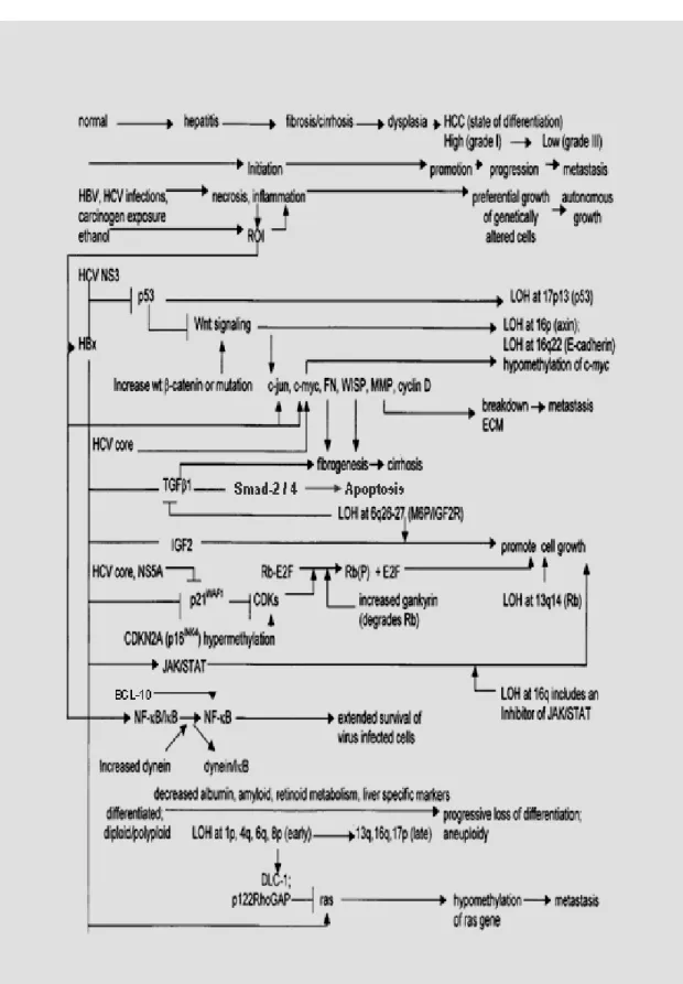
Overall, the consequences of these changes suggest that the pathogenesis of HCC is accompanied by a progressive loss of differentiation, normal cell adhesion, and extracellular matrix, as well as constitutive activation of selected proliferative and survival signal transduction pathways. Figure 1 presents an overview of the common changes that contributes to the pathogenesis of HCC.
Figure 1. Proposed model of multistep hepatocarcinogenesis by Feitelson et al (2002).
1.5 The p53 Tumor Suppressor
p53 is a tumor suppressor mutated in more than 50% of all human tumors, and its germ line mutations in Li-Fraumeni syndrome patients are known to pre-dispose to a variety of cancers (Hansen and Oren, 1997; Levine, 1997). The p53 gene can be classified as either a “gatekeeper” or a “caretaker” tumor suppressor gene. As an inducer of apoptosis, p53 appears to act as a gatekeeper, however the critical role of p53 in maintaining genomic integrity has earned it the nickname “guardian of the genome” (Lane, 1992) .
1.5.1 Activation of p53
Excessive wild-type p53 activity results in several cellular responses, most prominently cell cycle arrest and apoptosis. Such severe outcomes become very undesirable and harmful if occurring in normal, resting cells. Therefore p53 activation should be kept under tight control.
Under normal physiological conditions, p53 is likely to be in its latent form and does not interfere with significant cellular processes; since p53 knock-out mice in most cases undergo appropriate development and maturation (Donehower et al., 1992). On the other hand, a variety of conditions cause a rapid transition of latent p53 form into a physiologically active form. The common characteristic of these environmental insults are that they create a cellular stress condition, like deoxyribonucleic acid (DNA) damage (Kastan et al., 1991), microtubule disruption (Cross et al., 1995), matrix detachment (Wu and Schonthal, 1997; Park et al., 1999a), hypoxia (Graeber et al., 1996), ribonucleotide depletion (Wahl et al., 1997), oxidative stress (Forrester et al., 1996) or oncogene activation (Lane, 1992; Hermeking and Eick, 1994).
Activation of the p53 in response to stress conditions occurs primarily through alteration in p53 protein. Change in the rate of p53 expression plays a minor role, if any, in such induction. Activation of p53 was first thought to be achieved at transcriptional level
either by a transcriptional repressor sequence present at the 3’-untranslated region of p53 mRNA (Fu et al., 1996) or by p53 itself which binds to its own mRNA (Mosner et al., 1995; Fontoura et al., 1997), however it has been well-documented that post-translational modifications are the major mechanism that cause accumulation of active p53 in response to stress. The p53 is usually a very labile protein and its turnover rate is as short as a few minutes (Rogel et al., 1985). Therefore significant increase in p53 levels requires stabilization of p53 protein.
A key component in the regulation of p53 is the Mdm2 protein. Mdm2 is a product of an oncogene, whose oncogenic activation was observed in several human cancers (Lozano and Montes de Oca Luna, 1998; Freedman et al., 1999). Mdm2 inhibits p53 activity either by binding to transactivation domain of p53 (Lu and Levine, 1995), or by interacting with p53 protein to act as an active transcription repressor (Thut et al., 1995). However, it is now clear that Mdm2 is a p53-specific E3 ubiquitine-protein ligase, which covalently attaches ubiquitine groups to p53 for its complete elimination by proteolytic degradation. On the other hand p53 binds to the mdm2 gene and activates its transcription. The p53-Mdm2 negative feedback/ autoregulatory loop serves as a tightly controlled shift mechanism for activation and inactivation of p53 according to the presence or absence of triggering stress factors (Wu et al., 1993; Honda and Yasuda, 1999). It has also been reported that c-jun N-terminal kinase (JNK) has a possible role in p53 ubiquitination and degradation (Fuchs et al., 1998).
Covalent modifications of p53 – particularly phosphorylation- also play an important role in activation of p53 protein upon stress. p53 becomes phosphorylated at serine residues in both amino- and carboxy-terminal regions in vivo in response to various types of stress, and many stress-activated kinases such as casein kinase I and II (CKI / II) can phosphorylate p53 in vitro (Milne et al., 1992; Hall et al., 1996). Double-stranded DNA-activated protein kinases (Lees-Miller et al., 1990; Shieh et al., 1997), ATM and ATR (Banin et al., 1998; Canman et al., 1998), cyclin-dependent kinase(cdk) activating kinase (CAK) (Ko et al., 1997), cdk2 and cdc2 (Bischoff et al., 1990; Price et al., 1995) and protein kinase C (PKC) also phosphorylate p53 protein (Baudier et al., 1992).
p53 acts as a transcriptional activator which relies on sequence- specific DNA binding activity of p53 (Weintraub et al., 1991; Donehower et al., 1992; Crook et al., 1994; Levine, 1997). It has been reported that p53 acetylation and gylcosylation activate the latent sequence-specific DNA-binding activity of p53 (Hupp et al., 1992; Shaw et al., 1996; Sakaguchi et al., 1998) .
1.5.2 Biochemical and Physiological Functions of p53
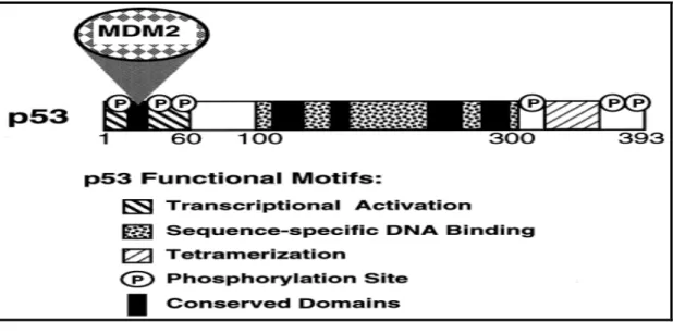
p53 is a sequence-specific transcriptional factor, and most of p53 functions are carried out by its target genes (Amundson et al., 1998; el-Deiry, 1998). The p53 protein is capable of binding to specific DNA sequences with its central core domain (Figure 2). The common feature of these downstream genes is that they contain one or more p53 consensus binding sites in their regulatory regions. The p53 consensus binding site contains two or more copies of a 10 bp half-site 5'-PuPuPuC(A/T)(T/A)GpyPyPy-3' (el-Deiry et al., 1992). The amino-terminal region of p53 functions as a transcriptional activation domain, and the carboxy-terminal region appears to be required for p53 to form dimers and tetramers with itself (Cho et al., 1994; Ko and Prives, 1996).
Genotoxic and other stress signals may trigger the increase of p53 protein, which has three major functions: growth arrest, DNA repair and apoptosis (cell death). The growth arrest stops the progression of cell cycle, preventing replication of abnormal DNA. In the mean time, p53 may activate the transcription of genes involved in DNA repair. On the other hand, p53 could also induce apoptosis, which is the "last resort" to avoid proliferation of abnormal cells.
p53 has been shown to activate transcription of anumber of genes with roles in the control of the cell cycle, including p21WAF1/CIP1 which encodes a regulator of Cdk activity
(el-Deiry et al., 1993), gadd45; a growth arrest DNA damage-inducible gene (Kastan et al., 1992), mdm2, as note above, encodes a protein that is a known negative regulator of p53 (Juven-Gershon and Oren, 1999) and 14-3-3σ; (a regulator of G2/M progression) (Hermeking et al., 1997).
Figure 2 p53 functional motifs.
Domains of p53 involved in transcriptional activation, sequence-specific DNA binding, tetramerization, and binding by the MDM2 protein are indicated. The five distinct regions of p53 that are highly conserved between p53 proteins of diverse species are indicated. In addition, the locations of several sites in the protein that are phosphorylated (P) and that regulate p53 function are indicated
p53 can also induce apoptosis by activating some apoptotic genes including Bax (a pro-apoptotic Bcl-2 related protein) (McCurrach et al., 1997), Apaf-1 (apoptotic protease activating factor 1) (Kannan et al., 2001), Puma (p53 upregulated modulator of apoptosis) (Nakano and Vousden, 2001), NoxA (a Bcl-2 homology 3 (BH3)-only member of the Bcl-2 family of proteins) (Oda et al., 2000). Involvement of p53 in DNA repair processes has been proposed in several reports (Kastan et al., 1992; Lakin and Jackson, 1999; Lozano and Elledge, 2000; Khanna and Jackson, 2001; MacLachlan et al., 2002) and a solid evidence came from a report showing that p53 induces transcription of p53R2 (p53 induced ribonucleotide reductase small subunit-like protein) in response to ultraviolet and gamma-irradiation and adriamycin treatments (Tanaka et al., 2000).
While the specific mechanisms of p53 repression are not well understood, several candidate targets of p53 repression have been suggested, including the gene for the microtubule-associated protein MAP4 (Murphy et al., 1996; Murphy et al., 1999), the multi drug resistance associated protein 1 (MRP1) (Wang and Beck, 1998) and the gene for FKBP25, an FK506/rapamycin-binding protein (Ahn et al., 1999).
1.5.3 p53 and Oncogenic Stress
Deregulated expression of oncoproteins (like adenovirus E1A, Ras, Myc and β-catenin) cause p53 accumulation and activation, often in an p14ARF-dependent manner (de Stanchina et al., 1998; Stott et al., 1998; Damalas et al., 2001; Palmero et al., 2002) . At the biochemical level, excess activity of oncoproteins leads to enhanced transcription of p14ARF gene, at least some of which is mediated by the E2F transcriptional factor (Bates et
al., 1998) . The induced p14ARF protein binds to the Mdm2 protein and this binding sequesters Mdm2 in the nucleolus, where it can not target p53 for ubiquitinylation and p14ARF directly inhibits the E3 activity of Mdm2 (Honda and Yasuda, 1999; Tao and Levine, 1999; Weber et al., 1999; Lohrum et al., 2000; Weber et al., 2000). Therefore oncogenic stress activated p14ARF inhibits ubiquitin-dependent p53 degradation, and as a consequence p53 accumulation with subsequent stimulation of p53 mediated cellular responses take place. This is believed to serve as a failsafe mechanism that defends cells against the tumorogenic outcomes of oncogene activation (Sherr, 1998).
1.5.4 The p53 Tumor Suppressor in HCC
p53 is functionally inactivated by structural mutations, viral proteins, and endogenous cellular mechanisms in greater part of human cancers (Hollstein et al., 1991; Greenblatt et al., 1994). The majority of base substitutions target DNA sequences that encode the highly conserved central domain of the protein that mediates sequence-specific DNA binding and transcriptional activation (Murakami et al., 1991b; Hollstein et al., 1994). Loss of p53 function also occurs through allelic deletions at chromosome 17p13 where the gene is located. A detailed analysis of p53 gene mutations indicates that the sites and features of DNA base changes differ among the variety of human tumor types (Hollstein et al., 1991; Hollstein et al., 1994).
In the case of HCC, LOH at chromosome 17p13 has been detected in 24-65% of tumors, and approximately 28% of HCC cases harbor p53 mutations (Ozturk, 1999; Buendia, 2000). Although in most HCC tumors Knudson’s “two hit” model is observed in
p53 inactivation (besides 17p LOH, mutations can occur in the remaining allele) (Oda et al., 1992; Nishida et al., 1993), a considerable percentage of HCC have either a single wild-type p53 allele or a heterozgous p53 mutation (Rashid et al., 1999). Noteworthy studies have shown that there is a significant correlation between p53 mutation and 13q LOH where Rb gene is located at late tumor-stage (Murakami et al., 1991a; Unsal et al., 1994) and between p53 mutation and LOH of chromosome 4p (Rashid et al., 1999), suggesting that the corresponding tumor suppressor genes might be combined forces in HCC transformation.
Mutational screening studies in sporadic HCCs have shown that p53 gene mutations display a heterogenic pattern regarding the tumor’s geographical origins (Ozturk, 1991; Buetow et al., 1992; Unsal et al., 1994). In this regard, a significant worldwide study by Ozturk and colleagues put forward a close correlation between presence of G:C to T:A transversion leading to an Arg to Ser mutation at codon 249 of p53 and high risk of AFB1 intake (Ozturk, 1991). Now it is principally confirmed and well established that codon 249 p53 mutations in HCC is mostly seen in some regions of Africa and Asia where the AFB1 content of diets is high and chronic HBV infection is highly epidemic (Bressac et al., 1991; Hsu et al., 1991; Coursaget et al., 1993; Hosono et al., 1993; Li et al., 1993; Diamantis et al., 1994; Unsal et al., 1994) .
The p53 protein is now well established as a crucial player in the regulation of cell cycle arrest, apoptosis, differentiation, angiogenesis, and senescence. Therefore inactivation of this key regulator molecule presumably facilitates malignant progression by promoting extended hepatocyte survival and acquisition of mutations through genomic instability. For that reason, frequent involvement of p53 inactivation either via viral protein inactivation early in carcinogenesis, or via mutation or/and LOH much later during HCC tumor progression would not be surprising.
1.6 The Canonical WNT/Wg (Wingless) Signaling
The canonical Wnt/Wingless signaling pathway is involved in a large variety of developmental processes, including segment polarity in Drosophila, axis specification in Xenopus, mesoderm induction in Caenorhabditis elegant development of vertebrate limbs and urogenital system (Wodarz and Nusse, 1998). Recently, inappropriate reactivation of the Wnt pathway in adult tissues has shown to play a central role in the etiology of human cancers (Kondoh et al., 2001; Smalley and Dale, 2001; Taipale and Beachy, 2001; Moon et al., 2002).
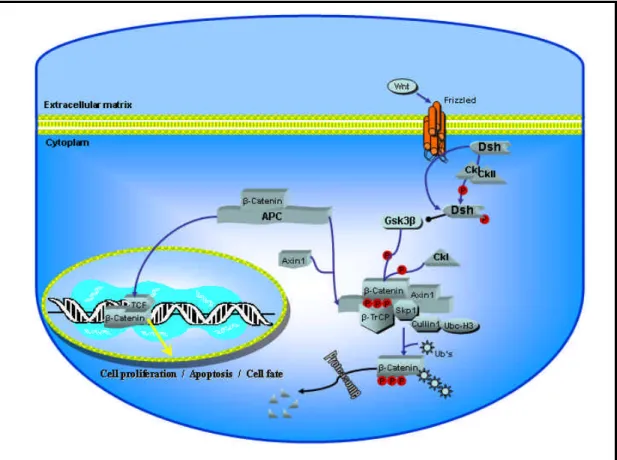
In most instances, the Wnt pathway is initiated by secreted Wnt protein (paracrine factors; at least 15 members of this family in vertebrates), which binds to a class of seven-pass transmembrane protein encoded by the frizzled (Fz) gene (Yang-Snyder et al., 1996; He et al., 1997). By the activation of Fz, the cytoplasmic protein Dishevelled (Dsh) is recruited to the membrane (Axelrod et al., 1998; Boutros et al., 2000) and becomes phosphorylated (Lee et al., 1999a). Phoshorylated Dsh associates with Axin1 and causes the disintegration of multiprotein complex containing Axin 1, glycogen synthase kinase 3β (GSK-3β) the APC tumor suppressor and β-catenin, and prevents glycogen GSK-3β from phosphorylating critical targets (Itoh et al., 1998; Kishida et al., 1999; Smalley et al., 1999). Although, the exact mechanism of Dsh-mediated inactivation of this complex is not understood yet, it has been shown that GBP/Frat-1 inhibit GSK-3β- Axin 1 binding (Yost et al., 1998; Farr et al., 2000; Salic et al., 2000). The GSK-3β substrates include the Axi1, APC and β-catenin (Rubinfeld et al., 1996; Yost et al., 1996; Yamamoto et al., 1999).
Disintegration of multiprotein complex in response to Wnt signal results in cytoplasmic accumulation of hypo-phosphorylated free cytoplasmic-β-catenin, which then translocates into the cell nucleus. Nuclear β-catenin interacts with T cell factor/lymphocyte enhancer binding factor (TCF/LEF) family of transcriptional factors and stimulate transcription of a variety of target genes (Behrens et al., 1996; Molenaar et al., 1996)
which modulate cell fate, proliferation, and apoptosis (Mann et al., 1999; Chen et al., 2001; Kawakami et al., 2001; Kofron et al., 2001; Olson, 2001; van Gijn et al., 2001).
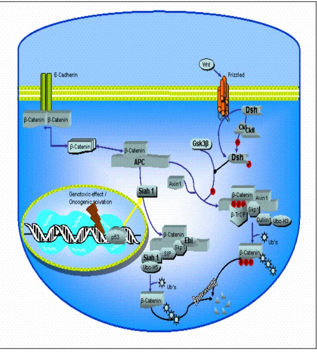
In the absence of Wnt signal, β-catenin is destabilized by the multiprotein complex (mentioned above). Axin serves as a scaffolding component for binding of APC, GSK-3β and β-catenin. Interaction between Axin and GSK-3β facilitates phosphorylation of N-terminal located serine/theronine residues of β-catenin by GSK-3β/CK-I dual kinase system (Polakis, 2000; Amit et al., 2002; Liu et al., 2002) leading to degradation initiated by ubiquitin-mediated proteolysis of β-catenin via β-TRCP (an E2 ubiquitin ligase) (Aberle et al., 1997; Hart et al., 1999; Latres et al., 1999; Liu et al., 1999; Sadot et al., 2002).
Cleary, the stability of free cytoplamic β-catenin is the heart of the canonical Wnt pathway and most of the effects of Wnt signaling are mediated by nuclear β-catenin and TCF.
The canonical Wnt pathway is highly conserved between Drosophila and vertebrates, and the Figure 3 showing the core elements of the canonical WNT pathway is undoubtedly an oversimplification, because different cells use this pathway in different ways.
1.7. Canonical WNT Signaling in Cancer
1.7.1 Ligand and Receptor
The Wnt ligands are secreted glycoproteins that can be categorized according to their ability to promote neoplastic transformation (Wodarz and Nusse, 1998). Oncogenic potential of Wnt was first described by Nusse and Varmus almost 20 years ago inciting intense investigation into the role of Wnt genes in human cancer (Nusse and Varmus, 1982). Subsequent studies covered the way for assembling a signaling pathway and consequently identification of candidate cancer-causing genes within the same pathway.
Figure 3 Canonical WNT pathway.
Core elements of WNT/β-catenin pathway are shown, illustrating how activation of Frizzled receptor by the Wnt ligand leads to activation of β-catenin. This activates gene expression leading to diverse cellular responses in both embryonic development and in adults.
Regarding the ligand-receptor interaction, at least 11 vertebrate frizzled genes have been identified. However except for few published data (Tanaka et al., 1998; Gazit et al., 1999; Sheldahl et al., 1999), there is no direct evidence for involvement of Frizzled in neoplastic transformation.
1.7.2 Dishevelled (Dsh)
Dishevelled is a phosphoprotein that becomes hyperphosporylated on serine /threonine residues when wnt signaling is activated. It is located downstream of the receptor (e.g. frizzled) and upstream of β-catenin (Noordermeer et al., 1994). Under normal physiological conditions, Dsh is localized in the cytoplasm, by the activation of Wnt signaling it is recruited to the membrane (Yang-Snyder et al., 1996). Overexpression of
Dsh in the absence of wnt activation leads to accumulation of β-catenin (Yanagawa et al., 1995). However, its involvement in human cancer has not been reported. One possible connection to Dsh in human cancer can be via its interaction and phosphorylation, thereby activation by casein kinase II (CK II). It has been reported that in CKII transgenic mice develop lymphomas (Seldin and Leder, 1995; Willert et al., 1997; Song et al., 2000).
1.7.3 Glycogen Synthase Kinase 3ββ (GSK-3ββ)
The serine/threonine kinase GSK-3β is an integral component and negative regulator of Wnt signaling in vertebrates (Dominguez et al., 1995; He et al., 1995). GSK-3β carries out its regulatory effect by phosphorylating several substrates in the Wnt pathway (Rubinfeld et al., 1996; Ikeda et al., 1998; Itoh et al., 1998; Li et al., 1999; Yamamoto et al., 1999). The down-regulatory nature of GSK-3β could be qualified as putative tumor suppressor in neoplastic transformation. However GSK-3β displays no mutation in colorectal cancers, which are known to activate Wnt pathway (Bienz and Clevers, 2000). The participation of GSK-3β in human cancers can be linked to its inactivation by Frat-1, a GSK-3β binding protein antagonistic to Wnt signaling in mammalian cells (Li et al., 1999; Thomas et al., 1999). Recently, Frat-1 was found to be involved in to cancer progression in transgenic mice model and human cancer-associated upregulation of Frat-1 has been reported (Jonkers et al., 1999; Saitoh and Katoh, 2001; Saitoh et al., 2001).
1.7.4 Axin 1
Axin was identified as an inhibitor of Wnt signaling was positioned downstream of Dsh and GSK-3β, and upstream of β-catenin and shown that the Axin protein can bind to GSK-3, APC, β-catenin and Dsh (Peifer and Polakis, 2000). The apparent lack of enzymatic function and its direct interaction with other critical component of the Wnt pathway suggest that Axin1 functions as a scaffold to assemble critical Wnt pathway component in close proximity. In this regard, Axin 1 facilitates the phosphorylation of
β-catenin by GSK-3β and CKII (Polakis, 2000; Amit et al., 2002; Liu et al., 2002). The tumor suppressor role, thereby biallelic inactivation of Axin1 has been verified by Satoh et al. in human hepatocellular cancers and cell lines (Satoh et al., 2000). Tumor suppressor nature of Axin was also shown by other studies (Dahmen et al., 2001; Lustig et al., 2002; Taniguchi et al., 2002).
1.7.5 The Adenomatous Polyposis Coli (APC)
The adenomatous polyposis coli protein can bind to β-catenin and promotes its degradation in vertebrates (Su et al., 1993; Munemitsu et al., 1995; Rubinfeld et al., 1995). Loss of APC is observed in the majority of human colon cancers and leads to elevated β-catenin levels (Korinek et al., 1997; Morin et al., 1997; Rubinfeld et al., 1997). This and many other observations suggest that the main function of APC as a tumor suppressor is to control β-catenin levels. Despite the massive amount of data implicating APC in a β-catenin regulation, its exact role in Wnt pathway is not firmly established. As negative regulator of β-catenin, APC phosphorylation by GSK-3β is needed for its association with β-Catenin (Cavallo et al., 1997; Willert and Nusse, 1998). Recently, it has been proposed that APC is a nuclear-cytoplasmic shuttling protein, and can act as a β-catenin chaperone, providing a specific transport route for nuclear β-catenin to the cytoplasm (Henderson, 2000; Henderson et al., 2002).
1.7.6 Beta Catenin (ββ-catenin)
β-catenin is a 92 kDa multifunctional protein and initially considered to be a cell-cell adhesion protein. It binds to the intracell-cellular domain of cadherins, transmembrane proteins that mediate calcium-dependent cell-cell adhesion. β-catenin links cadherins to the actin cytoskeleton (DeMarais and Moon, 1992). β-catenin also behaves as an essential component of the Wnt/Wingless signaling pathway. Another line of evidence strongly suggesting a role for catenin in signal transduction stems from the observation that
β-catenin interacts directly with the epidermal growth factor receptor (EGFR) and that EGF induces an immediate tyrosine phosphorylation of β-catenin (Hoschuetzky et al., 1994; Shibamoto et al., 1994). Similarly, tyrosine phosphorylation of β-catenin has been observed after stimulation of cells with hepatocyte growth factor (HGF) (Shibamoto et al., 1994). More recently, c-met receptor of HGF was shown to associate with β-catenin, and upon binding of HGF ligand, β-catenin dissociates and translocates to nucleus in a Wnt-independent manner. (Monga et al., 2002)
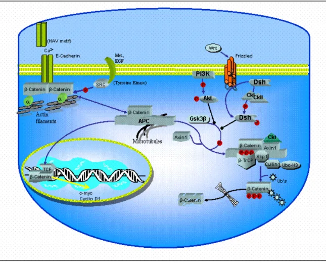
The cellular level of the β-catenin protein, as well as its subcellular localization are very tightly regulated (Figure 4). When not associated with cell-cell junctions, β-catenin forms a large complex with the protein encoded by APC, GSK-3β, and Axin 1 (Behrens et al., 1998). In the absence of Wnt signal, constitutive phosphorylation of β-catenin by CKI/GSK-3β dual-kinase system lower its levels via ubiquitin-mediated proteolysis (Aberle et al., 1997; Kitagawa et al., 1999; Liu et al., 2002). Activation of the Wnt pathway upon binding of the Wnt ligand leads to inactivation GSK-3β that give rise to stabilization of and accumulation of hypo-phosphorylated free-β-catenin, which then translocates into the cell nucleus. Nuclear β-catenin interacts with TCF/LEF family of transcriptional factors and stimulate transcription of a variety of target genes (Hulsken et al., 1994; Molenaar et al., 1996). The nature of the target genes of the β-catenin/LEF complex remain mostly unknown, except for the recently documented c-myc , c-jun, fra-1 and cyclin D1 genes in colorectal cancers (He et al., 1998; Mann et al., 1999; Shtutman et al., 1999; Tetsu and McCormick, 1999).
β-catenin seems to be an essential component of intercellular junctions as well as wnt growth factor signaling pathways. The free cytosolic β-catenin is considered to represent the signaling pool of β-catenin (Sadot et al., 2001; Simcha et al., 2001). In addition, modulation of APC/-catenin association has been suggested to play a role in the regulation of both cadherin-based cell-cell adhesion and cell motility(Hermiston and Gordon, 1995b; Hermiston and Gordon, 1995a; Adams et al., 1996; Nathke et al., 1996)
Figure 4 ββ-catenin is a key molecule in E-cadherin-mediated cell-cell adhesion and in the WNT signaling pathway.
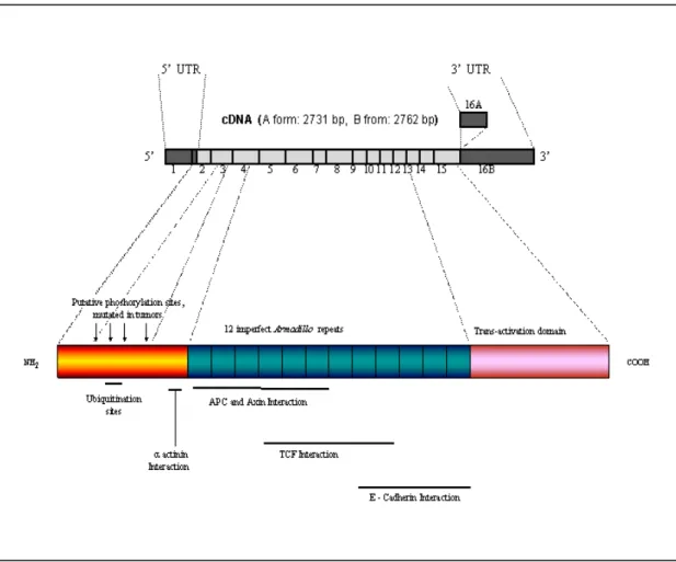
The primary structure of the β-catenin protein reveals that it contains a 130 amino acid amino-terminal domain, an armodillo domain composed of 12 imperfect repeats of 42 amino acids (arm repeats), and a carboxy-terminal domain of 100 amino acids (Figure 5). Several proteins bind to β-catenin through the central region (arm repeats) and this positively-charged groove may serve as a binding motif including TCF/LEF, APC and adherent junction protein E-cadherin. (Rubinfeld et al., 1995; Yost et al., 1996; Huber et al., 1997).
The carboxyl terminal domain functions as a transcriptional activator when fused to the GAL4 DNA-binding domain (van de Wetering et al., 1997), whereas the amino terminus of β-catenin is known to be important for regulating the stability of β-catenin. This region of β-catenin, also called destruction box, contains a series of serine and threonine
residues, which may be phosphorylated. Phosphorylation of serine/threonine residues at positions 29, 33, 37, 41 and 45 by GSK-3β in complex with Axin and APC appears to be a prerequisite for ubiquitination (Aberle et al., 1997; Orford et al., 1997). Recently, another Axin-associated kinase, Casein kinase I (CKI) has been shown to be both necessary and sufficient to initiate the β-catenin phosphorylation-degradation cascade (Liu et al., 2002). Complex of Axin and CKI induces β-catenin phosphorylation at a single site: serine 45, and creates a priming site for subsequent GSK-3β phosphorylation cascade finally hitting the S33/S37 of β-catenin (Amit et al., 2002; Ding and Dale, 2002; Gao et al., 2002; Liu et al., 2002). FWD1 (mouse homologoue of Slimb/βTrCP), an F-box/WD40-repeat protein, specifically forms a multi-molecular complex with β-catenin, Axin, GSK-3β and APC and facilitate ubiquitination and degradation of β-catenin. FWD1 also serves as an intracellular receptor for phosphorylated β-catenin (Kitagawa et al., 1999).