Influence of cigarette smoking on white matter in patients with
clinically isolated syndrome as detected by diffusion tensor imaging
Gamze Durhan
Sevda Diker
Arzu Ceylan Has
Jale Karakaya
Asli Tuncer Kurne
Kader Karli Oguz
C
linically isolated syndrome (CIS), an inflammatory demyelinating disorder of the cen-tral nervous system, is the first clinical episode with features predictive of multiple sclerosis (MS) (1). Many environmental risk factors for conversion from CIS to MS were shown in recent studies (1, 2). Among these, cigarette smoking was shown to increase the risk of developing MS even in passive smokers (3), worsen clinical disability, and exacerbate disease progression (4) .Diffusion tensor imaging (DTI) gives the opportunity to evaluate the tissue integrity, mainly the white matter (WM). Through changes in metrics such as fractional anisotropy (FA) and mean diffusivity (MD), DTI enables the assessment of axonal organization and myelin attenu-ation in addition to the WM lesions evaluated by conventional magnetic resonance imaging (MRI) sequences (5). The brains of MS patients have demonstrated that an increase in MD and a decrease in FA values were far beyond fluid attenuation inversion recovery (FLAIR) hyperin-tense lesions, commonly used as a clinical parameter of MS (6, 7). Brain parenchyma with no apparent lesion, namely, the normal-appearing white matter (NAWM), has drawn much at-tention due to the presence of not only histopathologic and radiologic abnormalities but also a close association with clinical disability and disease progression (8). Changes in the NAWM may also be responsible for the clinicoradiologic discrepancy observed in many patients (6). In addition, several MRI studies have shown significantly altered FA measures in specific brain regions in healthy smoker individuals (9, 10). Although a number of studies have investigated the impact of smoking on patients with MS (4, 11), to the best of our knowledge, no study has studied the influence of smoking on the WM in patients with CIS using DTI.
In this study we sought to determine the effects of smoking on NAWM, lesions, and per-ilesional WM of patients with CIS. The secondary objective was to examine whether
diffu-From the Department of Radiology (G.D.), Ministry of Health Ankara Training and Research Hospital, Ankara, Turkey; the Departments of Neurology (S.D., A.T.K.) and Radiology (K.K.O.) Hacettepe University Medical School, Ankara, Turkey; the National Magnetic Resonance Research Center (UMRAM) (A.C.H., K.K.O. [email protected]), Bilkent University, Ankara, Turkey; the Department of Biostatistics (J.K.), Hacettepe University, Ankara, Turkey.
Received 11 September 2015; accepted 8 October 2015. Published online 24 March 2016. 10.5152/dir.2015.15415
Diagn Interv Radiol 2016; 22:291–296 © Turkish Society of Radiology 2016
NEURORADIOLOGY
ORIGINAL ARTICLE
PURPOSE
Cigarette smoking has been associated with increased occurrence of multiple sclerosis (MS), as well as clinical disability and disease progression in MS. We aimed to assess the effects of smok-ing on the white matter (WM) in patients with clinically isolated syndrome (CIS) ussmok-ing diffusion tensor imaging.
METHODS
Smoker patients with CIS (n=16), smoker healthy controls (n=13), nonsmoker patients with CIS (n=17) and nonsmoker healthy controls (n=14) were included. Thirteen regions-of-interest in-cluding nonenhancing T1 hypointense lesion and perilesional WM, and 11 normal-appearing white matter (NAWM) regions were drawn on color-coded fractional anisotropy (FA) maps. Le-sion load was determined in terms of number and volume of WM hyperintensities.
RESULTS
A tendency towards greater lesion load was found in smoker patients. T1 hypointense lesions and perilesional WM had reduced FA and increased mean diffusivity to a similar degree in smoker and nonsmoker CIS patients. Compared with healthy smokers, smoker CIS patients had more ex-tensive NAWM changes shown by increased mean diffusivity. There was no relationship between diffusion metrics and clinical disability scores, duration of the disease and degree of smoking exposure.
CONCLUSION
Smoker patients showed a tendency towards having greater number of WM lesions and dis-played significantly more extensive NAWM abnormalities.
292
• May–June 2016 • Diagnostic and Interventional Radiology Durhan et al. sion tensor changes relate to the durationof disease, clinical disability and degree of smoking exposure.
Methods
Participants
The local institutional review board ap-proved the study. All participants gave writ-ten informed consent.
A total of 60 right-handed subjects (33 CIS patients, 27 healthy volunteers; female (F)/male (M) ratio, 37/23; age range, 20–57 years; mean age, 31.8±8.0 years) participat-ed in this study. Thirty-three CIS patients, admitted to our institution between 2012 and 2014 were included in the study. All patients fulfilled the McDonald diagnostic criteria for MS but had only one clinical ep-isode involving optic neuritis, cervical and/ or thoracic myelitis or brainstem/cerebellar syndrome. None of the patients converted to clinically definite MS during the study. Twenty-seven healthy volunteers were re-cruited using e-mail advertisement. Same individuals also constituted the partici-pants of another MRI study on brain volume alterations. Exclusion criteria were presence of any systemic or neurologic disease other than CIS, receiving steroid therapy, having an attack within four weeks of the MRI scan, having a lifetime history of dependence disorders other than nicotine, neurologic illness other than CIS, and contraindications to MRI.
Two neurologists (S.D., A.T.) with four and 15 years of experience, blinded to the MRI findings, evaluated all patients and control subjects, and recorded expanded disability status scale scores, age at onset of smoking,
pack-years of cigarettes and duration of the disease.
The subjects were categorized into four groups according to the smoking habit and presence of CIS: smoker patients with clini-cally isolated syndrome (CISS: n=16; F/M ra-tio, 8/8; mean age, 34.3±8.9 years), smoker healthy controls (HS: n=13; F/M ratio, 6/7; mean age, 33.7±7.4 years), nonsmoker patients with clinically isolated syndrome (CISNS: n=17; F/M ratio, 13/4; mean age, 29.7±6.9 years), and nonsmoker healthy controls(HNS: n=14; F/M ratio, 10/4; mean age, 29.8±7.9 years). Nonsmoker partici-pants had smoked no more than five ciga-rettes in their lifetime.
Image acquisition
Imaging protocol included T1-weight-ed three-dimensional (3D) high resolu-tion images with 0.9 mm isotropic voxels (MPRAGE) (TR/TE, 2600/3.1 ms; matrix, 224×256; NEX, 1; TA, 4.06; number of slic-es, 176; slice thickness, 1.00 mm; distance factor, 50%; voxel size, 1 mm; in plane reso-lution, 1 mm) and 3D double inversion-re-covery sampling perfection with appli-cation optimized contrast using different flip angle evolution (DIR-SPACE) (TR/TE, 7500/325 ms; matrix, 192×192; NEX, 1; TA, 6.09; number of slices, 144; slice thickness, 1.12 mm without intersection gap; voxel size, 1 mm; in plane resolutions, 1 mm) imaging on 3.0 T MRI scanner (Magnetom, Tim, Siemens). This imaging sequence was selected because it enables confident le-sion detection and delineation by mul-tiplanar imaging and cerebrospinal flu-id-attenuation (12, 13). Patients received intravenous gadopentetate dimeglumine (0.01 mmol/kg) contrast material. Postcon-trast axial T1-weighted 3D MPRAGE coro-nal fat saturated images (TR/TE, 550/15 ms; matrix, 256×256; NEX, 2) were obtained at 5 min after injection in all patients to de-tect active demyelinating lesion.
Isotropic high-resolution diffusion ten-sor imaging (DTI) of the whole brain (sin-gle-shot EPI; TR/TE, 8020/83 ms; maximum b value, 1000s/mm²; 60 independent di-rections; FOV, 256 mm; matrix, 128×128; 64 axial sections with 2 mm thickness without intersection gap; voxel size, 2×2×2 mm) was also selected to recognize the WM integrity.
Image processing and analysis
Immediately after image acquisition, a neuroradiologist with 14 years of
experi-ence (K.K.O.) reviewed the images to rule out any non-demyelinating lesion or to detect active demyelinating lesions. From this review, the investigator checked the radiologic eligibility of the patients for the study.
To determine lesion load in terms of num-ber and volume, BrainVoyager QX 2.6 for Linux (http://www.brainvoyager.com) was used. Two radiologists with 5 and 14 years of experience (G.D. and K.K.O., respectively) recorded the coordination of every hyperin-tense WM lesion on 3D DIR SPACE imaging in consensus. The lesions were ranged by a bounding box in which all the intensities of voxels were fixed to a threshold for a clear visual distinction of lesions and parenchy-ma (http://support.brainvoyager.com/vol- ume-space/107-volume-rendering/314-us-ers-guide-masking-and-cutting.html). This was done by radiologists in consensus. In this way, the number and total volume of interests were calculated.
FSL (http://fsl.fmrib.ox.ac.uk/fsl/fslwiki/) was used for DTI data analysis. All scans were corrected for head motion, and eddy currents using the affine registration. b0 volumes of each subject were extracted and averaged. By fitting a main diffusion tensor in each voxel with FSL DTI Fit tool, the FA and MD maps were calculated. All FA, MD, pre- and postcontrast 3D T1 MPRAGE and DIR-SPACE images were registered to Montreal Neurological Institute template by FLIRT command, a part of FSL. The mean FA and MD of the regions-of-interest (ROIs), specified below were computed for each subject.
A total of 13 ROIs were manually drawn on color-coded FA maps, with a careful review of the co-registered pre- and postcontrast 3D T1-weighted images, 3D DIR-SPACE and T2 turbo spin-echo for accurate localization of ROIs of nonenhancing T1 hypointense lesion without edema in the left frontal WM (if unavailable, then right frontal WM). A perilesional WM 5 mm away from that le-sion and 11 NAWM regions of 5–6 mm2 were
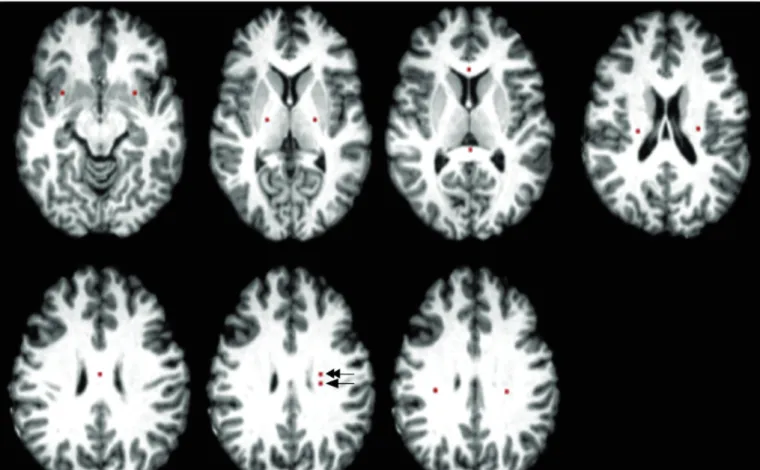
selected. NAWM ROIs were drawn from the genu, splenium and body of the corpus callosum, bilateral corona radiata, superior longitudinal fasciculus, posterior limb of internal capsule, and uncinate fasciculi on color-coded FA maps using Johns Hopkins University WM tractography, and the Inter-national Consortium for Brain Mapping DTI-81 WM atlases in FSL. DIR-SPACE sequence was used to avoid lesions in the determina-tion of the NAWM ROIs (Fig. 1).
Main points
•
Clinically isolated syndrome (CIS), an inflammatory demyelinating disorder of the central nervous system (CNS), is a first clinical episode with features predictive of multiple sclerosis.•
In previous studies, cigarette smoking has been shown to increase the risk of conversion from CIS to MS, worsen clinical disability, and exacerbate disease progression.•
Compared with nonsmoker patients, smoker patients with CIS show tendency towards increased white matter lesions, in our study.•
Smoker patients with CIS exhibit more extensive normal-appearing white matter abnormalities than nonsmoker patients on diffusion tensor images.Statistical analysis
Numerical variables were evaluated for normality of data distribution using the Kolmogorov-Smirnov test. Descriptive statistics were expressed as mean±stan-dard deviation. Independent samples t-test was performed to compare the means of two groups. The Mann Whitney U test was used to compare two groups of nonparametric data. One-way anal-ysis of variance (One-way ANOVA) was performed to compare the differences between groups. Spearman’s rho correla-tion coefficient was used to examine the relationship between two variables. Mul-tiple linear regression was carried out to assess the association between two or more independent variables and a sin-gle continuous dependent variable. The analysis was also used to assess whether confounding exists. The effect of group on dependent variables was examined by multiple linear regression model, after ad-justment for age and sex. A P value ≤ 0.05 was accepted as significant. Data analysis was performed by IBM SPSS Statistics 21.0 software (IBM Corp.).
Results
The demographic and clinical features of the patients are summarized in Table 1. There was no significant difference be-tween CISS and HS, nor between CISNS and HNS in terms of age and sex. Furthermore, age- and sex-corrected analyses were used. Exposure to smoking, which was assessed by mean pack-years, mean cigarettes/day,
age at onset of smoking and duration of smoking, was similar in CISS andHS. CISS and CISNS showed no difference in duration of disease (10.8 vs. 9.2 months, P > 0.05) and expanded disability status scale (0.06 vs. 0.18, P > 0.05).
The review of DIR-SPACE and postcon-trast 3D T1-weighted images showed no edematous and/or contrast-enhanced
de-Figure 1. Placement of regions of interest (ROIs). ROIs of normal appearing white matter (NAWM) were drawn from the genu, splenium and body of the
corpus callosum, bilateral corona radiata, superior longitudinal fasciculus, posterior limb of internal capsule, and uncinate fasciculi. Lesion (arrow) and perilesional (double arrow) ROIs were drawn on the left frontal white matter.
Table 1. Baseline demographic characteristics, clinical data, and smoking history
CISS (n=16) CISNS (n=17) HS (n=13) HNS (n=14) P
Age (years) 34.3±8.9 29.7±6.9 33.7±7.4 29.8±7.9 0.226
Sex (Female/male), n 8/8 13/4 6/7 10/4 0.223
Pack-year 9.3±10.1 - 7.7±4.7 - 0.904
Cigarettes per day 14.3±10.6 - 12.9±6.6 - 0.760
Duration of smoking (years) 11.6±8.5 - 12.6±8.8 - 0.680
Age at onset of smoking (years) 20.6±5.4 - 21.0±5.8 - 0.716 Time lapse from clinical episode (months) 10.8±16.5 9.2±7.0 - - 0.313
EDSS 0.06 0.18 - - 0.600
Data are presented as mean±SD, unless otherwise noted.
CISS, smoker patients with clinically isolated syndrome; CISNS, nonsmoker patients with clinically isolated syndrome; HS, smoker healthy controls; HNS, nonsmoker healthy controls; EDSS, expanded disability status scale.
myelinating lesions. ROIs of T1 hypointense lesions and perilesional WM could be placed over the left frontal WM in all patients.
CISS had a greater number and volume of WM hyperintense lesions than CISNS on DIR-SPACE imaging (15 vs. 11, P = 0.20 and 5.2 cm³ vs. 2.4 cm³, P = 0.24), although the difference did not reach statistical signifi-cance.
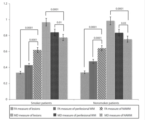
Significantly decreased FA and increased MD values were found in perilesional WM and lesions compared with those of NAWM in both CISS (mean values: FA=0.33,
MD=0.96 for lesions; FA=0.43, MD=0.84 for perilesional WM; FA=0.62, MD=0.77 for NAWM) andCISNS (mean values: FA=0.34, MD=0.98 for lesions; FA=0.47, MD=0.83 for perilesional WM; FA=0.64, MD=0.75 for NAWM) (P < 0.05, for all). Compared with perilesional WM and NAWM, lesions showed significantly decreased FA and in-creased MD in CISS andCISNS (P < 0.05, for all). However, FA and MD measures from the lesions and perilesional WM did not dif-fer significantly between CISS andCISNS (P > 0.05, for all) (Fig. 2).
Regions of NAWM with significant FA and/or MD differences in the patient and control groups are presented in Table 2. Compared with Hs, CISS showed elevated MD in the splenium of corpus callosum, bi-lateral corona radiate, and superior longitu-dinal fasciculus (P < 0.05, for all). Among the nonsmokers, a higher MD in the right coro-na radiata was found in CISNS than HNS (P = 0.02). There was no brain region where CISS andCISNS had significantly lower MD values than their matched controls. Compared with HNS, CISNS showed increased FA values
in three regions of the NAWM including the body of corpus callosum, left superior lon-gitudinal fasciculus and left posterior limb of the internal capsule (P < 0.05, for all). None of the NAWM regions showed signif-icant FA change in HS vs. CISS patients (P > 0.05, for all).
Compared with HNS, HS showed no region of FA alteration, while revealing significant-ly decreased MD values in the left corona radiata and superior longitudinal fasciculus (P < 0.05, Table 2).
In CIS patients, the mean MD value in the left superior longitudinal fasciculus was significantly higher in smokers than nonsmokers (0.78±0.03×10ˉ³ mm²/s vs. 0.75±0.04×10ˉ³mm²/s, P = 0.01; Table 2).
Correlation analysis failed to reveal any significant relationship between DTI mea-sures and the duration of disease,
expand-ed disability status scale, and exposure of smoking as assessed by pack-year calcula-tions, cigarettes/day, age at onset, and du-ration of smoking (P > 0.05).
Discussion
We aimed to investigate smoking-related WM alterations using DTI in patients with CIS in comparison with healthy controls matched for smoking habit. We calculated FA and MD as the most commonly used DTI measures in chronic T1 hypointense le-sions, perilesional WM, and NAWM far from the lesions (14). On ex vivo MRI, chronic, nonenhancing T1 hypointense lesions, so called “black holes,” show hypocellularity, reduced axonal density, decreased myelin and greater matrix disruption compared with non-black hole lesions histopatholog-ically (15). These lesions usually show the greatest WM injury, as also supported by findings of magnetic resonance spectros-copy and magnetization transfer imaging (16). In agreement with previously pub-lished reports (5, 17), we found the most significant changes in FA and MD values in T1 hypointense lesions, followed by perile-sional WM and NAWM of the patients com-pared with NAWM of the healthy controls. FA, generally regarded as a measure of WM
integrity, is closely related with axonal cali-ber and the surrounding myelin. MD gives a measure of the average molecular motion depending on cellular size, compartmental fluid imbalance, and degradation products (14, 18). With the abovementioned histo-pathologic abnormalities in myelin and ac-companying primary or secondary axonal loss, reduced FA and elevated MD were ex-pected changes in the WM of our patients with demyelinating disease.
Our study revealed a trend for a greater lesion load on double inversion recovery sequence in smoking patients, although it was not statistically significant. Further-more, smoker patients had evidently more extensive NAWM changes than nonsmok-ers (in five ROIs vs. one ROI) compared with matched healthy controls. Additional dete-riorating effects of the toxic contents of to-bacco smoke such as cyanide, nitric oxide, and free radicals might explain the greater vulnerability of the NAWM in CISS. The as-sociations between thiocyanate, the main metabolite of cyanide and demyelination in the central nervous system of animals (19), and between nitric oxide and axonal degeneration, blocks in axonal conduction, especially in demyelinated axons (20) are well known. Through increased axonal
de-Figure 2. Comparison of mean fractional anisotrophy (FA) and mean diffusivity (MD) measurements
from T1 hypointense lesions, perilesional white matter (WM) and normal appearing white matter (NAWM). Statistical significance level is given between the bars for each comparison.
1.2 1 0.8 0.6 0.4 0.2 0 Smoker patients 0.0001 0.0001 0.0001 0.0001 0.0001 0.0001 0.01 0.03
FA measure of lesions FA measure of perilesional WM FA measure of NAWM MD measure of lesions MD measure of perilesional WM MD measure of NAWM
generation, smoking has been blamed for a worse clinical course in MS (21).
With no apparent change in FA, de-creased MD in the left corona radiata and superior longitudinal fasciculus of HS may be due to nicotine-related alterations in the WM. The nicotine-induced cytotoxic swelling suggested by Gazdzinski et al. (22) and proinflammatory effects of ciga-rette smoke on tissue as shown by elevat-ed peripheral leukocytes, and recruitment of polymorphonuclear cells, monocytes, macrophages, and increased fibrinogen may be partly responsible for cellular ede-ma and reduced MD levels in the HS of the
current study (23). Some recent studies have hypothesized on the neurotropic and
promyelinating effects of smoking and have found higher FA in brain regions such as the prefrontal WM, cingulum, and cor-pus callosum in smokers compared with nonsmokers (9, 24). Yet, another study on heavy smokers reported decreased FA in the anterior corpus callosum (25). The vari-able degrees of addiction of smoking in the participants might be one important factor in these contradictory research findings. Al-though limited dependence and exposure to nicotine revealed no significant change, heavy smoking exposure showed marked FA reductions in WM (9, 24). Whether FA or MD is more sensitive and an earlier mea-sure of WM disintegrity remains controver-sial in the literature (26, 27).
We failed to show a significant correlation of DTI measures with exposure of smoking or duration of the disease in our relatively homogeneous patient group of CIS. This might have resulted from limited cigarette smoking exposure and relatively short du-ration of disease as well as the younger age of our patients.
Interestingly, CISNS in comparison with
HNS revealed increased FA in several regions of the NAWM. Increased intracellular water resulting in cellular swelling, fiber reorga-nization or remyelination can account for an increase in FA (14, 28). Similar findings have been previously shown in the NAWM of patients with chronic recurrent inflam-matory optic neuropathy, in T1-hyperin-tense lesions and normal appearing tissue in patients with MS. The authors attributed these findings to remyelination and glial activation (29, 30). Low expanded disability status scale and absence of conversion to MS during the two years of the study may have been partly related to remyelination in the NAWM, occurring as a compensato-ry mechanism. More heterogeneous histo-pathologic processes rather than uniform demyelination or axonal degeneration may be present in the NAWM of patients with CIS.
The limitations of the current study war-rant discussion. First, the number of subjects was relatively small. Second, we could not avoid gender disproportion in the smoking and nonsmoking groups because women usually show less tendency to smoke in our population. To minimalize this limitation we used age- and sex-corrected analysis. Finally, it was not possible to compare the conversion rate of CIS to MS in smokers vs. nonsmokers due to the relatively short time period of the study. Therefore, studies with more individuals, more detailed character-ization of smoking behavior and a longer follow-up are needed.
In conclusion, CIS and smoking appear to cause variable changes on the WM. Smoking patients showed a tendency towards greater numbers of demyelinating WM lesions and significantly more extensive NAWM abnor-malities on DTI. Our findings support the ad-verse effects of smoking on the WM, which in turn may be related with the clinical course of CIS and conversion to MS.
Acknowledgements
The authors thank the National Mag-netic Resonance Imaging Research Centre (UMRAM) for providing the MRI facility and
Table 2. FA and MD readings in normal-appearing white matter regions across the groups
FA readings
NAWM regions CISS/HS CISNS/HNS HS/HNS CISS/CISNS
CC genu 0.84/0.84 0.86/0.85 0.84/0.85 0.84/0.86 CC body 0.68/0.68 0.71/0.65a 0.68/0.65 0.68/0.71 CC splenium 0.87/0.88 0.87/0.86 0.88/0.86 0.87/0.87 R corona radiata 0.46/0.49 0.49/0.53 0.49/0.53 0.46/0.49 L corona radiata 0.49/0.53 0.51/0.53 0.53/0.53 0.49/0.51 R SLF 0.56/0.59 0.54/0.56 0.59/0.56 0.56/0.54 L SLF 0.54/0.58 0.59/0.55b 0.58/0.55 0.54/0.59 R uncinate fasciculus 0.51/0.58 0.51/0.53 0.58/0.53 0.51/0.51 L uncinate fasciculus 0.45/0.49 0.52/0.52 0.49/0.52 0.45/0.52 R PLIC 0.69/0.62 0.68/0.65 0.62/0.65 0.69/0.68 L PLIC 0.68/0.62 0.73/0.65b 0.62/0.65 0.68/0.73 MD readings
CISS/HS CISNS/HNS HS/HNS CISS/CISNS
CC genu 0.74/0.62 0.70/0.68 0.62/0.68 0.74/0.70 CC body 0.82/0.74 0.80/0.78 0.74/0.78 0.82/0.80 CC splenium 0.75/0.65a 0.76/0.68 0.65/0.68 0.75/0.76 R corona radiata 0.77/0.69b 0.74/0.71c 0.69/0.71 0.77/0.74 L corona radiata 0.77/0.68b 0.75/0.74 0.68/0.74c 0.77/0.75 R SLF 0.77/0.72d 0.77/0.75 0.72/0.75 0.77/0.77 L SLF 0.78/0.69a 0.75/0.74 0.69/0.74b 0.78/0.75a R uncinate fasciculus 0.78/0.73 0.80/0.77 0.73/0.77 0.78/0.80 L uncinate fasciculus 0.80/0.74 0.77/0.75 0.74/0.75 0.80/0.77 R PLIC 0.74/0.69 0.73/0.73 0.69/0.73 0.74/0.73 L PLIC 0.76/0.69 0.71/0.73 0.69/0.73 0.76/0.71
Data represent mean FA and MD (×10ˉ³ mm²) measurements in four matched groups.
FA, fractional anisotropy; MD, mean diffusivity; NAWM, normal-appearing white matter; CISS, smoker patients with clinically isolated syndrome; HS, smoker healthy controls; CISNS, nonsmoker patients with clinically isolated syndrome; HNS, nonsmoker healthy controls; CC, corpus callosum; R, right; L, left; SLF, superior longitudinal fasciculus; PLIC, posterior limb of the internal capsule.
Yıldıray Gökhalk for his help in data acqui-sition.
Conflict of interest disclosure
The authors declared no conflicts of interest.
References
1. Miller DH, Chard DT, Ciccarelli O. Clinically iso-lated syndromes. Lancet Neurol 2012; 11:157– 169. [CrossRef]
2. Mandia D, Ferraro OE, Nosari G, Montomoli C, Zardini E, Bergamaschi R. Environmental fac-tors and multiple sclerosis severity: a descrip-tive study. Int J Environ Res Public Health 2014; 11:6417–6432. [CrossRef]
3. Hedstrom AK, Baarnhielm M, Olsson T, Alfreds-son L. Exposure to environmental tobacco smoke is associated with increased risk for multiple sclerosis. Mult Scler 2011; 17:788–793. [CrossRef]
4. Di Pauli F, Reindl M, Ehling R, et al. Smoking is a risk factor for early conversion to clinical-ly definite multiple sclerosis. Mult Scler 2008; 14:1026–1030. [CrossRef]
5. Filippi M, Cercignani M, Inglese M, Horsfield MA, Comi G. Diffusion tensor magnetic reso-nance imaging in multiple sclerosis. Neurology 2001; 56:304–311. [CrossRef]
6. Ciccarelli O, Werring DJ, Wheeler-Kingshott CA, et al. Investigation of MS normal-appear-ing brain usnormal-appear-ing diffusion tensor MRI with clin-ical correlations. Neurology 2001; 56:926–933. [CrossRef]
7. Yu CS, Lin FC, Liu Y, Duan Y, Lei H, Li KC. Histo-gram analysis of diffusion measures in clinical-ly isolated syndromes and relapsing-remitting multiple sclerosis. Eur J Radiol 2008; 68:328– 334. [CrossRef]
8. Filippi M, Comi, G., Rovaris, M. Normal-appear-ing white and grey matter damage in multiple sclerosis. 1sted. Milano: Springer, 2004; 3–8. 9. Paul RH, Grieve SM, Niaura R, et al. Chronic
cig-arette smoking and the microstructural integri-ty of white matter in healthy adults: a diffusion tensor imaging study. Nicotine Tob Res 2008; 10:137–147. [CrossRef]
10. Gons RA, van Norden AG, de Laat KF, et al. Cig-arette smoking is associated with reduced mi-crostructural integrity of cerebral white matter. Brain 2011; 134:2116–2124. [CrossRef] 11. Salzer J, Hallmans G, Nystrom M, Stenlund H,
Wadell G, Sundstrom P. Smoking as a risk factor for multiple sclerosis. Mult Scler 2013; 19:1022– 1027. [CrossRef]
12. Kober T, Granziera C, Ribes D, et al. MP2RAGE mul-tiple sclerosis magnetic resonance imaging at 3 T. Invest Radiol 2012; 47:346–352. [CrossRef] 13. Wattjes MP, Lutterbey GG, Gieseke J, et al.
Dou-ble inversion recovery brain imaging at 3T: diagnostic value in the detection of multiple sclerosis lesions. AJNR Am J Neuroradiol 2007; 28:54–59.
14. Beaulieu C. The basis of anisotropic water diffu-sion in the nervous system - a technical review. NMR Biomed 2002; 15:435–455. [CrossRef] 15. van Walderveen MA, Kamphorst W, Scheltens P,
et al. Histopathologic correlate of hypointense lesions on T1-weighted spin-echo MRI in mul-tiple sclerosis. Neurology 1998; 50:1282–1288. [CrossRef]
16. van Walderveen MA, Barkhof F, Pouwels PJ, van Schijndel RA, Polman CH, Castelijns JA. Neuro-nal damage in T1-hypointense multiple sclero-sis lesions demonstrated in vivo using proton magnetic resonance spectroscopy. Ann Neurol 1999; 46:79–87. [CrossRef]
17. Oguz KK, Kurne A, Aksu AO, Taskiran A, Karabu-lut E, Karabudak R. A comparative assessment of cerebral white matter by magnetization transfer imaging in early- and adult-onset multiple scle-rosis patients matched for disease duration. J Neurol 2010; 257:1309–1315. [CrossRef] 18. Pierpaoli C, Jezzard P, Basser PJ, Barnett A, Di
Chi-ro G. Diffusion tensor MR imaging of the human brain. Radiology 1996; 201:637–648. [CrossRef] 19. Van Houten WH, Friede RL. Histochemical
stud-ies of experimental demyelination produced with cyanide. Exp Neurol 1961; 4:402–412. [CrossRef]
20. Redford EJ, Kapoor R, Smith KJ. Nitric oxide donors reversibly block axonal conduction: demyelinated axons are especially susceptible. Brain 1997; 120:2149–2157. [CrossRef]
21. Sundstrom P, Nystrom L. Smoking worsens the prognosis in multiple sclerosis. Mult Scler 2008; 14:1031–1035. [CrossRef]
22. Gazdzinski S, Durazzo TC, Studholme C, Song E, Banys P, Meyerhoff DJ. Quantitative brain MRI in alcohol dependence: preliminary evidence for effects of concurrent chronic cigarette smoking on regional brain volumes. Alcohol Clin Exp Res 2005; 29:1484–1495. [CrossRef] 23. Shirani A, Tremlett H. The effect of smoking
on the symptoms and progression of multiple sclerosis: a review. J Inflamm Res 2010; 3:115– 126. [CrossRef]
24. Hudkins M, O’Neill J, Tobias MC, Bartzokis G, London ED. Cigarette smoking and white mat-ter microstructure. Psychopharmacology 2012; 221:285–295. [CrossRef]
25. Lin F, Wu G, Zhu L, Lei H. Heavy smokers show abnormal microstructural integrity in the ante-rior corpus callosum: a diffusion tensor imaging study with tract-based spatial statistics. Drug Alcohol Depend 2013; 129:82–87. [CrossRef] 26. Werring DJ, Clark CA, Barker GJ, Thompson AJ,
Miller DH. Diffusion tensor imaging of lesions and normal-appearing white matter in multi-ple sclerosis. Neurology 1999; 52:1626–1632. [CrossRef]
27. Cubon VA, Putukian M, Boyer C, Dettwiler A. A diffusion tensor imaging study on the white matter skeleton in individuals with sports-re-lated concussion. J Neurotrauma 2011; 28:189– 201. [CrossRef]
28. Le Bihan D. Looking into the functional archi-tecture of the brain with diffusion MRI. Nat Rev Neurosci 2003; 4:469–480. [CrossRef] 29. Colpak AI, Kurne AT, Oguz KK, Has AC, Dolgun
A, Kansu T. White matter involvement beyond the optic nerves in CRION as assessed by dif-fusion tensor imaging. Int J Neurosci 2015; 125:10–17. [CrossRef]
30. Calabrese M, Rinaldi F, Seppi D, et al. Cortical diffusion-tensor imaging abnormalities in mul-tiple sclerosis: a 3-year longitudinal study. Ra-diology 2011; 261:891–898. [CrossRef]