Case report and the surgical treatment of two cases with pulmonary
atresia in which pulmonary arteries are perfused by coronary arteries
Pulmoner arter kanlanması koroner arterler yoluyla olan
pulmoner atrezili iki olgu sunumu ve cerrahi tedavisi
1Department of Pediatric Cardiology, Başkent University Faculty of Medicine, Ankara, Turkey 2Department of Cardiovascular Surgery, Başkent University Faculty of Medicine, Ankara, TurkeyKahraman Yakut, M.D.,1 Kürşad Tokel, M.D.,1 Birgül Varan, M.D.,1 İlkay Erdoğan, M.D.,1 Murat Özkan, M.D.2
Özet– Pulmoner atrezi (PA), ventriküler septal defekt (VSD) ile birlikte basit bir kapak atrezisinden, akciğer yatağının aortadan ayrılan kollateral arterler ile kanlandığı gerçek pulmoner arterlerin bulunmadığı veya ağır hipoplazik ol-duğu tabloya kadar çok geniş spektrum halinde görülebil-mektedir. Pulmoner arterin ve dallarının boyutu ile birlikte eşlik eden diğer karmaşık kalp lezyonlarının varlığı tedavi yönteminde ve tüm düzeltme seçeneklerinin belirlenme-sinde önemlidir. Basit formlarında Fallot tetralojisi onarımı tarzında tam düzeltme ameliyatı yapılırken, spektrumun di-ğer ucundaki hastalarda çok sayıda girişim gerekmekte ve yaşam kalitesi oldukça değişken olmaktadır. Pulmoner arter ve dallarının boyutu, eşlik eden kollateral arterlerin varlığı tedavi yönetiminde belirleyicidir. Burada ilk olgu 40 günlük, ikinci olgu iki günlük iken muayene sırasında üfürüm fark edilmesi üzerine yapılan ekokardiyografik incelemede VSD, pulmoner atrezi tanısı konmuş olan ve pulmoner arterlerin koroner arter aracılığı ile kanlanmasının sağlandığı iki olgu-yu sunmak istedik. İkinci olgumuzda tanı kateter anjiyografi ile kesinleşti. İlk olgumuzda ancak cerrahi sırasında kesin tanı konulabildi. Bu çalışmayı sunmaktaki amacımız pul-moner atrezili hastalarda pulpul-moner arter boyutu, pulpul-moner konfluansın varlığı, kanlanmasının nereden sağlandığı ve eşlik eden kollateral arterlerin tedavi yönetimindeki önemini vurgulamaktır.
Summary– Pulmonary atresia (PA) and ventricular septal defect (VSD) can occur in a variety of ways, from simple valve atresia to a condition in which circulation to the pul-monary bed occurs through collateral arteries separated from the aorta and there are no real pulmonary arteries, or they are present but hypoplastic. The size of the pulmonary arteries and concomitant complex cardiac lesions are impor-tant in making decisions about treatment and correctional alternatives. While complete correction surgeries in the style of a correction of tetralogy of Fallot are performed in simpler cases, many very invasive procedures are also performed and the resulting quality of life is very variable. The size of the pulmonary artery and its branches and the presence of accompanying collateral vessels are determining factors in the management of the disease. In this report, 2 cases of VSD and PA, in which the circulation of the pulmonary arter-ies was through the coronary arterarter-ies, diagnosed as a result of echocardiography performed following murmurs heard during examination, are described. The first patient was 40 days old and the second was 2 days old. In the second case, diagnosis was confirmed by catheter angiography, and in the first case, the final diagnosis was made during surgery. The aim of this report is to emphasize the importance of pul-monary artery size, the presence of pulpul-monary confluence, the origin of circulation, and concomitant collateral arteries when considering treatment methods.
P
ulmonary atresia (PA) is a complex congenital heart disease with very variable morphological characteristics which may be seen in association with ventricular septal defect (VSD) or intact ventricular septum.[1–3] In congenital heart diseases associatedwith PA surgical treatment still continues to be a
problematic issue, satisfac-tory results can be obtained using materials provided by developing technological facilities, and novel surgical
techniques. If PAs are hypoplastic, palliative
interven-Received:March 31, 2017 Accepted: August 21, 2017
Correspondence: Dr. Kahraman Yakut. Başkent Üniversitesi Tıp Fakültesi, Çocuk Kardiyoloji Bilim Dalı, Ankara. Tel: +90 312 - 203 68 68 / 1382 e-mail: e-posta: [email protected]
© 2018 Turkish Society of Cardiology
Abbreviations:
MAPCA Major aortopulmonary collateral artery PA Pulmonary atresia VSD Ventricular septal defect
tions which will allow the development of PA so as to permit conduction of complete correction surgeries as soon as possible. With palliative methods it is possible to gain time for the development of pulmonary artery, and its branches, and perform complete correction surgeries at a later time. Although debates concern-ing the most optimal surgical approach still continue, with increasing information, more comforable life is ensured for the patients. When diagnosis of PA is established, echocardiography, catheter angiography, and in case of need, computed tomography are impor-tant imaging modalities in determining dimensions of pulmonary artery, and its branches, presence of con-fluence (if any), source of its blood supply, dimen-sions, and location of the associated aortopulmonary collaterals, in specification of surgical, and interven-tional treatment methods, and decreasing postopera-tive morbidity, and mortality. Developments in diag-nostic, and interventional methods, novel materials offered by technological facilities, accumulation of information about treatment modalities contribute significantly to survival time, and quality of life of the patients.
In this article we aimed to present rarely seen two cases with PA together with their surgical treatment whose pulmonary arterial perfusion was provided through coronary arteries.
CASE REPORT
Case 1– During routine examination of a
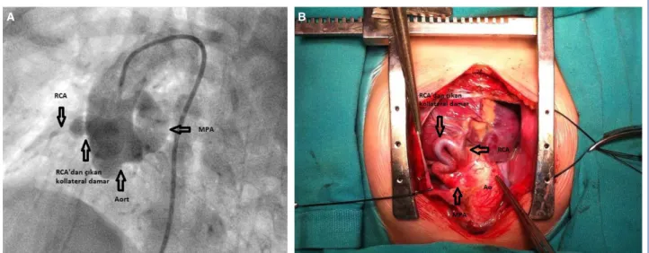
2-month-old male patient at another center, heart murmurs were heard. On echocardiographic examination, he received diagnoses of PA, VSD, and major aortopul-monary collateral artery (MAPCA) so he was sent to our clinic. When he was 10-month old, catheter angiography was performed preoperatively to eval-uate main pulmonary artery, right, and left branches of pulmonary arteries, and collateral arteries. On an-giography, a pulmonary artery arising from ventricles was not detected, while a collateral artery stemming from aortic root perfused pulmonary artery, Besides, common pulmonary artery, right, and left branches of pulmonary arteries had smaller calibres. Origins, and courses of right, and left coronary arteries were not anomalous, and two MACPAs which were not related to pulmonary arteries, and their branches but deliv-ered contrast substance to the vascular beds of right, and left coronary arteries were detected. The patient was brought into the operating room with the decision to perform left Blalock-Taussig (BT) shunt. During surgery, it was determined that common pulmonary artery was not perfused by a collateral artery stem-ming from the aortic root, but its blood supply was provided by a tortuous collateral artery arising from the right coronary artery (Fig. 1a, b – Video 1, 2*).
Besides two MAPCAs unrelated to pulmonary artery, and its branches, delivered blood to the lungs Mod-ified BT shunt was performed between left brachio-cephalic artery, and left pulmonary artery. Collateral artery stemming from the right coronary artery was closed.
Figure 1. (A) Angiogram demonstrating blood circulation of the main pulmonary artery provided by a tortuous collateral vessel stemming from the right coronary artery. (B) Intraoperative view of the blood circulation of the main pulmonary artery provided by a tortuous collateral vessel stemming from right coronary artery.
In addition, two MAPCAs unrelated to pulmonary artery, and its branches, and unfit for unifocalization were also closed. Cardiac output of the patient who was monitorized in the intensive care unit decreased on postoperative 2. day, and the patient exited the next day.
Case 2– A 2-day-old newborn who had undergone
echocardiographic examination at another center be-cause of heart murmurs heard during its PE which necessitated catheter angiography due to persistently low oxygen saturation. The baby was referred to our center with the diagnoses of pulmonary atresia, VSD, and MAPCA for surgical intervention. The patient was referred to our clinic when it was 5-month-old, and underwent preoperative angiographic examina-tion to determine, and evaluate the development, and the source of blood supply of main pulmonary artery, right, and left pulmonary arteries. On angiograms, any evidence suggesting pulmonary artery originat-ing from ventricles was not observed, and also main pulmonary artery, right-left pulmonary arteries were hypoplastic. Blood supply of pulmonary artery was provided by a collateral artery arising from the right coronary artery which communicated with the main pulmonary artery in the supraclavicular region. Be-sides MAPCAs delivered blood into pulmonary
arte-rial bed, and these MAPCAs were not associated with conflent pulmonary arteries. The patient underwent Sano shunt when it was 8-month-old, and collateral vessel between the right coronary artery, and pul-monary artery was closed intraoperatively.
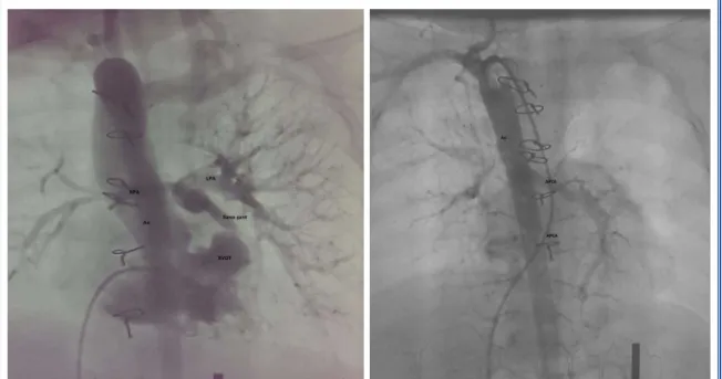
Since in this case peripheral pulmonary arterial bed was hypoplastic, MAPCAs were not closed with the intention to unifocalize them at a later date. Because of stenosis of outlet of the Sano shunt developed when the patient was 2 years of age, previously placed graft was replaced. Catheter angiographies performed dur-ing follow-up period revealed that pulmonary arterial bed, and collateral arteries did not develop adequately for complete correction surgery (Fig. 2a, b).
DISCUSSION
Though considered as a term defining only pulmonary valvular pathology, pulmonary atresia also demon-strates its effects on all cardiac structures mainly left ventricle. In VSD patients with PA, very variable pul-monary artery circulation, and anatomy are the most effective determinant of natural course of this pathol-ogy, and treatment approach. Pulmonary circulation can be achieved with patent ductus arteriosus, and collaterals between coronary, and pulmonary arteries. Size of pulmonary artery, and its branches, presence
Figure 2. (A) Catheter angiogram demonstrating inadequately developed pulmonary arterial bed for complete correction, and unifocalization surgeries, and two collateral arteries stemming from aorta. (B) Catheter angiogram demonstrating inadequately developed pulmonary arterial bed for complete correction, and unifocalization surg-eries, and two collateral arteries stemming from aorta.
of confluence, and suitability of collaterals for unifo-calization are important factors in the preference for complete correction or palliative surgeries.[1–3]
Post-operative survival, and morbidity may be related to the size of the remaining collaterals, and closure or unifocalization of these collaterals may be required before, and after surgery. Therefore in these patients confluence, and diameters of pulmonary arteries, lo-cation, and clinical significance of collaterals should be determined using echocardiography, and catheter angiography. In our two cases, preoperatively, catheter angiography was performed. Angiography report of Case 1, indicated that pulmonary artery was perfused from a collateral vessel stemming from a point near aortic root. However during surgery it was determined that blood circulation of pulmonary artery was provided by a collateral artery stemming from right coronary artery. Angiography of Case 2, could demonstrate that pulmonary arterial circulation was provided by a tortuous artery originating from coronary artery. Preoperative identification of the ori-gin of these collaterals both increases success rate of surgery, and also shortens operative time. Although in our Case 1 any relationship between collaterals, and coronary arteries was not detected preoperatively, demonstration of collaterals stemming from a point near aortic root guided the surgical team.
In VSD patients with pulmonary atresia, pul-monary perfusion is entirely dependent on systemic circulation. Blood circulation of pulmonary artery is provided by MAPCAs mostly originating from aorta or ductus arteriosus. Pulmonary artery circulation is rarely provided by coronary arteries or plexuses formed by bronchial collateral arteries. Collateral ar-teries mostly stem from aorta. Since collateral arar-teries may rarely stem from coronary arteries, as was seen in our cases, aortography which also visualizes coronary arteries is recommended.[4,5]
While planning complete correction or palliative shunt surgeries, preoperative identification of large collaterals which might cause excess blood flow, and determination of their clinical importance are neces-sary prerequisites for decreasing rates of morbidity, and mortality.[1–3,6,7] In both of our cases collateral
ar-teries arising from descending aorta, and delivering blood into lungs were angiographically demonstrated. It has been reported that for achievement of an im-proved quality of life of the patients, at least 14–15
pulmonary artery segments should communicate with the right ventricle, and central pulmonary artery di-ameter should be ≥50% wider than normal. In our Case 1, since pulmonary arterial bed was adequate, and collateral arteries were not suitable for unifocal-ization, two collateral arteries were closed during op-eration. In our Case 2, pulmonary arterial bed was hy-poplastic. Inadequacy of pulmonary arterial bed was predicted, so development of artery was awaited for unifocalization procedure. Many centers do not pre-fer shunting in newborns, and infants, however it has been reported that so as to achieve forward blood flow into pulmonary arteries at an early age, interposing 6–8 mm-wide homografts between the right ventricle, and pulmonary artery may further contribute to the development of pulmonary arterial bed. In our Case 2, we preferred Sano shunt based on the prediction that hypoplastic precapillary pulmonary arterial bed might develop faster with forward blood flow.
In cases with VSD, pulmonary atresia, dimensions of pulmonary artery, and its branches are the most im-portant entities which determine the applicability of complete correction surgeries. Therefore adequacy of diameters of these vessels, and their branches should be evaluated using echocardiography, and angiogra-phy.[1,2,6,7] Since pulmonary arteries, and their branches
are hypoplastic, complete correction surgery could not be performed in both of our cases. As a palliative treatment, modified BT shunt was preferred in Case 1, and Sano shunt in Case 2.
Conclusion
In patients with VSD and pulmonary atresia, de-termination of dimensions, perfusion capability, pres-ence, and origin of collateral arteries is important for the treatment, and prognosis. When performing imag-ing studies, it should not be forgotten that though col-lateral arteries may rarely stem from coronary arteries, ascending aorta together with coronary arteries should be evaluated during these radiological examinations.
*Complementary video file of this article can be found in online version of the journal.
Peer-review: Externally, and independent.
Informed consent: Written, and undersigned informed
consent forms were obtained from the parents of the pa-tients related to the publication of the case, and relevant images.
Conflict of interest: None declared.
Author contributions: Concept – K.T., B.V., M.O.;
De-sign – K.T., B.V., I.E.; Supervision – K.T., B.V., I.E., M.O.; Materials – K.Y.; Data collection &/or processing – K.Y., M.O.; Analysis and / or interpretation – K.Y., M.O., I.E.; Literature search – K.Y.; Writing – K.Y.; Critically revised by – K.T.
REFERENCES
1. Reddy VM, McElhinney DB, Amin Z, Moore P, Parry AJ, Te-itel DF, et al. Early and intermediate outcomes after repair of pulmonary atresia with ventricular septal defect and major aortopulmonary collateral arteries: experience with 85 pati-ents. Circulation 2000;101:1826–32.
2. Tchervenkov CI, Roy N. Congenital Heart Surgery Nomenc-lature and Database Project: pulmonary atresia-ventricular septal defect. Ann Thorac Surg 2000;69:S97–105.
3. Zhu J, Meza J, Kato A, Saedi A, Chetan D, Parker R, et al. Pul-monary flow study predicts survival in pulPul-monary atresia with ventricular septal defect and major aortopulmonary collateral
arteries. J Thorac Cardiovasc Surg 2016;152:1494–503.e1. 4. Cheung EW, Richmond ME, Turner ME, Bacha EA, Torres
AJ. Pulmonary atresia/intact ventricular septum: influence of coronary anatomy on single-ventricle outcome. Ann Thorac Surg 2014;98:1371–7.
5. Walsh MA, Asoh K, Van Arsdell GS, Humpl T. Critical care outcomes in pulmonary atresia and intact ventricular sep-tum undergoing single-ventricle palliation. Cardiol Young 2010;20:290–6.
6. Santos FC, Croti UA, Marchi CH, Hassem Sobrinho S. Doub-le Aortic Arch Associated with Pulmonary Atresia with Vent-ricular Septal Defect. Braz J Cardiovasc Surg 2016;31:63–5. 7. Carotti A, Trezzi M. Pulmonary atresia with ventricular septal
defect and major aortopulmonary collateral arteries: primary repair. Multimed Man Cardiothorac Surg 2016;2016. pii: mmv040.
Keywords: Congenital heart disease; coronary artery; pulmonary
atresia; surgical treatment.
Anahtar sözcükler: Doğumsal kalp hastalığı; koroner arter;