Magneto-Optics of Excitons Interacting with
Magnetic Ions in CdSe/CdMnS Colloidal
Nanoplatelets
Elena V. Shornikova,
*
Dmitri R. Yakovlev,
*
Danil O. Tolmachev, Vitalii Yu. Ivanov, Ina V. Kalitukha,
Victor F. Sapega, Dennis Kudlacik, Yuri G. Kusrayev, Aleksandr A. Golovatenko, Sushant Shendre,
Savas Delikanli, Hilmi Volkan Demir, and Manfred Bayer
Cite This:ACS Nano 2020, 14, 9032−9041 Read Online
ACCESS
Metrics & More Article Recommendations*
sı Supporting InformationABSTRACT: Excitons in diluted magnetic semiconductors
represent excellent probes for studying the magnetic properties of these materials. Various magneto-optical effects, which depend sensitively on the exchange interaction of the excitons with the localized spins of the magnetic ions can be used for probing. Here, we study core/shell CdSe/(Cd,Mn)S colloidal nanoplatelets hosting diluted magnetic semiconductor layers. The inclusion of the magnetic Mn2+ions is evidenced by three
magneto-optical techniques using high magneticfields up to 15 T: polarized photoluminescence, optically detected magnetic
resonance, and spin-flip Raman scattering. We show that the holes in the excitons play the dominant role in exchange interaction with magnetic ions. We suggest and test an approach for evaluation of the Mn2+concentration based on the spin−
lattice relaxation dynamics of the Mn2+ spin system.
KEYWORDS: diluted magnetic semiconductors, CdSe nanoplatelet, colloidal nanocrystals, magneto-optics, spin-flip Raman scattering,
optically detected magnetic resonance
I
ncorporation of magnetic ions in colloidal nanocrystals (NCs) opens exciting opportunities for the engineering of spintronics devices.1−4 The underlying idea to exploit the strong sp−d exchange interactions of electrons and holes with the localized spins of magnetic ions originates from the physics of diluted magnetic semiconductors (DMSs).5 This research direction was establishedfirst for bulk DMS materials and later was successfully extended for epitaxially grown DMS hetero-structures, including quantum wells and quantum dots.6,7 In colloidal nanostructures, it is still at an early stage, while several important results have been already achieved. The giant Zeeman splitting was demonstrated by measuring the magnetic circular dicroism,8−11 including the photoinduced magnetism in Ag+ -doped CdSe NCs,12 and evidenced by polarized photo-luminescence (PL) in external magnetic fields.13−18 The exchange interaction of excitons with Mn2+ ions was provenby optically detected magnetic resonance (ODMR).19Magnetic polaron formation was reported,3,20−22 and the influence of Mn2+spinfluctuations was considered.23
Magneto-optical studies of the exciton emission, its giant Zeeman splitting and polarization, are a valuable tool for investigation of DMS nanostructures. There is, however, a
limitation for the parameters of DMS NCs to provide efficient exciton PL. The Mn2+ ions have an absorption band at the energy of about 2.1 eV associated with the internal transition
6A
1→4T1; the corresponding4T1→6A1emission is located at
about 2.0 eV. This means that in (Cd,Mn)Se NCs, the exciton resonance should be considerably detuned from this energy, because the efficient energy transfer to the Mn2+ ions would
otherwise provide a nonradiative recombination channel for the excitons, quenching their emission. For this reason, (Cd,Mn)Se spherical NCs with large diameters were synthesized in order to keep the exciton emission energy below 2.1 eV.11,13,21
Nanoplatelets (NPLs) are an emerging class of colloidal nanocrystals, which are atomicallyflat with several monolayer thickness, resembling free-standing quantum wells.24NPLs with magnetic Mn2+ ions were synthesized in 2015,16 providing
Received: May 14, 2020
Accepted: June 25, 2020
Published: June 25, 2020
Article
www.acsnano.org
Downloaded via BILKENT UNIV on February 12, 2021 at 12:44:36 (UTC).
remarkable opportunities for wave function engineering.11,25−27 The Mn2+ ions were incorporated in the NPL cores28 or shells.16,26
In this paper, we study the magneto-optical properties of core/shell CdSe/Cd1−xMnxS NPLs, which arise from excitons interacting with the magnetic ions. Three experimental approaches are used: (i) polarized PL in external magnetic fields, (ii) optically detected magnetic resonance of the Mn2+
ions, and (iii) spin-flip Raman scattering. We measure the spin− lattice relaxation dynamics of the Mn2+spin system and suggest an approach for evaluation of the Mn2+concentration.
Four NPL samples were grown for this study, see refs16,29, and30andSupporting Information S1for details. All of them have a 2-monolayer thick CdSe core and 4-monolayer thick shells cladding the core. The reference sample #0 has nonmagnetic CdS shells, and the other three DMS NPLs have Cd1−xMnxS shells with Mn2+ concentrations x ranging from
0.009 to 0.029. The sample parameters are given inTable 1.
Note that the Mn2+ concentrations obtained by inductively
coupled plasma mass spectrometry (ICP-MS) measurements differ from the values that we evaluate from the spin−lattice relaxation dynamics. We are convinced that the latter values are more reliable and we use them in the paper.
RESULTS AND DISCUSSION
Specifics of DMS Heterostructures. The band structure of the CdSe/Cd1−xMnxS NPLs is shown schematically inFigure
2a. The CdSe core with zincblende lattice (Figure S1) has the bandgap energy EgCdSe= 1.75 eV
31
and is sandwiched between shells with EgCdS= 2.50 eV. Note that the EgCdSe= 1.84 eV used in
refs 19 and 26 corresponds to the wurtzite lattice. The conduction and valence band offsets between CdSe and CdS are not precisely known. However, the valence band offsets reported in literature are large, so that the hole is believed to be
confined in the CdSe core. The reported conduction band offsets range from 300 to 0 meV, and this value depends on the crystal structure, NC size, lattice strain, and temperature. Due to the quite weak, if present at all, confinement, the electron wave function leaks into the CdS shell (for references, see ref32). The electron and hole wave functions are centered in the nonmagnetic CdSe core and only partially penetrate into the DMS shell. For this reason, all exchange effects in the studied DMS NPLs are expected to be reduced compared to bulk DMSs with the same Mn2+ concentration. Quantum mechanical calculations give an estimate of the electron wave function fraction in the shell about 60% and the hole fraction about 30%, seeSupporting Information S5.
There are several factors that need to be taken into account for evaluation of the strength of the exciton and carrier exchange interactions with the Mn2+spins in CdSe/Cd1−xMnxS NPLs:
(i) Penetration of the electron and hole wave functions into the DMS shells. Note that in bulk II−VI DMSs, the exchange interaction of holes is 4−8 times stronger than that of the electrons.6,7 Correspondingly, the holes provide the dominating contributions to the magneto-optical effects, like the giant Zeeman splitting of exciton states, Faraday rotation, formation of exciton magnetic polarons, etc.
(ii) Modification of the electron exchange constant α by strong quantization. α is reduced with increasing confinement and can even change its sign.33,34
(iii) Variations of the magnetic properties of the Mn2+ spin
system, which are controlled by the Mn2+−Mn2+ interactions and are different in bulk DMSs and in thin DMS layers or at the interfaces between DMS and nonmagnetic layers, because of different statistics of neighboring Mn2+spins.35,36
Therefore, it is a challenging task to account properly for the contributions of these factors to the magneto-optical properties of the studied NPLs. As a result, one can not use most of the established approaches in DMS physics for evaluation of the Mn2+content by means of magneto-optical techniques.
Time-Integrated and Time-Resolved Photolumines-cence.Figure 1a shows PL spectra of CdSe/CdS (sample#0) and CdSe/Cd0.991Mn0.009S (sample#1) NPLs. Both spectra are
very similar to each other, so that implementation of a small Mn2+concentration does not change the PL. The emission lines of both samples are centered at 2.127 eV (red arrow) and have Table 1. Parameters of the Studied CdSe/CdS and CdSe/
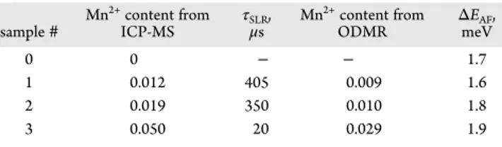
Cd1−xMnxS NPLs sample# Mn2+content from ICP-MS τSLR , μs Mn 2+content from ODMR ΔEAF , meV 0 0 − − 1.7 1 0.012 405 0.009 1.6 2 0.019 350 0.010 1.8 3 0.050 20 0.029 1.9
Figure 1. (a) PL spectra of CdSe/CdS (sample#0, pink) and CdSe/Cd0.991Mn0.009S (sample#1, green) NPLs in B = 0 T. (b) PL decay traces of
sample#0 (pink) and sample #1 (green) NPLs, measured at their PL maxima of 2.127 eV. (c) PL decay curves of CdSe/Cd0.991Mn0.009S NPLs
(sample#1) at various detection energies shown by the arrows in panel (a) with the same color code. Inset: Spectral dependence of the average decay time⟨τ⟩ in sample #1. (d,e) PL decay at 2.127 eV measured in magnetic fields B = 0 T (green) and 15 T (red) in samples #1 and #0. All measurements performed atT = 4.2 K.
full widths at half-maximum of about 100 meV, which is typical for core/shell NPLs.37 As the PL is close to the 4T
1 →6 A1
transition of the Mn2+ions at 2.1 eV, ourfirst task is to identify the origin of the emission from CdSe/Cd0.991Mn0.009S NPLs and to prove that it is dominated by exciton recombination. The similarity of the PL spectra of nonmagnetic and DMS NPLs gives afirst hint for that.
The recombination dynamics can be also used for the identification of the origin of the emission. It is known that at liquid helium temperatures, the decay of the Mn2+emission via
the4T1→6A1transition is very slow, occurring on time scales in
the 10−500 μs range in bulk DMSs,38,39 like (Cd,Mn)Te, (Zn,Mn)Te, and (Zn,Mn)S, and is 270 μs in (Cd,Mn)Se colloidal quantum dots.40The exciton recombination dynamics is by a few orders of magnitude faster, happening for neutral excitons in the range of 1 ns to 1μs, depending on the relative involvement of bright and dark exciton states or of a few nanoseconds for charged excitons (trions).37,41,42
The time-resolved recombination dynamics measured at the PL maxima in samples#0 and #1 are shown inFigure 1b. In both cases, the decay of the PL intensity takes place within 300 ns being 3 orders of magnitude faster than the recombination dynamics of Mn2+ ions. This allows us to conclude that the dominating part of the emission in CdSe/Cd0.991Mn0.009S NPLs is provided by exciton recombination and the Mn2+emission is very weak, if present at all. The two other DMS samples have similar properties.
As it is common for the colloidal nanocrystals, the recombination dynamics in the studied NPLs do not show a monoexponential decay. For example, the decays at the PL maxima, shown inFigure 1b, require a fit with a three-term exponential function for a good description: I(t) = A1exp(−t/ τ1) + A2exp(−t/τ2) + A3exp(−t/τ3). The three decay times are
τ1= 3 ns,τ2= 11 ns, andτ3= 56 ns for sample#0 and τ1= 3 ns,
τ2= 12 ns andτ3= 45 ns for sample#1. Note that they are close
to each other in these nonmagnetic and DMS NPLs.
The spectral dependence of the PL dynamics in CdSe/ Cd0.991Mn0.009S NPLs is given inFigure 1c. The general trend is that the decay times increase with decreasing emission energy. This is clearly seen in the inset ofFigure 1c, where the spectral dependence of the average decay time ⟨τ⟩ is given. ⟨τ⟩ is calculated as⟨τ⟩ = τ1ν1+τ2ν2+τ3ν3, whereνi= Aiτi/(A1τ1+ A2τ2+ A3τ3). The average decay time increases from 2 up to 70
ns from the high- to the low-energy tail. The nonmagnetic sample demonstrates the similar spectral dependence (Figure S2), which proves that it is not due to the Mn2+emission. Such behavior is typical for ensembles of colloidal nanocrystals with an efficient Förster energy transfer.43,44
Further more, the recombination dynamics are weakly affected by external magnetic fields. This is shown in Figure 1d,e, where the PL decays at the emission maximum are compared for B = 0 and 15 T.
Note that the character of the recombination dynamics at low temperatures in colloidal NPLs and its dependence on magnetic field allows one to identify whether the emission is contributed by neutral or by charged excitons.37,41,42For example, at T = 4.2 K, the trion emission in CdSe/CdS NPLs with thick shells is monoexponential with a decay time of 3 ns and is independent of magneticfield. Contrary to that, the decay of neutral excitons in CdSe NPLs has a biexponential decay with a very fast initial component of 20 ps and a long component of 80 ns, which shortens with increasing magneticfield.
The recombination dynamics in the NPLs studied in this paper do not clearly correspond to either neutral or charged exciton behavior, but are rather superpositions of both. Additionally, for resonant excitation, we clearly observe emission from dark excitons (Figure 4a). The bright-dark exciton energy splitting,ΔEAF, ranges between 1.6 and 1.9 meV
(see below). Therefore, at T = 4.2 K, the bright state has about 1% population in thermal equilibrium and should contribute to the emission. We also detect electron spinflips, which means that some of the NPLs are negatively charged, that is, they may contain negatively charged excitons (Figure 4a). From all these findings, we conclude that the PL is contributed by a recombination of neutral (bright and dark) and charged excitons. More details are given in theSupporting Information S4.
Polarized Photoluminescence in the Magnetic Field. This technique, which exploits the exciton (trion) spin polarization on their Zeeman split sublevels, is a sensitive tool to measure small spin splittings comparable with the thermal energy kBT, where kB is the Boltzmann constant.37,45,46 The
experiment is relatively easy in realization, but requires liquid helium temperatures and strong magneticfields of 10−15 T or even up to 30−65 T to gather sufficient information on the linear dependence of the circular polarization degree of PL, Pc(B), on
Figure 2. (a) Schematic diagram of the electron and hole wave functions (blue and red contours) in CdSe/Cd1−xMnxS NPLs for the conduction
valence band offset 0.15 eV. (b) PL spectra of the σ+
(red) andσ−(blue) polarized components in CdSe/CdS NPLs (sample#0, bottom) and CdSe/Cd0.991Mn0.009S NPLs (sample#1, top) at T = 4.2 K, B = 3 T (left), and 15 T (right). (c) Magnetic field dependence of the DCP in sample
magneticfields in weak fields, until it reaches saturation in high magneticfields, Pcsat. The degree of circular polarization (DCP)
is defined as Pc= (I+− I−)/(I++ I−), where I+and I−are the PL
intensities of theσ+ and σ− circularly polarized components, respectively. The magnetic field is applied in the Faraday geometry, that is, parallel to the emission wave vector direction.
Figure 2b shows polarized PL spectra of the nonmagnetic sample#0 (bottom) and Mn-doped sample #1 (top), measured in magnetic fields B = 3 and 15 T. One can clearly see the difference between DCP in nonmagnetic and DMS NPLs. First, they have opposite signs. In CdSe/CdS NPLs Pc < 0, the absolute value increases with growing magnetic field about monotonically and saturates above 12 T (Figure 2c). At B = 15 T, it reaches Pc=−0.47. This behavior is similar to what was
reported for thick-shell CdSe/CdS NPLs (see Figure 3c in ref
37), where the emission was provided by negatively charged excitons.
In the DMS sample Pc> 0, it increases fast, reaching a plateau
value of +0.22 at B = 4 T, and then slowly decreases in higher magneticfields. The Pcsign reversal in II−VI DMS materials, compared to the nonmagnetic reference, is clear evidence of the exchange interaction of charge carriers with the Mn2+ions. It is
provided by the signs of the exchange constants in the conduction (α > 0) and valence bands (β < 0).7More details are given in theSupporting Information S6.
Analysis of Polarized Photoluminescence. The exciton and trion DCP can be written as
τ τ τ = − + Δ P B P E B k T ( ) tanh ( ) 2 c csat s Z B (1)
whereΔEZ(B) is the Zeeman splitting,τ is the lifetime, τsis the
spin relaxation time, and Pcsat is the saturation degree of
polarization, which depends on the specifics of the spin level structure and NPL orientation in the ensemble.
In nonmagnetic samples, the intrinsic exciton Zeeman splitting is
μ
ΔEZ,X( )B =g B
X B (2)
where gXis the exciton g-factor andμBis the Bohr magneton. Accounting for the specifics of the bright and dark excitons is considered inSupporting Information S6.
In DMS samples, an additional term, Eexch,X(B), describing the
exciton exchange interaction with the Mn2+ spins has to be
added
μ
ΔEZ,X( )B =gX BB +Eexch,X( )B (3) Note that Eexch,X(B) is controlled by the exchange interaction
of both electron and hole composing the exciton with the Mn2+
ions and therefore depends on the overlap of the electron and hole wave functions with the (Cd,Mn)S shells.
For the negative trion, being composed of two electrons and one hole, the Zeeman splitting is determined by the hole splitting:
Figure 3. (a) Dependence of theσ+(red) andσ−(blue) circularly polarized components of PL on magneticfield in Faraday geometry for CdSe/
Cd0.991Mn0.009S NPLs (sample#1). Pexc= 4 W/cm2andT = 1.8 K. The peaks show the PL intensities for application of microwaves (ODMR
signals). Their intensities are multiplied by a factor of 10. (b) Normalized ODMR signals for the two circularly polarized components of the PL. The ODMR resonance has a line width ofΔB ≈ 40 mT and is located at B = 2.130 T, which corresponds to a g-factor of 1.999. The σ+component
decreases at resonance conditions (red), while theσ−component increases (blue). (c) Temporal evolution of theσ+PL component after
switching on/off the microwaves for resonance excitation at B = 2.130 T. The red line is an exponential fit with a time constant τSLR= 405μs. (d)
Schematic diagram of the interactions between the Mn2+and exciton (trion) spin systems. (e) Spin−lattice relaxation time as a function of the
Mn2+contentx; adapted from ref35. Blue line is a guide for the eye, and red circles are experimental data measured in the present work (see
μ
ΔEZ,h( )B = −3gh BB (4) where ghis the hole g-factor. In DMS samples
μ
ΔEZ,h( )B = −3gh BB+Eexch,h( )B (5) Eexch,h(B) is determined by the exchange interaction of the hole with the Mn2+ions. Here we use the definition of the hole g-factor sign that is commonly used for colloidal nanocrystals.37,47 In the frame of this convention, the intrinsic hole Zeeman splitting provided by the negative hole g-factor (gh< 0) is in competition with the hole exchange splitting determined byβ < 0. On the other hand, for the conduction band electron, both the intrinsic Zeeman splitting (ge> 0) and the exchange one with
α > 0 add to each other.
The electron g-factor is ge= +1.70 in CdSe/CdS NPLs (see
below). The hole g-factor gh = −0.7 was measured in high magneticfields.37For smallΔEAF, as in our case, the g-factor of
the bright exciton is gXA=−ge− 3gh= +0.4. This value matches well with gXA= +0.32 measured in the 4-monolayer thick bare
core NPLs by absorption spectroscopy in high magneticfields.48 For the dark exciton, gXF= ge− 3gh= +3.8. One can see fromeq 1
that the negative DCP found in experiment requires gX> 0, that is, can be achieved by the bright and the dark excitons. In case of the negative trion, Pc< 0 requires gh< 0, seeeqs 1and4, which is indeed the case for CdSe/CdS NPLs. To summarize, the negative DCP observed in nonmagnetic CdSe/CdS NPLs can be provided by the dark and bright excitons and the negative trions.
In DMS NPLs, the polarization is positive, which requires a negative sign of the Zeeman splittingΔEZ. One can see fromeqs 3and5that this can be the case when the intrinsic and exchange terms have different signs as well as when for the exciton case |Eexch,X(B)| > |gXμBB| and the trion case |Eexch,h(B)| > |3ghμBB|. In
the trion case, the fulfillment of this condition is solely provided by the hole exchange interaction with the Mn2+ ions, that is, requires a sufficiently large penetration of the hole wave function into the DMS shells. In CdSe, geandα are both positive, and
therefore, the Zeeman splitting for the conduction band electron can not be inverted. As a result, for the exciton case, the inversion of the DCP sign can also be provided only by the hole exchange with the Mn2+spins. To support this conclusion, we provide in theSupporting Information S6results of model calculations for the bright and dark excitons and for the negative trions for various penetrations of the hole wave functions into the DMS shells.
Optically Detected Magnetic Resonance. The ODMR technique combines the advantage of resonant excitation of spin states by microwave radiation with the high sensitivity of optical detection of the induced changes. It is especially useful for the investigation of semiconductor nanostructures, whose small volume is not sufficient to provide sufficiently strong signals for the electron paramagnetic resonance technique. Additionally, the possibility of spectrally selective detection on specific optical resonances, for example, the exciton or impurity related emission, allows one to obtain a clear identification of the addressed electronic transitions.
In the case of diluted magnetic semiconductors, the resonant microwave heating of the Mn2+ ions increases the Mn2+ spin
temperature TMn and, consequently, reduces the Mn2+ spin
polarization⟨SMn⟩. These changes can be detected optically via the excitons or trions interacting with the Mn2+spins, seeFigure 3d. The application of the ODMR technique to quantum well structures based on (Zn,Mn)Se DMSs is discussed in refs35,49,
and50. There it was shown that the resonant heating of the Mn2+ spin system can be detected by several effects: (i) the decrease of the exciton giant Zeeman splitting, resulting in a spectral shift of the exciton emission line, (ii) the decrease of the circular polarization degree induced by the magneticfield, and (iii) the redistribution of the emission intensity between the exciton line and the Mn2+ emission band. Recently, ODMR measured at 10 GHz microwave radiation via polarized PL was reported for CdSe/Cd1−xMnxS NPLs.19
Figure 3a shows the intensities of the σ+ and σ− PL components of the CdSe/Cd0.991Mn0.009S NPLs (sample#1),
measured versus magneticfield without and with microwaves. As discussed above, theσ+component has a higher intensity due to
the stronger thermal population of the excitons (trions) on the associated Zeeman sublevels split in magneticfield. Without microwaves, the intensities of these components change smoothly with magneticfield, following the DCP trend shown inFigure 2c. In the presence of 59.6 GHz microwave radiation, two sharp resonances are observed at B = 2.130 T. The resonant decrease of theσ+intensity and the correlated increase of theσ−
intensity evidence heating of the Mn2+ spin system, which accordingly decreases the exciton (trion) giant Zeeman splitting and the exciton (trion) DCP.35,50The PL intensity variations normalized to the relative PL intensities without microwaves (I) are shown in more detail inFigure 3b. They represent broad peaks with a width ofΔB = 40 mT and are centered at B = 2.130 T corresponding to a g-factor of 1.999± 0.005. This g-factor matches with the Mn2+ value of gMn = 2.01, reported for
ZnS:Mn2+and CdTe:Mn2+in electron spin resonance
measure-ments.51,52
The spin−lattice relaxation (SLR) dynamics of the Mn2+spin
system can be measured by modulating the microwave radiation between on and off and time-resolved detection of the changes induced thereby, reflecting cooling or heating of the Mn2+spins.
An example of such a measurement for CdSe/Cd0.991Mn0.009S
NPLs is shown inFigure 3c. Here, the red line is an exponential fit with the characteristic spin−lattice relaxation time τSLR= 405
μs. Similar measurements performed for samples #2 and #3 give 350 and 20μs, respectively (Table 1).
As we discussed above, most of the magneto-optical approaches that are commonly used for evaluation of the Mn2+concentration in bulk DMSs cannot be directly applied to CdSe/Cd1−xMnxS NPLs. We suggest that a quite accurate evaluation can be achieved from the spin−lattice relaxation time τSLR. It is known that the τSLR of the Mn2+ ions in II−VI
semiconductors has a very strong dependence on the Mn2+
concentration, which covers about 5 orders of magnitude from 1 ms down to 10 ns with increasing x from 0.004 up to 0.11, see
Figure 3e, where the data from Figure 8.10 in ref 35 are reproduced. This strong dependence arises from the quenching of the orbital momentum of the d-electrons in the Mn2+ion, that is, it has zero orbital momentum (L = 0) and does not interact with the phonon system. The only mechanisms that provide spin−lattice relaxation for the Mn2+ions are given by the Mn2+− Mn2+interactions, which obviously are strongly dependent on
the number of neighboring Mn2+ions and the distances between them, which in turn strongly change with increasing Mn2+
concentration. The red circles in Figure 3e mark the times that we measured for the CdSe/Cd1−xMnxS NPLs. Their
comparison with the literature data shown by the symbols allows us to evaluate the Mn2+concentration for the studied samples. As one can see fromTable 1, the Mn2+concentration measured
samples#1 and #2, but differs for sample #3. We emphasize that the suggested approach for evaluation of the Mn2+concentration
is very reliable and could be widely used for nanostructures. Note that in layers with a few monolayer thickness, Mn2+ions have less Mn2+neighbors than in bulk, which results in longer SLR dynamics. We studied this effect in (Cd,Mn)Te digital alloys grown by molecular beam epitaxy.36We found that in the layers with a thickness of 3 monolayers and larger, the SLR times are the same as in bulk, but in 1 monolayer thick layers, the time can be longer by a factor of 5, which corresponds to a factor 2 underestimation of the Mn2+concentration.
Spin-Flip Raman Scattering. The SFRS spectroscopy is a sophisticated tool to investigate the Zeeman splittings of carriers (electrons or holes), excitons, or magnetic ions. It provides detailed information about their spin structure and spin interactions. The polarization properties of the SFRS lines deliver information on the symmetries of the involved states and allow one to identify the responsibleflip mechanisms. In SFRS, the Zeeman splitting is obtained from the Raman shift, which is equal to the energy shift between the laser photon energy and the energy of the scattered light. The technique was successfully used for investigation of the exchange interactions of carriers and excitons with the magnetic ions in DMS bulk samples6,53and quantum well structures.54−56 We showed recently that nonmagnetic CdSe and CdSe/CdS NPLs can be studied by SFRS,37,57but this technique had not been used so far for DMS colloidal nanocrystals.
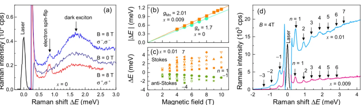
Figure 4a shows Raman spectra of the CdSe/CdS NPLs (sample#0) for resonant excitation of the exciton state at Eexc= 2.165 eV. Here positive values of the Raman shift ΔE correspond to a Stokes shift of the scattered photons to lower energies. At zero magneticfield, there is a relatively broad line with a full width at half-maximum of 0.5 meV, whose maximum is shifted by 1.7 meV. This shift does not change in the applied magneticfield of B = 8 T. We assign it to the energy splitting between the bright and dark exciton statesΔEAF.
41
The dark exciton line was observed in all studied samples with ΔEAF ranging between 1.6 and 1.9 meV (Table 1). This supports our assumption that the dark excitons contribute to the emission from the CdSe/Cd1−xMnxS NPLs.
At B = 8 T, a narrow line associated with the electron spin-flip, shifted by 0.82 meV from the laser, is seen in the Raman
spectrum (Figure 4a). Its shift depends linearly on magneticfield (Figure 4b, green circles). Afit with |ΔE| = |ge|μBB gives the g-factor value|ge| = 1.70 ± 0.02, which is close to the electron g-factor measured in CdSe and CdSe/CdS NPLs.37,57,58Note that ge> 0 in bulk CdSe and in these structures.
Figure 4d shows Raman spectra of CdSe/Cd0.991Mn0.009S and CdSe/Cd0.99Mn0.01S NPLs measured at B = 4 T with resonant
excitation of the exciton at Eexc= 2.151 eV, T = 1.6 K. They are
obviously very different compared to the spectra inFigure 4a. No electron spin-flip is detected; instead, a set of equidistant lines is observed. Up to seven lines in the Stokes and up to three lines in the anti-Stokes energy range can be resolved. All these lines shift linearly with magneticfield (seeFigure 4c), following the equationΔE = ngMnμBB, where n is an integer number. An accurate evaluation of gMn= 2.01± 0.03 is obtained from the fit
of the line with n = 1, shown inFigure 4b by orange squares. This g-factor matches well with the Mn2+g-factor of 2.01.51,52
Hence, we conclude that the measured Raman signals have to be attributed to spinflips of the Mn2+ ions interacting with the photogenerated exciton. We measured the spectral dependence of the Raman signal intensities. The maximal signal is reached when the laser is in resonance with the exciton. This shows that the exciton serves as an intermediate scattering state, which resonantly enhances the Raman cross-section.
The Raman signal is detected in all four combinations of circular polarizations of excitation and detection. The relative intensities (I++/I+−/I−+/I−−) of the Mn2+ SFRS lines depend strongly on the Mn2+concentration. Here Iijmeansσi-polarized excitation andσj-polarized detection. These intensities on the
Stokes side for n = 1 at B = 4 T are given by (1/0.95/0.99/0.95) for CdSe/Cd0.991Mn0.009S (sample#1), (1/0.75/0.84/0.62) for
CdSe/Cd0.99Mn0.01S (sample#2), and (1/0.33/0.42/0.18) for
CdSe/Cd0.971Mn0.029S (sample#3). One can conclude that the optical selection rules become more distinct with increasing Mn2+concentration and the Mn2+lines become more dominant forσ+polarized excitation and detection.
The observation of multiple spinflip Mn2+lines was reported
also for (Cd,Mn)Te-based quantum wells, where up to 15 spin-flip lines were observed.54
A mechanism for these flips was suggested in ref54and a corresponding model description was developed in refs34,59. The key point of this model is the anisotropic spin of the heavy-hole in a two-dimensional Figure 4. (a) Raman spectrum for the circularly copolarized configuration in Faraday geometry for the nonmagnetic CdSe/CdS NPLs (sample #0). Eexc= 2.165 eV,Pexc= 0.2 W/cm2,T = 1.6 K, B = 0 and 8 T. (b) Magnetic field dependence of the absolute Stokes energy shifts of the n = 1
Mn2+resonance in CdSe/Cd0.991Mn0.009S (sample#1, orange squares) and of the electron spin flip in CdSe/CdS NPLs (sample #0, green circles)
in Faraday geometry. Lines are linearfits to the data. (c) Magnetic field dependence of the relative Raman shifts of all detectable Mn2+ resonances in CdSe/Cd0.99Mn0.01S NPLs in Voigt geometry. (d) Mn2+spin scattering spectra for the copolarized configuration in Faraday
geometry for the CdSe/Cd0.991Mn0.009S (sample#1, pink) and CdSe/Cd0.99Mn0.01S (sample#2, blue) NPLs. Eexc= 2.151 eV,Pexc= 1.6W/cm2,B
nanostructure. In an external magneticfield, the Mn2+spins are
polarized along the field direction. When the spin of the photogenerated hole is not parallel to the magneticfield (for simplicity the case when it is perpendicular to thefield can be considered), the Mn2+spins are influenced by the external field
B and by the hole exchange field Bexch. The total magnetic
moment of all Mn2+spins within the hole localization volume,
IMn, precesses about the totalfield B + Bexch. When the exciton
recombines, i.e. scatters, the projection of IMnon B differs from the initial value by a multiple of the energy of the n = 1 Mn2+ spin-flip. Note that the electron with an isotropic spin does not support this mechanism. Therefore, we can conclude that in the studied DMS NPLs the holes have sufficient wave function overlap with the shell Mn2+spins for providing multiple SFRS. This is in line with our conclusions from the DCP data analysis. NPLs have a close analogy to quantum wells and the model approach suggested for quantum wells can be directly applied also here. The only specifics, which need to be accounted for, are the varying orientations of the NPLs in an ensemble measure-ment. As we have noticed above, the condition for observation of multiple Mn2+-flips is the noncollinearity of B and B
exch. This
means that in quantum wells the effect should be absent in the Faraday geometry, where the magneticfield is parallel to the structure growth axis. In DMS NPLs we observe the same amount of higher order spin scattering resonances in Faraday and Voigt geometry. We explain this result by the random orientation of the NPLs in the studied ensembles, leading to the situation that in any configuration a fraction of NPLs fulfills the condition for multiple Mn2+-flips. It is worthwhile to note that
the model developed for the exciton, namely for the hole in the exciton, can be readily applied for the negatively charged exciton, as the hole spin acts similar on Mn2+ions in both cases. CONCLUSIONS
In conclusion, we have demonstrated the exchange interaction of excitons (trions) with the Mn2+ ions in CdSe/Cd1−xMnxS
core/shell nanoplatelets by means of polarized PL, optically detected magnetic resonance and spin-flip Raman scattering. One can conclude that these structures can be studied in detail by these experimental approaches that were established for diluted magnetic semiconductors. In particular, assessment of the dynamics for spin−lattice relaxation gives accurate estimates for the Mn2+ ion concentration. Our studies may help to
functionalize colloidal DMS nanocrystals as magnetic or magneto-optical markers.
METHODS
Magneto-optical measurements. The NPLs were dropcasted on a substrate and mounted in a titanium sample holder on top of a three-axis piezo-positioner and placed in the variable temperature insert (4.2−70 K) of a liquid helium bath cryostat equipped with a superconducting solenoid (magnetic fields up to 15 T). The measurements were performed in the Faraday geometry (light excitation and detection parallel to the magneticfield direction). The PL was excited with a diode laser (photon energy 3.06 eV, wavelength 405 nm) in continuous-wave or pulsed mode (pulse duration 50 ps, pulse repetition rate 500 kHz) with a weak average excitation density of 0.5 mW/cm2. The PL was dispersed with a 0.5-m spectrometer and
detected either by a liquid-nitrogen-cooled charge-coupled-device (CCD) camera or a Si avalanche photodiode connected to a conventional time-correlated single-photon counting system. The instrumental response time was about 200 ps. The PL circular polarization degree was analyzed by a combination of a quarter-wave plate and a linear polarizer.
Spin-flip Raman scattering. The samples were mounted strain free inside the variable temperature insert of a magnet cryostat, which provided magneticfields up to 10 T. The temperature was set to 1.6 K. The backscattering experiments were performed in Faraday geometry (θ = 0°) or in tilted geometries up to θ = 90°, corresponding to the Voigt geometry, where the magneticfield and the normal to the sample substrate enclose the angle θ. The NPLs were excited by a single frequency dye laser (Matisse DS), whose actual wavelength was measured and monitored by afiber-coupled wavelength-meter device. The laser power was stabilized by a liquid-crystal variable attenuator. Unless specified otherwise, the power was set to about 0.2 W/cm2at the
sample surface. In order to ensure a stable detection position on the sample surface, each sample was covered by a mask having a hole of 1 mm diameter and the central part with 100× 100 μm2size of the
illuminated sample area was selected by a cross slit. The NPL emission was spectrally dispersed by a double monochromator (U1000) equipped with a Peltier-cooled GaAs photomultiplier. The SFRS spectra were measured in close vicinity of the laser line with photon energy Eexc. The spin-flip signals were shifted from the laser energy by
the Zeeman splitting of the involved spin state, either to lower (Stokes shift, Eexc− |g|μBB) or higher energies (anti-Stokes shift, Eexc+|g|μBB).
Optically detected magnetic resonance. The ODMR techni-que used in this study was described in detail in ref49. The ODMR spectrometer consisted of a 60 GHz all-solid-state microwave oscillator (photon energy of 0.248 meV) with a tuning range from 59.05 to 60.55 GHz and an output power of up to 100 mW. The output power of the oscillator could be varied by up to 40 dB attenuation level. The oscillator could operate either in continuous-wave mode or in a periodically pulsed mode with an on−off transition time of about 3 ns at more than 60 dB dumping level. The sample was mounted in a cylindrical H011microwave cavity with a low Q factor of about 600. The
cavity had two orthogonal pairs of apertures with a conic cross-section for sample illumination and collecting the sample emission. The cavity was placed in the variable temperature insert of a magnet cryostat, the measurements were performed at T = 1.8 K. The sample in the cavity was excited by a 405 nm (Eexc= 3.06 eV) semiconductor laser in cw
mode with 0.5 mW power, focused into a spot with a diameter of 400 μm. The PL was collected in backscattering geometry and detected with a 0.5-m grating monochromator and a CCD camera. Magneticfields up to 7 T were applied in the Faraday geometry. For time-resolved ODMR measurements a photon counter based on an avalanche photodiode was used, the temporal resolution was 30 ns.
ASSOCIATED CONTENT
*
sı Supporting InformationThe Supporting Information is available free of charge at
https://pubs.acs.org/doi/10.1021/acsnano.0c04048.
Additional information on synthesis details, XRD data, spectrally resolved PL decays, exciton bright-dark splitting, calculation of the band structure in core/shell NPLs, modeling of the exciton exchange interaction with Mn2+ions, Zeeman splitting and polarization degree for
the bright and dark excitons and negative trions (PDF)
AUTHOR INFORMATION
Corresponding Authors
Elena V. Shornikova− Experimentelle Physik 2, Technische Universität Dortmund, 44227 Dortmund, Germany;
orcid.org/0000-0002-6866-9013; Email:[email protected]
Dmitri R. Yakovlev− Experimentelle Physik 2, Technische Universität Dortmund, 44227 Dortmund, Germany; Ioffe Institute, Russian Academy of Sciences, 194021 St. Petersburg, Russia; orcid.org/0000-0001-7349-2745;
Authors
Danil O. Tolmachev− Experimentelle Physik 2, Technische Universität Dortmund, 44227 Dortmund, Germany; Ioffe Institute, Russian Academy of Sciences, 194021 St. Petersburg, Russia; orcid.org/0000-0002-7098-8515
Vitalii Yu. Ivanov− Institute of Physics, Polish Academy of Sciences, PL-02-668 Warsaw, Poland; orcid.org/0000-0002-4651-8476
Ina V. Kalitukha− Ioffe Institute, Russian Academy of Sciences, 194021 St. Petersburg, Russia
Victor F. Sapega− Ioffe Institute, Russian Academy of Sciences, 194021 St. Petersburg, Russia
Dennis Kudlacik− Experimentelle Physik 2, Technische Universität Dortmund, 44227 Dortmund, Germany;
orcid.org/0000-0001-5473-8383
Yuri G. Kusrayev− Ioffe Institute, Russian Academy of Sciences, 194021 St. Petersburg, Russia
Aleksandr A. Golovatenko− Ioffe Institute, Russian Academy of Sciences, 194021 St. Petersburg, Russia; orcid.org/0000-0003-2248-3157
Sushant Shendre− LUMINOUS! Center of Excellence for Semiconductor Lighting and Displays, School of Electrical and Electronic Engineering, School of Physical and Materials Sciences, Nanyang Technological University, 639798, Singapore Savas Delikanli− LUMINOUS! Center of Excellence for
Semiconductor Lighting and Displays, School of Electrical and Electronic Engineering, School of Physical and Materials Sciences, Nanyang Technological University, 639798, Singapore; Department of Electrical and Electronics Engineering,
Department of Physics, UNAM− Institute of Materials Science and Nanotechnology, Bilkent University, 06800 Ankara, Turkey;
orcid.org/0000-0002-0613-8014
Hilmi Volkan Demir− LUMINOUS! Center of Excellence for Semiconductor Lighting and Displays, School of Electrical and Electronic Engineering, School of Physical and Materials Sciences, Nanyang Technological University, 639798, Singapore; Department of Electrical and Electronics Engineering,
Department of Physics, UNAM− Institute of Materials Science and Nanotechnology, Bilkent University, 06800 Ankara, Turkey;
orcid.org/0000-0003-1793-112X
Manfred Bayer− Experimentelle Physik 2, Technische Universität Dortmund, 44227 Dortmund, Germany; Ioffe Institute, Russian Academy of Sciences, 194021 St. Petersburg, Russia;
orcid.org/0000-0002-0893-5949
Complete contact information is available at:
https://pubs.acs.org/10.1021/acsnano.0c04048
Notes
The authors declare no competingfinancial interest. ACKNOWLEDGMENTS
The authors are thankful to A. V. Rodina for fruitful discussions. This work was supported by the Deutsche Forschungsgemein-schaft through the International Collaborative Research Center TRR 160 (Projects B1, B2, and C7) and by the Russian Foundation for Basic Research (Grant No. 19-52-12064 NNIO-a). D.R.Y. acknowledges the partial support of the Russian Science Foundation (Project No. 20-42-01008). S.S., S.D. and H.V.D. acknowledge partial support from the Singapore National Research Foundation under NRF−NRFI2016−08. A.A.G. acknowledges support of the Grants Council of the President of the Russian Federation. V.Yu.I. acknowledges
support of the Polish National Science Center (Grant No. 2018/ 30/M/ST3/00276). H.V.D. gratefully acknowledges support from TUBA. Yu.G.K. acknowledges the support of the Russian Science Foundation (Project No. 18-12-00352).
REFERENCES
(1) Efros, Al. L.; Rashba, E. I.; Rosen, M. Paramagnetic Ion-Doped Nanocrystal as a Voltage-Controlled Spin Filter. Phys. Rev. Lett. 2001, 87, 206601.
(2) Beaulac, R.; Archer, P. I.; Ochsenbein, S. T.; Gamelin, D. R. Mn2+
-Doped CdSe Quantum Dots: New Inorganic Materials for Spin-Electronics and Spin-Photonics. Adv. Funct. Mater. 2008, 18, 3873− 3891.
(3) Muckel, F.; Barrows, C. J.; Graf, A.; Schmitz, A.; Erickson, C. S.; Gamelin, D. R.; Bacher, G. Current-Induced Magnetic Polarons in a Colloidal Quantum-Dot Device. Nano Lett. 2017, 17, 4768−4773.
(4) Moro, F.; Fielding, A. J.; Turyanska, L.; Patanè, A. Realization of Universal Quantum Gates with Spin-Qudits in Colloidal Quantum Dots. Adv. Quantum Technol. 2019, 2, 1900017.
(5) Furdyna, J. K. Diluted Magnetic Semiconductors. J. Appl. Phys. 1988, 64, R29−R64.
(6) Furdyna, J. K.; Kossut, J. In Diluted Magnetic Semiconductors; Furdyna, J. K., Kossut, J., Eds.; Semiconductors and Semimetals; Academic Press: London, 1988; Vol. 25.
(7) Gaj, J. A.; Kossut, J. In Introduction to the Physics of Diluted Magnetic Semiconductors; Kossut, J., Gaj, J. A., Eds.; Springer Series in Materials Science; Springer Verlag: Berlin, Heidelberg, 2010.
(8) Hoffman, D. M.; Meyer, B. K.; Ekimov, A. I.; Merkulov, I. A.; Efros, Al. L.; Rosen, M.; Couino, G.; Gacoin, T.; Boilot, J. P. Giant Internal Magnetic Fields in Mn Doped Nanocrystal Quantum Dots. Solid State Commun. 2000, 114, 547−550.
(9) Norris, D. J.; Yao, N.; Charnock, F. T.; Kennedy, T. A. High-Quality Manganese-Doped ZnSe Nanocrystals. Nano Lett. 2001, 1, 3− 7.
(10) Archer, P. I.; Santangelo, S. A.; Gamelin, D. R. Direct Observation of sp−d Exchange Interactions in Colloidal Mn2+- and
Co2+-Doped CdSe Quantum Dots. Nano Lett. 2007, 7, 1037−1043. (11) Bussian, D. A.; Crooker, S. A.; Yin, M.; Brynda, M.; Efros, Al. L.; Klimov, V. I. Tunable Magnetic Exchange Interactions in Manganese-Doped Inverted Core-Shell ZnSe-CdSe Nanocrystals. Nat. Mater. 2009, 8, 35−40.
(12) Pinchetti, V.; Di, Q.; Lorenzon, M.; Camellini, A.; Fasoli, M.; Zavelani-Rossi, M.; Meinardi, F.; Zhang, J.; Crooker, S. A.; Brovelli, S. Excitonic Pathway to Photoinduced Magnetism in Colloidal Nano-crystals with Nonmagnetic Dopants. Nat. Nanotechnol. 2018, 13, 145− 151.
(13) Beaulac, R.; Archer, P. I.; Liu, X.; Lee, S.; Salley, G. M.; Dobrowolska, M.; Furdyna, J. K.; Gamelin, D. R. Spin-Polarizable Excitonic Luminescence in Colloidal Mn2+-Doped CdSe Quantum
Dots. Nano Lett. 2008, 8, 1197−1201.
(14) Long, G.; Barman, B.; Delikanli, S.; Tsung Tsai, Y.; Zhang, P.; Petrou, A.; Zeng, H. Carrier-Dopant Exchange Interactions in Mn-Doped PbS Colloidal Quantum Dots. Appl. Phys. Lett. 2012, 101, No. 062410.
(15) Turyanska, L.; Hill, R. J. A.; Makarovsky, O.; Moro, F.; Knott, A. N.; Larkin, O. J.; Patanè, A.; Meaney, A.; Christianen, P. C. M.; Fay, M. W.; Curry, R. J. Tuneable Paramagnetic Susceptibility and Exciton g-Factor in Mn-Doped PbS Colloidal Nanocrystals. Nanoscale 2014, 6, 8919−8925.
(16) Delikanli, S.; Akgul, M. Z.; Murphy, J. R.; Barman, B.; Tsai, Y.; Scrace, T.; Zhang, P.; Bozok, B.; Hernández-Martínez, P. L.; Christodoulides, J.; Cartwright, A. N.; Petrou, A.; Demir, H. V. Mn2+-Doped CdSe/CdS Core/Multishell Colloidal Quantum Wells Enabling Tunable Carrier-Dopant Exchange Interactions. ACS Nano 2015, 9, 12473−12479.
(17) Murphy, J. R.; Delikanli, S.; Scrace, T.; Zhang, P.; Norden, T.; Thomay, T.; Cartwright, A. N.; Demir, H. V.; Petrou, A. Time-Resolved
Photoluminescence Study of CdSe/CdMnS/CdS Core/Multi-Shell Nanoplatelets. Appl. Phys. Lett. 2016, 108, 242406.
(18) Najafi, A.; Tarasek, S.; Delikanli, S.; Zhang, P.; Norden, T.; Shendre, S.; Sharma, M.; Bhattacharya, A.; Taghipour, N.; Pientka, J.; Demir, H. V.; Petrou, A.; Thomay, T. CdSe/CdMnS Nanoplatelets with Bilayer Core and Magnetically Doped Shell Exhibit Switchable Excitonic Circular Polarization: Implications for Lasers and Light-Emitting Diodes. ACS Appl. Nano Mater. 2020, 3, 3151−3156.
(19) Strassberg, R.; Delikanli, S.; Barak, Y.; Dehnel, J.; Kostadinov, A.; Maikov, G.; Hernandez-Martinez, P. L.; Sharma, M.; Demir, H. V.; Lifshitz, E. Persuasive Evidence for Electron-Nuclear Coupling in Diluted Magnetic Colloidal Nanoplatelets Using Optically Detected Magnetic Resonance Spectroscopy. J. Phys. Chem. Lett. 2019, 10, 4437−4447.
(20) Beaulac, R.; Schneider, L.; Archer, P. I.; Bacher, G.; Gamelin, D. R. Light-Induced Spontaneous Magnetization in Doped Colloidal Quantum Dots. Science 2009, 325, 973−976.
(21) Rice, W. D.; Liu, W.; Pinchetti, V.; Yakovlev, D. R.; Klimov, V. I.; Crooker, S. A. Direct Measurements of Magnetic Polarons in Cd1−xMnxSe Nanocrystals from Resonant Photoluminescence. Nano
Lett. 2017, 17, 3068−3075.
(22) Lorenz, S.; Erickson, C. S.; Riesner, M.; Gamelin, D. R.; Fainblat, R.; Bacher, G. Directed Exciton Magnetic Polaron Formation in a Single Colloidal Mn2+:CdSe/CdS Quantum Dot. Nano Lett. 2020, 20,
1896−1906.
(23) Rice, W. D.; Liu, W.; Baker, T. A.; Sinitsyn, N. A.; Klimov, V. I.; Crooker, S. A. Revealing Giant Internal Magnetic Fields Due to Spin Fluctuations in Magnetically Doped Colloidal Nanocrystals. Nat. Nanotechnol. 2016, 11, 137−142.
(24) Ithurria, S.; Dubertret, B. Quasi 2D Colloidal CdSe Platelets with Thicknesses Controlled at the Atomic Level. J. Am. Chem. Soc. 2008, 130, 16504−16505.
(25) Furdyna, J. K.; Lee, S.; Dobrowolska, M.; Wojtowicz, T.; Liu, X. Band-Offset Engineering in Magnetic/Non-Magnetic Semiconductor Quantum Structures. In Introduction to the Physics of Diluted Magnetic Semiconductors; Kossut, J., Gaj, J. A., Eds.; Springer Series in Materials Science; Springer Verlag: Berlin, Heidelberg, 2010; pp 103−160.
(26) Muckel, F.; Delikanli, S.; Hernandez-Martinez, P. L.; Priesner, T.; Lorenz, S.; Ackermann, J.; Sharma, M.; Demir, H. V.; Bacher, G. sp−d Exchange Interactions in Wave Function Engineered Colloidal CdSe/ Mn:CdS Hetero-Nanoplatelets. Nano Lett. 2018, 18, 2047−2053.
(27) Zhang, P.; Norden, T.; Pientka, J. M.; Oszwałdowski, R.; Najafi, A.; Barman, B.; Tsai, Y.; Fan, W.-C.; Chou, Wu-C.; Han, J. E.; Žutić, I.; McCombe, B. D.; Petrou, A. Optical Control of Hole Wavefunction in Type-II Magnetic Quantum Dot Structures. J. Phys. Chem. C 2019, 123, 25934−25940.
(28) Davis, A. H.; Hofman, E.; Chen, K.; Li, Z.-J.; Khammang, A.; Zamani, H.; Franck, J. M.; Maye, M. M.; Meulenberg, R. W.; Zheng, W. Exciton Energy Shifts and Tunable Dopant Emission in Manganese-Doped Two-Dimensional CdS/ZnS Core/Shell Nanoplatelets. Chem. Mater. 2019, 31, 2516−2523.
(29) Delikanli, S.; Yu, G.; Yeltik, A.; Bose, S.; Erdem, T.; Yu, J.; Erdem, O.; Sharma, M.; Sharma, V. K.; Quliyeva, U.; Shendre, S.; Dang, C.; Zhang, D. H.; Sum, T. C.; Fan, W.; Demir, H. V. Ultrathin Highly Luminescent Two-Monolayer Colloidal CdSe Nanoplatelets. Adv. Funct. Mater. 2019, 29, 1901028.
(30) Shendre, S.; Delikanli, S.; Li, M.; Dede, D.; Pan, Z.; Ha, S. T.; Fu, Y. H.; Hernández Martínez, P. L.; Yu, J.; Erdem, O.; Kuznetsov, A. I.; Dang, C.; Sum, T. C.; Demir, H. V. Ultrahigh-Efficiency Aqueous Flat Nanocrystals of CdSe/CdS@Cd1−xZnxS Colloidal Core/Crown@
Alloyed-Shell Quantum Wells. Nanoscale 2019, 11, 301−310. (31) Adachi, S. Handbook on Physical Properties of Semiconductors; Springer US: Boston, MA, 2004.
(32) Javaux, C.; Mahler, B.; Dubertret, B.; Shabaev, A.; Rodina, A. V.; Efros, Al. L.; Yakovlev, D. R.; Liu, F.; Bayer, M.; Camps, G.; Biadala, L.; Buil, S.; Quelin, X.; Hermier, J.-P. Thermal Activation of Non-Radiative Auger Recombination in Charged Colloidal Nanocrystals. Nat. Nanotechnol. 2013, 8, 206−212.
(33) Merkulov, I. A.; Yakovlev, D. R.; Keller, A.; Ossau, W.; Geurts, J.; Waag, A.; Landwehr, G.; Karczewski, G.; Wojtowicz, T.; Kossut, J. Kinetic Exchange between the Conduction Band Electrons and Magnetic Ions in Quantum-Confined Structures. Phys. Rev. Lett. 1999, 83, 1431−1434.
(34) Merkulov, I. A., Rodina, A. V. Exchange Interaction between Carriers and Magnetic Ions in Quantum Size Heterostructures. In Introduction to the Physics of Diluted Magnetic Semiconductors; Kossut, J., Gaj, J. A., Eds.; Springer Series in Materials Science; Springer Verlag: Berlin, Heidelberg, 2010; pp 65−101.
(35) Yakovlev, D. R., Merkulov, I. A. Spin and Energy Transfer between Carriers, Magnetic Ions, and Lattice. In Introduction to the Physics of Diluted Magnetic Semiconductors; Kossut, J., Gaj, J. A., Eds.; Springer Series in Materials Science; Springer Verlag: Berlin, Heidelberg, 2010; pp 263−303.
(36) Kneip, M. K.; Yakovlev, D. R.; Bayer, M.; Karczewski, G.; Wojtowicz, T.; Kossut, J. Engineering of Spin-Lattice Relaxation Dynamics by Digital Growth of Diluted Magnetic Semiconductor CdMnTe. Appl. Phys. Lett. 2006, 88, 152105.
(37) Shornikova, E. V.; Biadala, L.; Yakovlev, D. R.; Feng, D.; Sapega, V. F.; Flipo, N.; Golovatenko, A. A.; Semina, M. A.; Rodina, A. V.; Mitioglu, A. A.; Ballottin, M. V.; Christianen, P. C. M.; Kusrayev, Yu. G.; Nasilowski, M.; Dubertret, B.; Bayer, M. Electron and Hole g-Factors and Spin Dynamics of Negatively Charged Excitons in CdSe/ CdS Colloidal Nanoplatelets with Thick Shells. Nano Lett. 2018, 18, 373−380.
(38) Müller, E.; Gebhardt, W. Position and Lifetime of Photo-luminescence in Cd1−xMnxTe and Zn1−xMnxTe. Exchange Dependent
Effects. Phys. Status Solidi B 1986, 137, 259−267.
(39) Schenk, H.; Wolf, M.; Mackh, G.; Zehnder, U.; Ossau, W.; Waag, A.; Landwehr, G. Influence of the Negative Thermal-Expansion Coefficient on the Luminescence Properties of (CdMnMg)Te. J. Appl. Phys. 1996, 79, 8704−8711.
(40) Beaulac, R.; Archer, P. I.; van Rijssel, J.; Meijerink, A.; Gamelin, D. R. Exciton Storage by Mn2+ in Colloidal Mn2+-Doped CdSe
Quantum Dots. Nano Lett. 2008, 8, 2949−2953.
(41) Shornikova, E. V.; Biadala, L.; Yakovlev, D. R.; Sapega, V. F.; Kusrayev, Y. G.; Mitioglu, A. A.; Ballottin, M. V.; Christianen, P. C. M.; Belykh, V. V.; Kochiev, M. V.; Sibeldin, N. N.; Golovatenko, A. A.; Rodina, A. V.; Gippius, N. A.; Kuntzmann, A.; Jiang, Ye; Nasilowski, M.; Dubertret, B.; Bayer, M. Addressing the Exciton Fine Structure in Colloidal Nanocrystals: the Case of CdSe Nanoplatelets. Nanoscale 2018, 10, 646−656.
(42) Shornikova, E. V.; Yakovlev, D. R.; Biadala, L.; Crooker, S. A.; Belykh, V. V.; Kochiev, M. V.; Kuntzmann, A.; Nasilowski, M.; Dubertret, B.; Bayer, M. Negatively Charged Excitons in CdSe Nanoplatelets. Nano Lett. 2020, 20, 1370−1377.
(43) Furis, M.; Hollingsworth, J. A.; Klimov, V. I.; Crooker, S. A. Time- and Polarization-Resolved Optical Spectroscopy of Colloidal CdSe Nanocrystal Quantum Dots in High Magnetic Fields. J. Phys. Chem. B 2005, 109, 15332−15338.
(44) Liu, F.; Rodina, A. V.; Yakovlev, D. R.; Golovatenko, A. A.; Greilich, A.; Vakhtin, E. D.; Susha, A.; Rogach, A. L.; Kusrayev, Yu. G.; Bayer, M. Förster Energy Transfer of Dark Excitons Enhanced by a Magnetic Field in an Ensemble of CdTe Colloidal Nanocrystals. Phys. Rev. B: Condens. Matter Mater. Phys. 2015, 92, 125403.
(45) Liu, F.; Biadala, L.; Rodina, A. V.; Yakovlev, D. R.; Dunker, D.; Javaux, C.; Hermier, J.-P.; Efros, Al. L.; Dubertret, B.; Bayer, M. Spin Dynamics of Negatively Charged Excitons in CdSe/CdS Colloidal Nanocrystals. Phys. Rev. B: Condens. Matter Mater. Phys. 2013, 88, No. 035302.
(46) Shornikova, E. V.; Golovatenko, A. A.; Yakovlev, D. R.; Rodina, A. V.; Biadala, L.; Qiang, G.; Kuntzmann, A.; Nasilowski, M.; Dubertret, B.; Polovitsyn, A.; Moreels, I.; Bayer, M. Surface Spin Magnetism Controls the Polarized Exciton Emission from CdSe Nanoplatelets. Nat. Nanotechnol. 2020, 15, 277−282.
(47) Efros, Al. L. Fine Structure and Polarization Properties of Band-Edge Excitons in Semiconductor Nanocrystals. In Semiconductor and
Metal Nanocrystals: Synthesis and Electronic and Optical Properties; Klimov, V. I., Ed.; Dekker: New York, 2003; pp 103−141.
(48) Brumberg, A.; Harvey, S. M.; Philbin, J. P.; Diroll, B. T.; Lee, B.; Crooker, S. A.; Wasielewski, M. R.; Rabani, E.; Schaller, R. D. Determination of the In-Plane Exciton Radius in 2D CdSe Nano-platelets via Magneto-Optical Spectroscopy. ACS Nano 2019, 13, 8589−8596.
(49) Ivanov, V. Yu; Godlewski, M.; Yakovlev, D. R.; Ryabchenko, S. M.; Karczewski, G.; Waag, A. Time-Resolved Optically-Detected Magnetic Resonance of II-VI Diluted Magnetic-Semiconductor Heterostructures. Phys. Status Solidi A 2007, 204, 174−178.
(50) Ivanov, V. Yu; Godlewski, M.; Yakovlev, D. R.; Kneip, M. K.; Bayer, M.; Ryabchenko, S. M.; Waag, A. Optically Detected Magnetic Resonance in (Zn,Mn)Se/(Zn,Be)Se Quantum Wells. Phys. Rev. B: Condens. Matter Mater. Phys. 2008, 78, No. 085322.
(51) Matarrse, L.M.; Kikuchi, C. Paramagnetic Resonance Absorption of Mn+2in Single Crystals of Zincblende. J. Phys. Chem. Solids 1956, 1,
117−127.
(52) Lambe, J.; Kikuchi, C. Paramagnetic Resonance of CdTe:Mn and CdS:Mn. Phys. Rev. 1960, 119, 1256−1260.
(53) Heiman, D.; Wolff, P. A.; Warnock, J. Spin-Flip Raman Scattering, Bound Magnetic Polaron, and Fluctuations in (Cd,Mn)Se. Phys. Rev. B: Condens. Matter Mater. Phys. 1983, 27, 4848−4860.
(54) Stühler, J.; Schaack, G.; Dahl, M.; Waag, A.; Landwehr, G.; Kavokin, K. V.; Merkulov, I. A. Multiple Mn2+-Spin-Flip Raman
Scattering at High Fields via Magnetic Polaron States in Semimagnetic Quantum Wells. Phys. Rev. Lett. 1995, 74, 2567−2570.
(55) Bao, J. M.; Bragas, A. V.; Furdyna, J. K.; Merlin, R. Control of Spin Dynamics with Laser Pulses: Generation of Entangled States of Donor-Bound Electrons in a Cd1−xMnxTe Quantum Well. Phys. Rev. B:
Condens. Matter Mater. Phys. 2005, 71, No. 045314.
(56) Smith, L. C.; Davies, J. J.; Wolverson, D.; Lentze, M.; Geurts, J.; Wojtowicz, T.; Karczewski, G. Dependence of Multiple Mn2+Spin-Flip
Raman Scattering in Quantum Wells on the Magnetic Field Direction. Phys. Rev. B: Condens. Matter Mater. Phys. 2008, 77, 115341.
(57) Kudlacik, D.; Sapega, V. F.; Yakovlev, D. R.; Kalitukha, I. V.; Shornikova, E. V.; Rodina, A. V.; Ivchenko, E. L.; Dimitriev, G. S.; Nasilowski, M.; Dubertret, B.; Bayer, M. Single and Double Electron Spin-Flip Raman Scattering in CdSe Colloidal Nanoplatelets. Nano Lett. 2020, 20, 517−525.
(58) Feng, D.; Yakovlev, D. R.; Dubertret, B.; Bayer, M. Charge Separation Dynamics in CdSe/CdS Core/Shell Nanoplatelets Addressed by Coherent Electron Spin Precession. ACS Nano 2020, 14, 7237−7244.
(59) Kavokin, K. V.; Merkulov, I. A. Multispin Raman Paramagnetic Resonance: Quantum Dynamics of Classically Large Angular Momenta. Phys. Rev. B: Condens. Matter Mater. Phys. 1997, 55, R7371−R7374.