Molecular Characterization of the
ATPase component of a putative ABC
type transporter in Moraxella catarrhalis
Jale Şahin
Abstract
Molecular Characterization of the ATPase component of a putative ABC
type transporter in Moraxella catarrhalis
Moraxella catarrhalis is one of the major causes of respiratory tract and middle ear
infections. Little is known about the pathogenesis of M. catarrhalis infection. A pathogenic bacterium requires adherence, invasion, establishment and dissemination with the host for a successful infection. The interaction between the host and the pathogen also includes uptake and secretion of substances that are facilitated by a family of proteins termed transporters. ATP-binding cassette (ABC) transporter is one of the active transport system common in prokaryotic and eukaryotic cells. In this study, we identified a gene encoding ATPase component of the ABC type transporter in M. catarrhalis and performed a nucleotide and deduced amino acid sequence analysis of this gene. We used Genomic Survey Sequence approach (GSS) to identify the gene from M. catarrhalis genomic library. The insert size of the positive clone is 934 bp and was sequenced by DNA walking. The ATPase domain of an ABC type transporter has conserved motifs: Walker A, Q loop, C loop (signature sequence), Walker B, D loop and Switch II. The ATPase domain is considered as the most conserved domain of the ABC type transporter due to presence of these motifs. Based on the homology and motif search, and amino acid sequence alignment, performed by using BLAST programs available on the internet, it was found that the deduced amino acid sequence encoded by this gene exhibited 50-80% identity with the sequences of the ATPase component of the ABC transporters of several pathogens.
Keywords: Moraxella catarrhalis, ABC type transporter, ATP-binding cassette transporter,
ÖZET
Moraxella catarrhalis’te bulunan muhtemel bir ABC taşıyıcısının
ATPase komponenti’nin Moleküler Karakterizasyonu
Moraxella catarrhalis, solunum yolu enfeksiyonları ve orta kulak iltihaplarının en
büyük etkenlerinden biridir. M. Catarrhalis’in hastalık yapıcı özelliği hakkındaki şu ana kadar edinilen bilgiler çok az.
Patojenik bir bakterinin konakladığı canlıda enfeksiyona yol açabilmesi için, konakladığı canlının hücrelerinin yüzeyine yapışması birinci koşuldur. Konak canlının hücrelerine yapışıp çoğalan patojen; ardından tüm vücuda yayılarak canlıda bir enfeksiyona sebep olabilir. Patojen ve konak canlı arasındaki etkileşimi sağlayan olaylardan bir tanesi de , iki canlı arasında, bazı protein taşıyıcıları (transporter) aracılığıyla, sağlanan alış verişdir. Bunlardan ATP-binding cassette (ABC) transporter sistemleri hem ökaryotlarda hem de prokaryotlarda aktif transportta yer alır. Bu tez çalışmasında, M. Catarrhalis’de bulunan muhtemel bir ABC transporter’ın ATP componentinin moleküler karakterizasyonu yapılmıştır. Bu proteinin DNA ve amino asit dizileri çıkarılmıştır. Bu genin karakterizasyonunda yapılan deneylerde esas alınan yaklaşım Genomic Survey Sequence (GSS) approach’tur. Elimizdeki pozitif klonda bulunan insert, DNA walking methodu kullanılarak sekanslanmıştır. Buna göre ATPase komponentinin yapısında bulunan korunmuş motifler şunlardır: Walker A, Q loop, C loop (signature sequence), Walker B, D loop and Switch II. ATPase komponentinin ABC taşıyıcılarında en korunmuş yapı olarak kabul edilmesinin sebebi yapısında bu motifleri barındırıyor olmasıdır. Amino asit dizisi, çokça kullanılan, güvenilir biyoinformatik
programlarından biri olan BLAST veri tabanında taranmıştır. Buna göre klonumuza ait amino asit dizisi bu veri tabanında bulunan diğer patojen bakterilerdeki ATPase komponentleriyle % 50-70 arası homoloji göstermiştir. Böylece, klonumuza ait amino asit dizisinin ABC
taşıyıcılarıyla olan yakınlığı istatistiksel verilerle ortaya konmuştur.
Anahtar Kelimeler: Moraxella catarrhalis, ABC protein taşıyıcısı, ATPase komponent, Walker A, Walker B.
ACKNOWLEDGEMENTS
I would like to express my sincere appreciateion to my thesis supervisor Associate Professor Kamruddin Ahmed, for his constant encouragement, guidance, valuable comments, providing relevant references and insight throughout the research.
I would like to express my thanks to Dr. Özlen Konu and Assist. Prof. Dr. Rengül Çetin Atalay, for their valuable help in bioinformatics analysis of the experimet data. In addition, I would like to address my special thanks to Dr. Özlen Konu for our conversations those kept me working with hope and joy throughout the research.
I would like to offer my sincere thanks to Yeliz Yuva, for her valuable friendship and help.
I would like to express my deep gratitude to my family, my sister Şule Şahin, my older brother Tolgahan Şahin, and my mum Sevda Şahin for their valuble support.
I would like to address my special thanks to my dad, Mehmet Şahin, for his valuble suggestions. Although he is death now, our talks we had when he was alive gave me strength to contunie all the time.
Table of Contents Dedication...iii Abstract...iv Özet...v Acknowledgements...vi Table of contents...1 List of tables...3 List of figures...4 Introduction... 9
ABC Type Transporters... 9
Structure... 11
Mechanism... 13
Moraxella catarrhalis ... 13
MATERIALS AND METHODS... 17
2.1 MATERIALS... 17
2.1.1 Chemicals... 17
2.1.2 Bacterial Strains ... 17
2.1.3 Enzymes... 17
2.1.4 Cloning vectors ... 17
2.1.5 Commercially Available Kits ... 19
2.1.6 Apparatus and Equipment... 19
2.1.7 DNA Size Marker ... 20
2.2 SOLUTIONS AND MEDIA... 20
2.2.1 Agarose Gel Electrophoresis Solutions ... 20
2.2.2 Microbiological Media and Antibiotics ... 20
2.2.3 Solutions for competent cell preparation: ... 21
2.3 METHODS ... 21
2.3.1 Growth and Maintenance of Bacteria ... 21
2.3.1.1 M. catarrhalis ... 21
2.3.1.2 E. coli ... 22
2.3.2 DNA Experiments... 22
2.3.2.1 Competent Cell preparation and E. coli transformation ... 22 2.3.2.1.1 Simple and Efficient Method (SEM) for Preparation
2.3.2.1.2 Transformation of E.coli... 22
2.3.2.2 Plasmid DNA Isolation ... 23
2.3.2.3 DNA Ligation Reactions... 23
2.3.2.4 Restriction Enzyme Digestion ... 23
2.3.2.5 Primers ... 23
2.3.2.6 Polymerase Chain Reaction (PCR)... 25
2.3.2.6.1 PCR assay ... 25
2.3.2.7 Genomic DNA Isolation from M. catarrhalis ... 26
2.3.2.8 Phenol-Chloroform Extraction... 27
2.3.2.9 Agarose Gel Electrophoresis... 27
2.3.2.10 Isolation of PCR Product from Agarose Gels... 27
2.3.2.11 DNA Sequence Analysis... 28
2.3.2.12 Bioinformatic tools used for nucleotide and amino acid sequence analysis ... 28
RESULTS ... 30
3.1 Isolation of the clones ... 30
3.2 Nucleotide sequence ... 30
3.3 Amino acid sequence ... 31
3.3.1... 32
3.3.1.1... 33
3.3.2 Motif search results... 35
3.4 PCR results... 36
DISCUSSION ... 38
Future perspectives ... 40
List of Tables Table 1……….……….……....…20 Table 2……….……….20 Table 3……….……….………21 Table 4………..…22 Table 5……….……….24 Table 6……….……….27 Table 7……….……….27
List of Figures Figure 1……….…8 Figure 2………..……..……14 Figure 3………..…....…..14 Figure 4………..…….…….15 Figure 5……….…...……...….28 Figure 6………...……….…28 Figure 7……….…..………….29 Figure 8………..….……….29 Figure 9……….…….……….….30 Figure 10………..………....30 Figure 11……….…...……….….31 Figure 12………...…………...31 Figure 13……….……….31 Figure 14……….……….32
Introduction
ABC Type Transporters:
Bacterial membrane transport occurs via pores, phosphoenolpyruvate (PEP)-dependent phosphotransferase(PTS)and peptide transporter systems such as ATP-binding cassette (ABC) type transporters. An ATP-binding cassette (ABC) type transporter is a protein having a
characteristic ATPase domain, which is responsible for specific, unidirectional matter transportation across a lipid bilayer by ATP hydrolysis. These ABC type transporters are among the most conserved and wide spread protein superfamily found in from microbe to man (Schmitt L 2000; Higgins CF 2001; Verdon G 1998;). For instance, nearly 5% of the
Escherichia coli genome encodes components of ABC transporters (Linton KJ 1998; Higgins
CF 2001; Verdon G 1998).
The ABC type transporters are also called as traffic ATPases (Higgins CF 1992). There are three reasons for this designation. First of all, the ATP-binding sites in all ABCs are very similar in amino acid sequence. This implies that the members of the family hydrolyze ATP during transport processes. Secondly, the family members can translocate a variety of substrates differs both in size and shape. Finally, the term traffic is used to state that the transport takes place in either direction (Doige CA 1993).
Bacterial ABC transporters generally consists of up to four separate protein subunits, whereas their eukaryotic counterparts are generally composed of a single polypeptide (Locher KP 2002). Mainly the ABC type transporters are made up of four domains. Two of them are called as transmembrane domains (TMDs). They form a pathway across the membrane through which solutes move. These domains consist of multiple membrane spanning segments
(putative α-helices) contain the substrate binding sites. On the other hand, the other two domains are highly conserved nucleotide –binding domains (NBDs), i.e. the ATPase domain. The NBDs are located at the cytoplasmic face of the membrane. They couple ATP hydrolysis and substrate translocation. (Schneider E 1998; Detmers F 2001; Locher KP 2002; Higgins CF 2001).
In general we can divide ABCs into three groups: Importers are mainly the prokaryotic substrate binding protein-dependent (BPD) transporters and they provide essential nutrients to
environment. Exporters are found in both prokaryotes and eukaryotes. They are involved in the extrusion of waste products, the secretion of extracellular toxins and the targeting of membrane components. The third class of ABC systems are not involved in transport. However, they are involved in cellular processes such as DNA repair, translation or regulation of gene expression. (Schneider E 1998; Schmitt L 2002; Higgins CF 2001; Dassa E 2001; Locher KP 2002).
The ABC type transporters have a wide variety of physiological functions, including import of essential nutrients in prokaryotes, export of waste products out of the cell in both prokaryotic and eukaryotic cells. Additionally, they are involved in a large variety of processes, including signal transduction, protein secretion, drug and antibiotic resistance, antigen presentation, bacterial pathogenesis and sporulation.
The bacterial permeases are involved in solute uptake. They have been extensively analyzed and some shown to have homologous protein components with ATP-binding
consensus sites (Fath MJ 1993). These permeases are members of an ABC superfamily, which includes the medically important eukaryotic multidrug resistance (MDR) protein and cystic fibrosis transmembrane regulator (CFTR) (Doige CA 1993). Members of this superfamily are very much alike in both sequence and structure; they have an ATP-binding motif (Doige CA 1993; Locher KP 2002). Moreover, in bacteria, multi-component permease consisting of one periplasmic substrate-binding protein, two hydrophobic cytoplasmic membrane protein with the ATP-binding motifs, forms a complex substrate binding protein-dependent transport system. This complex enables the bacteria to transport nutrients much more effectively (Schneider E 1998; Dassa E 2001; Mourez M 1997).
On the other hand, all of the eukaryotic ABC type transporters have their ATP-binding domain on a single polypeptide with the TMDs. Several of these ABCs have significant medical importance and since their discovery they have been studied very much in details. Among these, the ABC transporter MDR1 is responsible for the resistance of tumor cells to chemotherapy, and mutations in the ABC transporter, CFTR is the main cause of the most frequently occurring deadly inherited disease, cystic fibrosis (Gottesman MM 2001; Schmitt L 2000; Schmitt L 2002; Dassa E 2001; Verdon G 1998; Chang G 2001; Higgins CF 2001; Young J 1999). The Transporter associated with antigen processing protein (TAP) is another clinically important relevant ABC transporter, which is responsible for translocation of antigenic polypeptides from cytoplasm into endoplasmic reticulum (ER). In the ER they are being loaded onto major histocompatibility complex (MHC) class I molecules (Schmitt L 2000; Detmers F 2001; Dassa E 2001; Locher KP 2002).
As mentioned above, the highly conserved motifs of ATPase domainATPase domain are the common feature of all members in the ABC superfamily. Many of these motifs, such as P-loop or Walker A motif; the Walker B motif, a glutamine residue in the Q loop, and a
histidine residue in the Switch region, are directly involved in the binding and hydrolysis of ATP. In addition, ATPase domain has a D loop and a short polypeptide stretch (…LSGG…), which is so specific to this protein class that is generally called as “ABC signature sequence” (Gottesman MM 2001; Saurin W 1999; Doige CA, 1993; Locher KP 2002; Higgins CF 1992).
Structure:
The first high-resolution structure of an ATPase domain, HisP, the ATP-binding domain of histidine permease isolated from Salmonella typhimurium
(Figure 1a), was reported in 1998 (Higgins CF 1982; Schmitt L 2002). The structure of HisP enables the scientists to understand the properties of ABC transporters and of defective CFTR proteins (Verdon G 1998). The HisP showed a novel, two-domain architecture. Domain I, the catalytic domain, has an α/β structure and contains the nucleotide-binding site, which is formed by a Walker A motif that precedes helix 1 plus the first three residues of this helix. In addition to this frequently encountered sequence (which is observed in many ATPases and GTPases), arm I contains the Walker B motif, is located in strand 9, and a conserved histidine, which acts as a γ-phosphate sensor, in the so-called ‘switch II’ region between strand 10 and helix 7. A sequence alignment of the ABC domains for which structures have been solved is shown in Figure 1b (Schmitt L 2002).
A conserved glutamine-rich sequence (residues 154-162 in His P; bacterial consensus sequence LSGGQQQRV), called the ‘linker peptide, ‘signature motif’ or ‘ motif C, is located in arm II in the crystal structure, with part of it forming the first half of α-5 and with residues 154-158 only being partially exposed. Several mutations of this motif inactivate the permease activity indicating that the motif performs an essential function. It is possible that the motif is necessary for the integrity of the folded HisP molecule (Verdon G 1998).
Fig.1) ATPase domain: structure-sequence conservation. (a) Structure of HisP. Helices are shown in red, strands are shown in blue and ATP is shown in ball-and-stick representation. (b) Sequence alignments of all reported X-ray structures of ATP-binding domains. Conserved motifs are colored and labeled. Secondary structure elements indicated above the alignment are from HisP (Schmitt L 2002).
Mechanism:
It is generally assumed that all ATPase domains bind and hydrolyze ATP in a similar fashion and use a common mechanism to power the translocation of substrate through the membrane-spanning partner domains. According to the current view of the mechanism begins with binding of the substrate to the transporter. Importers in gram-negative bacteria generally require a periplasmic binding protein that delivers the substrate to the periplasmic side of the transporter, whereas exporters recruit their substrates directly from the cytoplasm or, in the case of hydrophobic substances, from the inner leaflet of the plasma membrane. The binding event is signaled to the nucleotide hydrolysis sites, where it is thought to increase the affinity for ATP, a prerequisite for a productive transport cycle. The two ATPase domains then carry out the “power stroke”, a highly cooperative ATP-binding and hydrolysis reaction that is concurrent with a substantial conformational change. This change is coupled to mechanistically critical rearrangements in the membrane-spanning domains associated with unidirectional substrate translocation, which is believed to occur through a tailored pathway at the domain interface. After the substrate has crossed the membrane, the transporter returns to the resting state through the dissociation of ADP and inorganic phosphate (Locher KP 2002).
Moraxella catarrhalis:
Moraxella (Branhamella) catarrhalis, formerly called Neisseria catarrhalis or Micrococcus catarrhalis is a gram-negative aerobic diplococcus that is frequently found as a
normal inhabitant of human respiratory tract. This bacterium has emerged as a pathogen over the last 20 years, (Johnson 1981). However, now it is considered as an important cause of respiratory tract infection (RTI) and otitis media together with Heamophilus influenzae and
Streptococcus pneumoniae.
M. catarrhalis had been known as a harmless commensal for a long time in this century.
The reason for how or why suddenly emerged, as a pathogen is not yet known. However, the followings might be considered as major reasons:
1. The bacterium may have changed such that it is now more virulent than in past decades. 2. Since M. catarrhalis has long been regarded as a nonpathogenic commensal, infections by the organism may have been occurring but have been overlooked for years.
Since the infections M. catarrhalis causes results in a high level of morbidity and mortality and a substantial financial burden, It is important to explore this organism. For instance, it is responsible for about 15-20 % of all otitis media cases in children, in USA (Enright 1997). This results in a substantial financial burden ($2 billion/year in US) on the health care system (Murphy 1996).
Although, it is accepted that M. catarrhalis is an established pathogen all over the world, its virulence factors remain to be discovered. Moreover, the pathogenesis of this
bacterium is not yet clear, either. In addition, substantial increase in β-lactamase producing M.
catarrhalis makes it difficult to treat with mostly used antibiotics (Martinez, 1998). Therefore,
it is urgent to find new preventive and therapeutic strategies to fight with the infections caused by this bacterium. In addition to all these, the complete genome sequence of M. catarrhalis is not known.
The outer membrane proteins (OMPs) of M. catarrhalis are similar to other gram-negative bacteria. Similar to them 10 to 20 OMPs are present with 6 to 8 of these proteins predominating (Murphy 1990). The outer membrane of M. catarrhalis contains 8 major OMPs that are named beginning with OMP A to OMP H. The molecular weight of these proteins ranges from 98 to 21 kDa. It is interesting that there is a significant homogeneity among OMPs of 50 strains. This means that an immune response against one strain will also recognize a subsequent strain (Bartos 1988).
The list of the major OMPs of M. catarrhalis is given below:
CopB:
One of the first OMP studied was CopB. It shows moderate degree of antigenic
conservation when compared to other strains tested (Karalus 2000). Evidence for saying that an antigen is conserved:
• Antibody reactivity
• Restriction Fragment Length Polymorphism (RFLP)
• Comparison of sequence of the gene
The protein is immunogenic as the antibody against it could be detected in convalescent sera of patients (Sethi 1995). Moreover, passive immunization of mice with the antibody against CopB resulted in enhanced clearance of M. catarrhalis from the lungs of these animals (Karalus 2000). Mutation and expression analysis show that CopB is involved in iron
Therefore bacteria developed strategies to transfer low concentration of iron from their environment.
OMP CD:
Sequence analysis and RFLP studies on OMP CD show high degree of conservation among different strains of M. catarrhalis. It is a heat-modifiable protein that was initially reported as OMP C and OMP D. Then it was realized that these two bands were the two forms of a single antigen. BLAST search shows that the protein has homology with Pseudomonas
aeruginosa porin protein (Murphy 1993). The antibody against OMP CD elicit
complement-mediated bactericidal activity against various strains of M. catarrhalis which indicate it may stimulate a protective immune response if used as a vaccine.
UspA1 and UspA2:
They were first named as High Molecular Weight OMP (HMW-OMP) since the protein forms oligomeric complexes. Later on it was realized that the complex was composed of two proteins and named as ubiquitous surface protein A1 and A2 (UspA1 and UspA2). They were characterized as a single protein not only because of a single band in Sodium Dodecyl Sulphate Polyacrylamide Gel Electrophoresis (SDS-PAGE ) but also because they share a common epitope. In fact they share a common 140-aminoacid region that is 93% identical (Cope 1999). In addition, passive immunization of mice with the antibody against CopB resulted in
enhanced clearance from the lungs of these animals (Karalus 2000).
Fimbriae:
Fimbriae have been described on the surface of M. catarrhalis by electron microscopy (Ahmed 1990). In M. catarrhalis, fimbriae are responsible for adherence and
hemagglutination, therefore thought to be an important virulence factor (Ahmed 1992a). It was also found that fimbriae are present on all fresh isolates of M. catarrhalis, and it decreases in number upon repeated in vitro passages (Ahmed 1992). Since fimbriae has not been isolated, little information available regarding the characterization of these structures.
Lipooligosaccharide(LOS):
Another prominent bacterial surface component of M. catarrhalis is the LOS. M.
catarrhalis LOS shares homology with LOS of other gram-negative bacteria. In addition, these
to bactericidal activity mediated by normal human serum (Zaleski 2000). There are three major LOS serotypes in M. catarrhalis: A, B and C. these structures encompass 95% all strains studied to date (Rahman 1996). Recently, it has been suggested that LOS may act as an adhesin of M.
catarrhalis (Hu W-G 2001).
As in all prokaryotes, ABC type transporters involve in many cellular transport
processes in M. catarrhalis. In some cases they might be responsible for bacterial resistance to antibiotics. By using the genomic survey sequence approach (GSS) (Rosinha GM, 2002) in this project the ATPase domain of an ABC type transporter in M. catarrhalis was identified.
MATERIALS AND METHODS
2.1 MATERIALS
2.1.1 ChemicalsAll chemicals were of analytical grade and supplied by Sigma-Aldrich Chemie GmbH (Germany) except for:
• Ampicillin was purchased from Roche Diagnostics GmbH (Germany).
• IPTG and X-Gal were purchased from MBI Fermentas Inc. (USA).
2.1.2 Bacterial Strains
For all purposes M. catarrhalis B-88-152 isolated from sputum of patients with RTI and for cloning purposes E. coli DH5α were used. The E.coli strain was used in library construction and propagation of plasmids. The manufacturer is Bio-Rad (Hercules, USA).
2.1.3 Enzymes
The restriction enzyme HindIII, the DNA modifying enzyme, T4 ligase and
Taq polymerase were purchased from MBI Fermentas Inc. (USA).
2.1.4 Cloning vectors
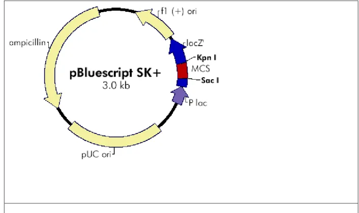
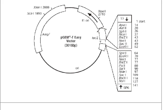
The pQE-31 (Qiagen Inc. USA) and pBluescript SK (Stratagene Inc. USA) vectors were used in colony hybridization experiments, as the pGEM T-Easy vector, Promega (USA) was used in cloning of PCR products.
Figure 2) The map of pBluescript cloning vector. It contains lacZ gene and suitable for
α-complementation.
Figure 3) The map of pQE expression vector. It contains lac operon and is suitable for
2.1.5 Commercially Available Kits
• Qiaex II Gel Extraction Kit (by Qiagen Inc., USA) was used to purify PCR product from agarose gels.
• MN Nucleospin Plasmid (Macherey-Nagel, GmbH& Co, Germany) was used in isolation of plasmids from E.coli.
• DYEnamic ET Terminator Cycle Sequencing Kit (Amersham Biosciences UK Limited, England) was used for sequencing reactions.
2.1.6 Apparatus and Equipment
Agarose gel electrophoresis were done by using horizontal mini gel apparatus Mupid-2 purchased from Advance Co., Ltd., Japan. Thermal cycler for PCR was a product of Perkin Elmer (USA).
Figure 4) pGEM®-T Easy Vector circle map. It contains lacZ gene and suitable for
2.1.7 DNA Size Marker
1kb DNA ladder from MBI FERMENTAS Inc. (USA) was used as DNA size marker.
2.2
SOLUTIONS AND MEDIA
2.2.1 Agarose Gel Electrophoresis Solutions50X Tris-Acetic acid-EDTA (TAE) Per Liter: 242 g Tris Base
57.1 ml Glacial Acetic acid 100 ml of 0.5 M EDTA
Ethidium bromide 10 mg/ml in water (stock solution)
30 ng/ml (working solution)
2.2.2 Microbiological Media and Antibiotics
Luria-Bertani Medium (LB) Per Liter: 10 g Bacto tryptone,
5 g Yeast extract 5 g NaCl
15 g Agar (for solid media)
Blood Agar %7 Rabbit blood in BHI agar
Brain Heart Infusion (BHI) Agar Per Liter: 52 g of BHI agar powder
BHI Broth Per Liter: 37 g of BHI broth powder
Mueller Hinton (MH) Broth Per Liter: 21 g of MHB powder
Ampicillin 100 mg/ml (stock solution)
2.2.3 Solutions for competent cell preparation:
SOB medium (100ml) 2% Bacto-tryptone
(Sterilized by autoclaving) 0.5 % yeast extract
10 mM NaCl 2.5 mM KCl 10 mM MgCl2
10 mM MgSO4
Transformation Buffer (TB) 10mM Pipes (Sterilized by filtration through 55mM MnCl2
0.45μm filter, stored at 4 °C) 15mMCaCl2
250mM KCl pH 6.7
SOC SOB with 20mM glucose
2.3
METHODS
2.3.1 Growth and Maintenance of Bacteria
2.3.1.1 M. catarrhalis
The stocks of the M. catarrhalis strains were done in MH broth containing 5% rabbit blood and stored at -20C until use. Prior to stocking, the bacteria were cultured on 7% rabbit blood agar plates. Then, the colonies were taken with a sterile cotton swab and transferred into the 1 ml of media contained in a cryotube. During experiments, after cultured on BHI agar, the bacteria were incubated within a box containing 5% CO2 at 37 °C. The CO2 generated by the
CO2 Gen Compact (Oxoid Limited, UK). All media were purchased from Merck Kga
2.3.1.2 E. coli
E. coli strains were stored in 50% sterile glycerol at -70 °C till it was used. The
stocking was done as following: Overnight grown cultures were mixed with sterile glycerol in a ratio of 1:1, mixed for homogenization and kept at -70 °C. During the experiments the E. coli were cultured either on LB agar or in LB broth. The broth cultures were incubated in a rotary shaking incubator at 37 °C with a speed of ~200 rpm. Similarly, LB agar plates were incubated overnight at 37 °C incubator.
2.3.2 DNA Experiments
2.3.2.1 Competent Cell preparation and E. coli transformation
2.3.2.1.1 Simple and Efficient Method (SEM) for Preparation of Competent Cells
A single colony of appropriate E. coli DH5α strain was inoculated into 15 ml of LB broth (containing the appropriate antibiotics which is ampicillin in this study) and grown overnight at 37 °C. Then, the starter culture was diluted to OD600 of 0.2-0.3 into 250 ml of
SOB medium and grown until the OD600 of the culture reaches to 0.6 at 18 °C with a speed of
200-250 rpm shaking. It is very important that the aeration of the culture during growth is well. For this grow the culture into a wide flask closed with a sterile cotton cab. After that, the culture was chilled on ice for 10 minutes, and centrifuged at 2500 X g for 10 minutes at 4 °C.
The pellet was resuspended in 80 ml of ice-cold TB, and an other 10 minute incubation on ice was done. The mixture was pelleted as mentioned above, resuspended gently in another 20 ml of TB. After that, DMSO was added to a final concentration of 7%, mixed gently and
incubated on ice for 10 minutes for the last time. Subsequently, the aliquots of this mixture were done and these aliquots were chilled in liquid nitrogen, and then stored at -70 °C up to 3 months without loss of transformation efficiency (Inoue et al. 1990).
2.3.2.1.2 Transformation of E.coli
An aliquot of frozen competent cells was thawed on ice. 200 μl of the cells was mixed with plasmid DNA with a concentration of less then 100 ng/μl in a 1.5 ml eppendorf tube and incubated on ice for 30 minutes. The cells were then exposed to 42 °C for a heat shock for 90
added and the cells were grown at 37 °C for 45 minutes with vigorous shaking at 250 rpm. The culture was then centrifuged and the pellet was resuspended in 100 μl of SOC, and spread on LB agar plates containing the appropriate antibiotic, which is ampicillin in this study.
2.3.2.2 Plasmid DNA Isolation
The plasmid, was isolated with MN Nucleospin Plasmid kit according to instructions of the manufacturer.
2.3.2.3 DNA Ligation Reactions
Ligation of the DNA fragments into plasmid vectors were done with 0.3-1.0 μg of linearized plasmid vector and 3-5 times molar excess of insert DNA in the presence
of 1-4 Weiss units of T4 ligase and 1X concentration of the standard ligation buffer supplied by
the T4 DNA ligase. And the mixture was incubated overnight at 16 °C. The reaction volume was 15µl.
2.3.2.4 Restriction Enzyme Digestion
In restriction enzyme digestion 5 μl of plasmid (1-10 μg/μl), 2 μl restriction enzyme buffer, and 1 μl restriction enzyme, HindIII, was mixed in small tubes and the reaction volume was adjusted to 20 μl with sterile ddH2O.
2.3.2.5 Primers
The primers for both sequencing and PCR reactions were designed by using the Primer3 program. The primers designed for PCR reactions were used in the sequencing reactions as well. The criteria for proper primer design are given below.
• Tm (the melting temperature of the primer) in the range of 52 º C to 65 º C
• Lack of dimerization capability
• Lack significant hairpin formation (>3 bp)
• Absence of secondary priming sites
Generally the lengths of the primers used in this study were 19bp or 20 bp. Moreover, their GC content is ranging from 40 % to 55 %. All of the primers designed worked properly (Table 1 and Table 2). All primers used in this study were custom synthesized and purchased from Iontek (Bursa, Turkey).
In addition, the primers designed for the first PCR reaction were constructed from the most conserved amino acid sequences of Walker A motif of several organisms, Neisseria
meningitidis, Pseudomanas aeruginosa, Haemophilus influenzae, Agrobacterium tumeficians,
and Brucella melitensis, that insert B5 has significant homology. In addition to the most
conserved amino acid sequence of the Walker A, gram negative codon usage was considered as ABC1 was designed; as the ABC2 primer was designed according to universal codon usage, and finally ABC3 was constructed by using universal codon usage, but in this case the primer has degeneracy (Table 2).
B5 2: 3’-TGGATGAGCTGCTTGATTTG-5’
ABC1: 5’-AACGGCAGCGGCAAAAGCAC-3’ ABC2: 5’-TGGCCTAGTGGTAGTGGTA-3’
ABC3: 5’-GGYCCYAGYGGYAGYGGYA-3’
Table 2) Primers used in PCR assays
KA2: 5’-TGCAGGTCTTGAAAATGCAG-3’ B5 2: 3’-TGGATGAGCTGCTTGATTTG-5’ T3: 5’-AATTAACCCTCACTAAAGGGA-3’ T7: 5’ TAATACGACTCACTATAGGG 3’
2.3.2.6 Polymerase Chain Reaction (PCR)
PCR was done in order to find out the Walker A motif in the putative ATPase domain of ABC transporter in M. catarrahalis. The reaction conditions and procedure for PCR are given below. In general all of the reagents were delivered in a 1.5 ml eppendorf tube and a master mix was prepared on ice. In addition, before and after giving the Taq polymerase, the master mix was mixed gently by vortexing.
The master mix was distributed into PCR tubes with a volume of 48 µl. Then, 2 µl of
M. catarrahalis genome template DNA was given into tubes. For negative control instead of
genomic DNA, 2 µl sterile ddH2O was added. The PCR products were run on 0.8 % or 1.0 %
agarose gel containing EtBr for visualization under UV lamp.
2.3.2.6.1 PCR assay
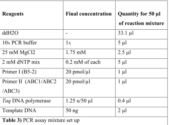
Three sets of PCR assays were done with three different forward primers (ABC1, ABC2 and ABC3), one for each set, and one reverse primer (B5-2), for all sets. The reaction volume in each PCR tube was 50 µl and the reaction cycle was 35.
Reagents Final concentration Quantity for 50 µl of reaction mixture ddH2O - 33.1 µl 10X PCR buffer 1X 5 µl 25 mM MgCl2 1.75 mM 2.5 µl 2 mM dNTP mix 0.2 mM of each 5 µl Primer I (B5-2) 20 pmol/µl 1 µl Primer II (ABC1/ABC2 /ABC3) 20 pmol/µl 1 µl
Taq DNA polymerase 1.25 u/50 µl 0.4 µl
Template DNA 50 ng 2 µl
2.3.2.7 Genomic DNA Isolation from M. catarrhalis
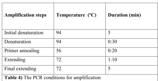
Amplification steps Temperature (ºC) Duration (min)
Initial denaturation 94 5
Denaturation 94 0:30
Primer annealing 56 0:20
Extending 72 1:10
Final extending 72 5
Table 4) The PCR conditions for amplification
Large scale genomic DNA isolation was performed by the standard methods of Current Protocols for gram-negative bacteria. 100 ml BHI broth was inoculated with 1 or 2 colonies of
M. catarrhalis. After overnight incubation the bacteria were harvested by centrifugation at
10000 rpm. The pellet was resuspended in 8.75 ml of 10X TE (100mM Tris-HCl, pH 7.5, 10
mM EDTA) by repeated pipetting and then transferred to a disposable 50 ml centritube. The cell suspension was frozen at -20 °C for 60 minutes. 1.0 ml of freshly prepared lysozyme (10 mg/ml in 0.25 Tris-HCl, pH 8.0) solution was added to the frozen cells and then thawed by mixing in a room-temperature water bath. Just after thawing, the cells were put on ice for 45 minutes. 0.25 ml of 20 % SDS, 50µl of 20mg/ml proteinase K and 10 µl of 10 mg/ml DNase-free RNase was added to the cell suspension. After mixing thoroughly the solution was incubated at 55 °C for 1 hour with occasional gentle mixing. Then 1.8 ml of 5 M NaCl was added and the tube was mixed thoroughly by inverting for 5 minutes. 1.5 ml of CTAB/NaCl solution was added and the tube was mixed thoroughly and then it was incubated at 65 C for 20 minutes with occasional shaking.
Approximately equal volumes of chloroform: isoamyl alcohol (49:1) was added, the tube was mixed thoroughly by inverting the tube for 5 minutes, and it was spin down 10 min at 3000 rpm. The chloroform phase was removed and the supernatant and the interphase were homogenized with a plastic pipette. The suspension was centrifuged 10 min at 3000 rpm again and the aqueous, viscous supernatant was transferred to a fresh centrifuge tube, leaving the interface behind. An equal volume of phenol:chloroform: isoamyl alcohol (25:24:1), was added and the solution was extracted thoroughly by inverting the tube and was centrifuged 10
minutes at 3000 rpm. The aqueous, viscous supernatant was transferred to a fresh centritube, leaving the interface behind. 0.6 volume of isopropanol was added to precipitate the nucleic acids. The DNA pellet was transferred to a fresh tube containing 70 % ethanol by hooking it onto the end of a glass Pasteur pipette. The DNA was washed with 70 % ethanol to remove the residual CTAB and recentrifuged to pellet it. The supernatant was removed carefully and the pellet was dissolved in TE.
2.3.2.8 Phenol-Chloroform Extraction
Phenol-chloroform extraction was done to purify DNA after each enzymatic
manipulation. First, an equal volume of phenol: chloroform: isoamyl alcohol (25:24:1) was added to the DNA solution containing the impurities. The optimum starting volume was about 100 μl. If the solution was less than this volume it was diluted with ddH2O accordingly. After
mixing 45 seconds by vortexing, the tube was centrifuged for 5 minutes at 13000 rpm. The upper (water phase) was transferred to a new eppendorf tube. Then 1/10 volume of 3M NaOAc was added. After mixing by inverting the tube 2.5 volume of cold absolute ethanol was added. After mixing by inverting the tube was kept in -80C for 20 minutes. Then the tube was
centrifuged for 10 minutes at 13000 rpm. After discarding the supernatant, the pellet was washed with 200 μl of 70% ethanol. After drying all the ethanol the pellet was dissolved in TE or water.
2.3.2.9 Agarose Gel Electrophoresis
DNA fragments of less than 1kb were generally separated on 1.0 % agarose gel, those greater than 1kb were separated on 0.8 % gels. Required amount of agarose gels were
completely dissolved in 1X TAE buffer by heating in microwave, then ethidium bromide was
added to a final concentration of 30 ng/ml. The DNA samples were mixed with one volume loading buffer and loaded onto gels. The gel was run at room temperature in 1X TAE at 50 V or
100 V.
2.3.2.10 Isolation of PCR Product from Agarose Gels
The kit given in materials part was used in isolation of PCR product. We followed the instructions of the manufacturer.
2.3.2.11 DNA Sequence Analysis
DNA sequence analysis of the positive clones was performed by the dideoxy chain termination method. Cycle sequencing reactions were set up in 0.2 ml eppendorf tubes. The sequencing procedure was performed by DYEnamic ET Terminator Cycle Sequencing Kit according to the instructions of the manufacturer. The cycle sequenase reactions were performed in Perkin Elmer GeneAmp PCR 9600 system thermal cycler with the following parameters: 25 cycles of denaturation (95 °C for 20 seconds), annealing (50 °C for 15 seconds) and extension (60 °C for 1 minutes). Electrophoresis was performed on the ABI PRISMTM 377 Genetic Analyzer (Perkin Elmer, USA) by loading the samples into the lanes of a vertical polyacrylamide slab gel. The separated DNA fragments were exposed to a laser which in turn would excite the flourescent dyes attached to the fragments. These data were then collected and analyzed by the help of ABI Sequencing Analysis Software (version 3.0.1).
In addition, for complete nucleotide sequencing new primers were designed and the procedure mentioned above was used in subsequent sequencing reactions.
Sample Template (500ng) (µl) Primer name Primer (2pmol/µl) (µl) Mix (µl) ddH2O (µl) B5 8 T3 2 8 2 B5 8 T7 2 8 2 B5 8 KA2 2 8 2 B5 8 B52 2 8 2 Control 1 Control 2 8 7
Table 5) Protocol for DYEnamic ET Terminator Cycle Sequencing Kit
2.3.2.12 Bioinformatic tools used for nucleotide and amino acid sequence analysis
For nucleotide sequence homology search “BLAST align two sequences” tool was used. For conserved domain homology search, “the conserved domain homology search” engine of BLAST was used. In addition, translation of the inserts’ nucleotide sequences into 6 phase amino acid sequence were done by the “justbio translator” tool and the complementary sequence of the inserts’ reverse sequences were obtained by using “justbio complementor
program”. Also, “justbio aligner” tool was used for two amino acid sequences alignments. Finally, for multiple amino acid sequence alignments “clustalW” program was used.
RESULTS
3.1 Isolation of the clones
In previous studies performed in our lab, a genomic library of M. catarrhalis was prepared in pBluescript SK (pBSK) and pQE 31 vectors after digesting the genomic DNA with
HindIII using standard molecular biology methods. Then transformation of the genomic library
was done in E. coli DH5-α strain. The library constructed by using the pQE 31 vector was screened by monoclonal antibody (mAb) against 55kDa protein, which is an adhesin protein, isolated from M. catarrhalis in our lab. In addition, the library constructed by using the pBSK vector was screened by an oligonucleotide probe deduced from N-terminal amino acid
sequence of 55kDa protein.
A total of 12 positive clones (B5, C1, 1B1, 1B2, 1B3, 3A, 3B, 5A, 6A, 8A, 8B1 and 8B2) out of 4000 colonies were identified based on X-gal detection method. The size of the inserts of the clones ranged from 750bp to 1500bp (Turan T 2002).
3.2 Nucleotide sequence
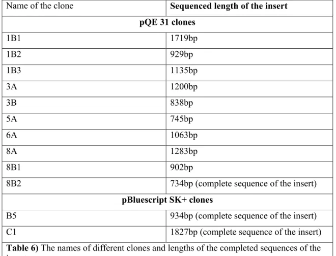
We attempted to obtain the nucleotide sequence of the inserts of all of the clones (Table 6). Inserts of clones, B5, C1 and 8B2 were sequenced completely. On the other hand, we obtained the partial sequences of the inserts of the rest of the clones. Almost 70 % to 80 % of these inserts were sequenced. Among these clones the insert of the B5 showed significant homology with the ATPase domain of the ABC type transporter.
Name of the clone Sequenced length of the insert pQE 31 clones 1B1 1719bp 1B2 929bp 1B3 1135bp 3A 1200bp 3B 838bp 5A 745bp 6A 1063bp 8A 1283bp 8B1 902bp
8B2 734bp (complete sequence of the insert)
pBluescript SK+ clones
B5 934bp (complete sequence of the insert)
C1 1827bp (complete sequence of the insert)
Table 6) The names of different clones and lengths of the completed sequences of the
inserts.
3.3 Amino acid sequence
LHYLP*LLWSLKD**AIKNSTDDILVK*DLHQNLHIFINLSKQLSSMSIKHEY*N PKHP*NFRGFCCLR*NQH*YFNR*ADNFIRSIRLWQDNFATHYCRS
[
KCRQRA GYILTVSNVTDIPVQHRGIGFVFQQYALFRHQTVAQNIAFGLTLLPRNTRPSA NTIDRRVDELLDLVQLSHTKTRYPHELSGGQRQRVALARALATEPKLLLLDE PFGALDAKVRKSLRDSLKDIQREIGITSILVTHDQEEAQAISDKIVIMNHGQIEQ IGTPSKLFAQPSSDFVVDFLGL]
AMSHRSKINRHMVFNLH*SQXTable 7) The converted amino acid sequence of the insert of clone B5. The motifs of
ATPase domain are found in the region shown dark within the brackets []. “ * ” Indicates the stop codons.
3.3.1
The conserved domain homology search was done in BLAST by using “conserved
domain homology” search tool in BLAST. The second phase amino acid sequence of the insert B5 showed 50 % to 70 % homology with the ATP-binding domain, the ATPase of the ABC transporters of several bacterial species including, N. meningitides, P.
aeruginosa, H. influenzae, H. pylori and S. typhimurium LT. Figures 8 and 9 below
show where 55% homology and 68% homology were found respectively. In both of the tables the query sequence is the insert B5 and the subject sequence is the corresponding consensus sequence. In addition, the CD-length shows the conserved domain length, bit score that each alignment has is the measure between the hit, the sequences that produce “significant” alignments to the query sequence, and the query. The Expectation value (e-value) is "an assessment of the statistical significance of the score”. Also, the e-value is showing how random a match is or how much chance is involved. As a result, a low e-value is an indication of a high homology.
CD-Length = 248 residues, only 55.2% aligned
Score = 136 bits (345), Expect = 1e-33
Query: 22 HRGIGFVFQQYALFRHQTVAQNIAFGLTLLPRNTRPSANTIDRRVDELLDLVQLSHTKTR 81 Sbjct: 71 GPDIGYVFQEDALLPWLTVLDNVALGLEL----RGKSKAEARERAKELLELVGLAGFEDK 126 Query: 82 YPHELSGGQRQRVALARALATEPKLLLLDEPFGALDAKVRKSLRDSLKDIQREIGITSIL 141 Sbjct: 127 YPHQLSGGMRQRVAIARALATRPKLLLLDEPFGALDALTREELQDELLRLWEETRKTVLL 186 Query: 142 VTHDQEEAQAISDKIVIMNHG 162
Sbjct: 187 VTHDVDEAVYLADRVVVLSNR 207
Figure 5) Homology between the insert B5 amino acid sequence (the query) and the
consensus amino acid sequence (subject). For a detailed explanation please see the text above.
CD-Length = 217 residues, only 68.7% aligned
Score = 119 bits (299), Expect = 2e-28
Query: 17 DIPVQHRGIGFVFQQYALFRHQTVAQNIAFGLTLLPRNTRPSANTIDRRVDELLDLVQLS 76 Sbjct: 69 DLRELRRRIGYVFQEPVLLFNGTVRENIAFGL----ELHGLSKEETRERVEEALELVGLH 124 Query: 77 -HTKTRYPHELSGGQRQRVALARALATEPKLLLLDEPFGALDAKVRKSLRDSLKDIQREI 135 Sbjct: 125 PDLLDRPPGQLSGGQRQRVAIARALLRDPKILLLDEPTSALDVESEAQILELLRELQEK- 183 Query: 136 GITSILVTHDQEEAQAISDKIVIMNHGQIEQIGTP 170
Sbjct: 184 GRTVLVVTHD-ELATRLADRILVLKNGKIVEQGTP 217
Figure 6) Homology between the insert B5 amino acid sequence (the query) and
the consensus amino acid sequence (subject). For a detailed explanation please see the text above.
3.3.1.1
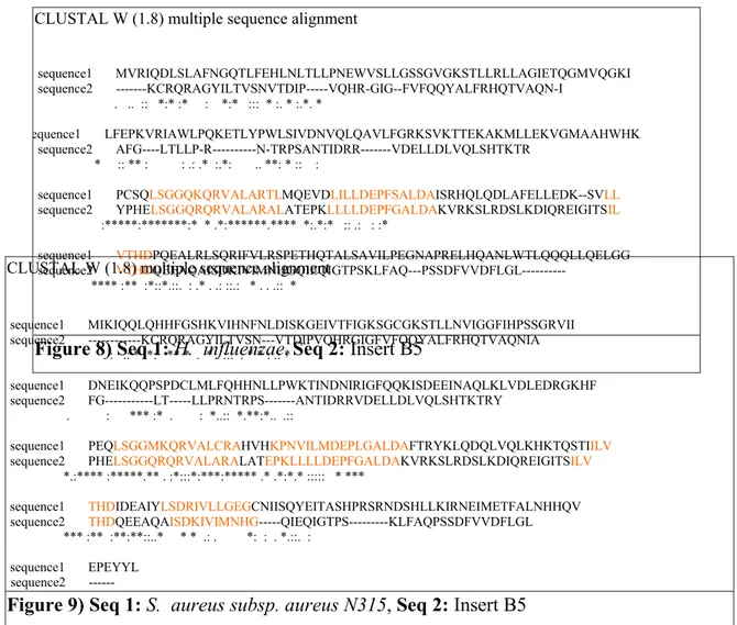
Specific examples showing the most homologous amino acid sequences
between the ATPase components of insert B5 amino acid sequence and several bacterial species: P. aeruginosa, H. influenzae, S. aureus subsp. aureus N315,
N. meningitidis serogroup B strain MC58, H. pylori . In each figure, asterisks
indicate identical residues, colons indicate conserved substitutions, and periods indicate semiconserved substitutions. In addition, the amino acids highlighted with orange indicates the regions those are most identical between the two sequences.
CLUSTAL W (1.8) multiple sequence alignment sequence1 MNAASRQPSTLPADALLAVDGVSLEYRTRSRVVRATQRVSFEVDPADRFVLLGPSGCGKS sequence2 ---KCRQRAGYILTVSN--- :. **..: : ::
sequence1 TLLKAVAGFITPSEGEIRLQGQAVRAPGPDRIVVFQEFDQLPPWKTVRQNVLFPLRVSGQ sequence2 ---VTDIPVQHRG----IG---FVFQQYALFR-HQTVAQNIAFGLTLLPR
:: :: :* .***:: : :** **: * * : :
sequence1 VTR---DEAERRADECLEKVGLAGFAEAYPHTLSGGMKARVAIARALAMQPKILLMDEPF sequence2 NTRPSANTIDRRVDELLDLVQLSHTKTRYPHELSGGQRQRVALARALATEPKLLLLDEPF ** : :**.** *: * *: *** **** : ***:***** :**:**:****
sequence1 AALDALTRRKMQEELLRLWEEVRFTLLFVTHSIEEALVVGNRILLLSPHPGRVRAEVHGH sequence2 GALDAKVRKSLRDSLKDIQREIGITSILVTHDQEEAQAISDKIVIMN-HG---QIEQIGT .**** .*:.:::.* : .*: :* ::***. *** .:.::*:::. * : * *
sequence1 PFGLHSLGGEPLQAAARRIHRLLFDEGGEPTAAATLDFADIRLAH sequence2 PSKLFAQ---PSSDFVVDFLGL----
* *.: *:: .:** .:
CLUSTAL W (1.8) multiple sequence alignment sequence1 MVRIQDLSLAFNGQTLFEHLNLTLLPNEWVSLLGSSGVGKSTLLRLLAGIETQGMVQGKI sequence2 ---KCRQRAGYILTVSNVTDIP---VQHR-GIG--FVFQQYALFRHQTVAQN-I . .. :: *:* :* : *:* ::: * :. * :.*. * sequence1 LFEPKVRIAWLPQKETLYPWLSIVDNVQLQAVLFGRKSVKTTEKAKMLLEKVGMAAHWHK sequence2 AFG----LTLLP-R---N-TRPSANTIDRR---VDELLDLVQLSHTKTR * :: ** : : .: .* :.*: .. **: * :: :
sequence1 PCSQLSGGQKQRVALARTLMQEVDLILLDEPFSALDAISRHQLQDLAFELLEDK--SVLL sequence2 YPHELSGGQRQRVALARALATEPKLLLLDEPFGALDAKVRKSLRDSLKDIQREIGITSIL :*****:*******:* * .*:******.**** *:.*:* :: .: : :* sequence1 VTHDPQEALRLSQRIFVLRSPETHQTALSAVILPEGNAPRELHQANLWTLQQQLLQELGG sequence2 VTHDQEEAQAISDKIVIMNHGQIEQIGTPSKLFAQ---PSSDFVVDFLGL--- **** :** :*::*.::. : .* . .: ::.: * . . .:: *
Figure 8) Seq 1: H. influenzae, Seq 2: Insert B5 CLUSTAL W (1.8) multiple sequence alignment
sequence1 MIKIQQLQHHFGSHKVIHNFNLDISKGEIVTFIGKSGCGKSTLLNVIGGFIHPSSGRVII sequence2 ---KCRQRAGYILTVSN---VTDIPVQHRGIGFVFQQYALFRHQTVAQNIA . : .: * :*: ** * . * . ::: . * * : .: * sequence1 DNEIKQQPSPDCLMLFQHHNLLPWKTINDNIRIGFQQKISDEEINAQLKLVDLEDRGKHF sequence2 FG---LT---LLPRNTRPS---ANTIDRRVDELLDLVQLSHTKTRY . : *** :* . : *..:: *.**:*.. .::
sequence1 PEQLSGGMKQRVALCRAHVHKPNVILMDEPLGALDAFTRYKLQDQLVQLKHKTQSTIILV sequence2 PHELSGGQRQRVALARALATEPKLLLLDEPFGALDAKVRKSLRDSLKDIQREIGITSILV *.:**** :*****.** . :*:::*:***:***** .* .*:*.* ::::: * ***
sequence1 THDIDEAIYLSDRIVLLGEGCNIISQYEITASHPRSRNDSHLLKIRNEIMETFALNHHQV sequence2 THDQEEAQAISDKIVIMNHG---QIEQIGTPS---KLFAQPSSDFVVDFLGL *** :** :**:**::..* * * .: . *: : . *.::. :
sequence1 EPEYYL sequence2
3.3.2 Motif search results CLUSTAL W (1.8) multiple sequence alignment sequence1 MSITIQNLNKHFGNFHALKNINLNVPTGKLVSLLGPSGCGKTTLLRIIAGLENADGGNIL sequence2 ---KC-RQR---AGYIL * : .* **
sequence1 FDGQDVTAKHVRERKVGFVFQHYALFRHMNVFDNVAFGLTVLPKSERPSKGQIRAKVEEL sequence2 TVS-NVTDIPVQHRGIGFVFQQYALFRHQTVAQNIAFGLTLLPRNTRPSANTIDRRVDEL . :** *:.* :*****:****** .* :*:*****:**:. *** . * :*:**
sequence1 LKLVQLSHLAKSYPHQLSGGQRQRIALARALAVEPKLLLLDEPFGALDAKVRKELRTWLR sequence2 LDLVQLSHTKTRYPHELSGGQRQRVALARALATEPKLLLLDEPFGALDAKVRKSLRDSLK *.****** . ***:********:*******.********************.** *:
sequence1 DIHHNLGVTSILVTHDQEEALEVSDEIVVMNHGKIEQTGSAEAIYRKPENAFVTEFLGET sequence2 DIQREIGITSILVTHDQEEAQAISDKIVIMNHGQIEQIGTPSKLFAQPSSDFVVDFLGL- **::::*:************ :**:**:****:*** *:.. :: :*.. **.:*** sequence1 DAFEGRIEKGFWHYNGFAWKLDAQYKWQEQTATGYIRPHEWQIAAEHETPMICAEIEKIH sequence2 --- sequence1 AVGALTHILVKHDKQDVHITLAGSDAARYPIAEGKELKLIPKQVYVFSQNELIEYSI sequence2
---Figure 10) Seq 1: N. meningitidis serogroup B strain MC58, Seq 2: Insert B5
CLUSTAL W (1.8) multiple sequence alignment sequence1 MKEIVTIENVSFNYHNRAVFKDFNLSIQEGDFLCVLGESGSGKSTLLGLILGLLKPSLGS sequence2 ---KCRQRAGYILTVSN--- : : * :* * . sequence1 VKIFNETLSNNAFLRQKIGYIAQGNSLFSHLNAMQNMTFCLNLQGINKQAAQKEAK---A sequence2 ---VTDIPVQHRGIGFVFQQYALFRHQTVAQNIAFGLTLLPRNTRPSANTIDRRVD ::: .. :: **:: * :** * .. **::* *.* *.:.: : .
sequence1 LALKMGLDESLMDKFPNELSGGQAQRVGIIRGIIHRPELILLDEPFSALDSFNRKNLQDL sequence2 ELLDLVQLSHTKTRYPHELSGGQRQRVALARALATEPKLLLLDEPFGALDAKVRKSLRDS *.: . ::*:****** ***.: *.: .*:*:******.***: **.*:*
sequence1 IKEIHQNSHATFIMVTHDESEAQKLATKTLEIKALKQEQ---
sequence2 LKDIQREIGITSILVTHDQEEAQAISDKIVIMNHGQIEQIGTPSKLFAQPSSDFVVDFLG :*:*::: * *:****:.*** :: * : :: : **
sequence1 - sequence2 L
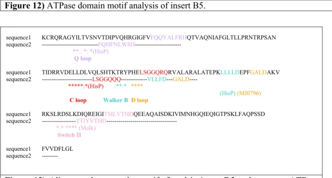
The ATPase domain motifs found in insert B5 amino acid sequence (Figure 12). The similarity of them with the motifs of most known, studied ATPase domains, HisP, MJ0796, Malk (Figure13) (Schmitt L 2002).
KCRQRAGYILTVSNVTDIPVQHRGIGFVFQQYALFRHQTVAQNIAFGLTLLPRNTRPSANTIDRRVDEL Q loop
LDLVQLSHTKTRYPHELSGGQRQRVALARALATEPKLLLLDEPFGALDAKVRKSLRDSLKDIQREIGI
C loop Walker BD loop
TSILVTHDQEEAQAISDKIVIMNHGQIEQIGTPSKLFAQPSSDFVVDFLGL
Switch II
Figure 12) ATPase domain motif analysis of insert B5.
sequence1 KCRQRAGYILTVSNVTDIPVQHRGIGFVFQQYALFRHQTVAQNIAFGLTLLPRNTRPSAN sequence2 ---FQHFNLWSH---
**:: *: *(HisP) Q loop
sequence1 TIDRRVDELLDLVQLSHTKTRYPHELSGGQRQRVALARALATEPKLLLLDEPFGALDAKV sequence2 ---LSGGQQQ---VLLFD---GALD----
*****:*(HisP) :**:* ****
(HisP) (MJ0796)
C loop Walker B D loop
sequence1 RKSLRDSLKDIQREIGITSILVTHDQEEAQAISDKIVIMNHGQIEQIGTPSKLFAQPSSD sequence2 ---TTIYVTHD--- *:* **** (Malk) Switch II sequence1 FVVDFLGL sequence2 ---
Figure 13) Alignment between the motifs found in insert B5 and common ATPase
domains, HisP, MJ0796, Malk.
3.4
PCR results
To get the Walker A motif in the PCR product we did, the expected band range was 700 bp to 1300 bp. After optimization of the PCR done we obtained two separate bands ranging from ~780bp (the lower band) to ~1200bp (the upper band) from the PCR assay prepared by using ABC1 and B5-2 primers (Figure14). These PCR products were cloned in pGEM®-T Easy vector and sequenced. The results of sequencing reactions did not give a significant homology with any motif in ATPase domain of ABC transporter.
~1200bp ~780bp
Figure14) PCR assay, forward
primer is ABC1. The bands obtained after optimization of the PCR assay.
DISCUSSION
In this study we attempted to sequence the inserts of different clones of
M. catarrrhalis with the intention of identification of some genes in this organism.
One of these, clone B5, has significant homology with the ATPase domain of ABC type transporters.
The ATPase domain of an ABC type transporter has conserved motifs: Walker A, Q loop, C loop (signature sequence), Walker B, D loop and Switch II. The ATPase domain is considered as the most conserved domain of the ABC type transporter due to presence of these motifs. The insert B5 has all these except the Walker A motif. Therefore, we speculated that the insert B5 is an ATPase component of a particular ABC type transporter in M. catarrhalis. However, the function of this putative ABC type transporter of M. catarrhalis is not known. The specificity of ABC type transporter comes from its TMDs not from the NBDs (Schmitt L 2002; Lage H 2003).
In microorganisms, the ABC type transporters are involved in both import and export processes (Dassa E 2001). Also, they are responsible for drug resistance in many bacterial species. For example, ABC type transporters mediate antibiotic resistance of some human pathogens, such as S. aureus and Staphylococcus epidermidis. The inhibition of ABC type transporter function in these organisms has been a subject of intensive study (Lage H 2003). However, the ABC type transporters, which insert B5 has significant homology with, are generally ion transporters. As a result of this, the putative ABC type transporter of M.
catarrhalis is not responsible for antibiotic resistance, but most probably is responsible for ion
transport. In addition, M. catarrhalis is a β-lactamase producer. Since it has such an effective antibiotic resistance mechanism, the probability of having ABC type transporters responsible for drug resistance is very low. In summary, we proposed that the putative ABC type
transporter, partially sequenced in this study, is an ion transporter, important for the survival of
M. catarrhalis. The functional studies of this putative ABC type transporter will be performed
after the full sequence of the ATPase domain is completed.
In the insert B5 amino acid sequence there were some stop codons. Actually the presence of these stop codons was an unexpected situation. Generally the stop codons, UAA, UAG, UGA, in universal genetic code may not be stop codons in bacterial genome. For example, Tetrahymena uses only one stop codon, UGA, to terminate protein synthesis. The
other stop codons, UAA and UAG are decoded as the glutamine codon (Nakamura Y 1998). In
M.catarrhalis the stop codon identified so far, is UAA (Du R-P 1998; Kakuda T 2003).
However, based on our literature search, which amino acid(s) was coded from the other stop codons, UAA and UAG, in M.catarrhalis has not been found yet. Since the nucleotide
sequence of insert B5 was translated into its amino acid sequence by a program, which is using universal genetic code, the program might give false stop signals. Another explanation for the extra stop codons in the amino acid sequence of insert B5 could be the following. During clone selection two different fragments from the M. catarrhalis genomic DNA library might have come together and inserted into the same vector. If this is the case for clone B5, the stop codons coming before the amino acid sequence consisting of the motifs might belong to the fragment that did not include the ATPase domain. In addition, although we have confirmed the nucleotide sequence of insert B5 by doing the sequencing reaction two times, the polymerase might not work properly and some parts of the sequence could include false bases.
Since the complete sequence of the ATPase domain is not known and the promoter sequence should be found before Walker A motif, the promoter sequence search will be performed after the full sequence of the ATPase component of the putative ABC type transporter in M. catarrhalis is completed.
In the insert B5, the length of the ATP-binding cassette domain is 188 amino acids. Our literature search showed that in several bacteria, length of the NBD domain of the ABC
transporters is about 215 amino acids (Linton KJ 1998). Therefore, it is necessary to find ~ 27 amino acids to obtain the complete sequence of the ATP-binding domain of the putative ABC transporter of M. catarrhalis.
The Walker A is the only motif that was remained to be identified. Since M. catarrhalis genome has not been fully sequenced yet, it was not possible to design a specific primer for this. Therefore, we followed another way to complete the putative ATPase domain. The primers were designed from the most conserved amino acid sequence of the Walker A motif of several species, N.
meningitidis, P. aeruginosa, H. influenzae, A. tumeficians, and B. melitensis. Since the Walker A
motifs of different species may show slight variations in some amino acids in the protein sequence (Holland IB 1999), in construction of the primers we took into consideration the most conserved amino acids found in the motif. In addition, in translation of the Walker A into nucleotide sequence we followed two different procedures, considering codon usages of the organisms. In the first approach, we considered the gram negative codon usage and prepared primer ABC1 according to this. As a second approach the ABC2 primer was constructed according to universal codon usage.
this case the primer had degeneracy in some bases. The reason for designing of these primers was to include all the possibilities to get a significant band at the end of the PCR assays. In these
experiments, the primers, ABC1, ABC2, ABC3 were used as forward primers and primer B5-2 was used as reverse primer. At the end of optimization of the PCR, we obtained two significant bands, ABC1a and ABC1b, 750bp and 1000bp in length respectively. These bands were significant because - based on the predicted length of our ATPase domain - the expected length of the PCR product ranged from 700 bp to 1000 bp. According to this, the bands obtained were promising that they could include the Walker A motif. After isolation of these PCR products from the agarose gel, we directly sequenced the PCR products. However, the direct sequencing of these PCR products did not give a satisfactory result. Then, we made TA cloning of the PCR products and sequenced the inserts. Unfortunately the restriction enzyme analysis, done after isolation of the plasmid, showed that TA cloning was not executed successfully.
Future perspectives
As a future study we will continue to do the sequencing of all clones and complete the nucleotide sequence of these clones. Moreover, we will perform TA cloning and subsequent sequencing reaction of the PCR products.
In order to go to 5’ end of the nucleotide sequence, another thing that could be done in future studies is RACE (Rapid Amplification of cDNA Ends). Moreover, the PCR assays could be continued till the Walker A region obtained. After obtaining the complete nucleotide
sequence of this ATPase domain, experiments to find its function could be performed. However, since mostly the domains of the ABC type transporters of bacteria were located in different parts of the genome (Schmitt L 2002) prior to the experiments, a careful search of which procedures will be followed should be done.
References:
Ahmed K, Rikitomi N, Nagatake T, Matsumoto K. “Electron microscopic observation of Branhamella catarrhalis.” Microbiol Immunol. 1990. 34: 967-975
Ahmed K. “Fimbriae of Branhamella catarrhalis as possible mediator of adherence to pharyngeal epithelial cells.” Acta Pathol Microbiol Immunol Scand 1992a. 100:1066-1072
Ahmed K, Rikitomi. “N and Matsumoto K. Fimbriation, hemagglutination and adherence properties of fresh clinical isolates of Branhamella catarrhalis” Microbiol Immunol.
1992. 36:1009-1017
Bartos, L., C., and T. F. Murphy. “Comparison of outer membrane proteins of 50 strains of Branhamella catarrhalis.” J Infect Dis. 1988. 158:761-5
Chang G.“Minireview: Multidrug resistance ABC transporters” FEBSLetters 555 (2003)
102-105
Chang G, Roth CB.”Structure of MsbA from E.coli: A homolog of the multidrug ressistance ATP-binding cassette (ABC) transporters” Science Vol.293 7 September 2001 p.1793-1800
Cope LD, Lafontaine RTSlaughter CA, HasemannJr CA, Aebi C, Henderson FW, Mc CrackenGH Jr, Hansen EJ. 1999. Characterization of the Moraxella catarrhalis UspA1 and UspA2 genes and their encoded products. J Bacteriol 181: 4026-4034
Dassa E, Bouige P. “The ABC of ABCs: a phylogenetic and functional classification of ABC systems in living organisims” Res. Microbiol. 152 (2001) 211-229
Detmers F, Lanfermeijer FC, Poolman B “Peptides and ATP binding cassette preptide transporters” Res. Microbiol. 152 (2001) 245-258
Doige CA, Ames GF “ATP-Dependent Transport Systems in Bacteria and Humans: Relevance to cystic Fibrosis and Multidrug” Annu. Rev. Microbiol. 1993. 47: 291-319
Du RP, Wang Q, Yang YP, Schryvers AB, Chong P, Klein MH, Loosmore SM. “Cloning and expression of the Moraxella catarrhalis lactoferrin receptor genes.” Infect Immun.
1998 Aug; 66(8): 3656-65.
Enright, M. C., and H. McKenzie. “M. catarrhalis-clinical and molecular aspects of a rediscovered pathogen.” J. Med. Microbiol. 1997. 46:360-371
Gottesman MM. and Ambudkar SV. “Overview: ABC Transporters and Human Disease” Journal of Bioenergetics and Biomembranes, Vol. 33, No. 6, December 2001 (2001)
Higgins CF. “ABC Transporters: physiology, structure and mechanism-an overview” Res. Microbiol. 152 (2001) 205-210
Higgins CF, Linton KJ. ”The XYZ of ABC transporters” Science 7 September 2001 Vol 293
Higgins CF. ”ABC transporters: From Microorganisms to Man” Annu. Rev. Cell Biol.1992.
8:67-113
Higgins CF, Haag PD, Nikaido K, Ardeshir F, Garcia G, Ames GF. “Complete nucleotide sequence and identification of membrane components of the histidine transport operon of S. typhimurium.” Nature 1982, 298:723-727.
Holland IB, Blight MA. “ABC-ATPases, adaptable energy generators fuelling
transmembrane movement of a variety of molecules in organisms from bacteria to humans.” J Mol Biol. 1999 Oct 22; 293(2): 381-99
Hu W-G, Chen J, McMichael JC and Gu X-X. “Functional characterization of a protective monoclonal antibody against serotype A and C lipooligosaccharides from M. catarrhalis.” Infect Immun 2001. 69: 1358-1363
Hyde SC, Emsley P, Hartshorn MJ, Mimmack MM, Gileadi U, Pearce SR, Gallagher MP, Gill DR, Hubbard RE, Higgins CF. “Structural model of ATP-binding proteins associated with cystic fibrosis, multidrug resistance and bacterial transport.” Nature 1990 Vol 346 p. 362-365
Johnson, M.A.,Drew, W.L. and Roberts, M. “Branhamella catarrhalis a lower respiratory tract pathogen?” J. Clin. Microbiol. (1981) 13:1066-9
Karalus R, Campagnari A. “M. Catarrhalis : a review of an important human mucosal pathogen.” Microb Infect. 2000. 2: 547-559
Kakuda T, Oishi D, Tsubaki S, Takai S. “Cloning and characterization of the fur gene from Moraxella bovis.” Microbiol Immunol. 2003. 47(6): 411-7.
Lage H. “ABC-transporters: implications on drug resistance from microorganisms to human cancers.” Int J Antimicrob Agents. 2003 Sep; 22(3): 188-99.
Linton KJ and Higgins CF. “The Escherichia coli ATP-binding cassette (ABC) proteins.” Molecular microbiology (1998) 28, 5-13
Locher KP, Borths E. “Minireview: ABC transporter architecture and mechanism: implications from the crystal structures of BtuCD and BtuF” FEBS Letters 564 (2004) 264-268
Locher KP, Lee AT, Rees DC. “The E. coli BtuCD structure: a framework for ABC transporter architecture and mechanism.” Science Vol 296 10 May 2002 p. 1091-1098
Martinez G, Ahmed K, Watanabe K, Tao M, and Nagatake T. “Changes in
antimicrobial susceptibility to Moraxella catarrhalis over a ten year period.” J Infect Chemother.
1998. 4:139-141
Mourez M, Hofnung M, Dassa E. “Subunit interactions in ABC transporters: a conserved sequence in hydrophobic membrane proteins of periplasmic permeases defines an important site of interaction with the ATPase subunits.” The EMBO Journal Vol.16 No. 11 pp.
3066-3077, 1997
Murphy, TF. “Studies of outer membrane proteins of Moraxella catarrhalis.” Am J. Med.
Murphy TF, Kirkham C, Lesse AJ. “The major heat modifiable outer membrane protein CD is highly conserved amongstrains of Moraxella catarrhalis.” Mol Microbiol 1993. 10; 87-97
Murphy, TF. “Branhamella catarrhalis: epidemiology, surface antigenic structure and immune response.” Microbiol Rev. 1996. 60; 267-79
Nakamura Y, Ito K. “How protein reads the stop codon and terminates translation.” Genes Cells, 1998 May; 3(5): 265-78.
Putman M, van Veen HW, and Konings W. “Molecular Properties of Bacterial
Multidrug Transporters” Microbiology and Molecular Biology reviews Dec. 2000, p. 672-693 Vol 64, No.4
Rahman M, Holme T. “Antibody response in rabbits to serotype specific-determinants in lipopolysaccarides from Moraxella catarrhalis.” J Med Microbiol. 1996. 44: 348-354
Rosinha GM, Freitas DA, Miyoshi A, Azevedo V, Campos E, Cravero SL, Rossetti O, Splitter G, Oliveira SC.”Identification and characterization of a Brucella abortus ATP-binding cassette transporter homolog to Rhizobium meliloti ExsA and its role in virulence and
protection in mice.” Infect Immun. 2002 Sep; 70(9): 5036-44.
Saurin W, Hofnung M, Dassa E “Getting In or Out: Early Segregation Between Importers and Exporters in the Evolution of ATP-Binding Cassette (ABC) Transporters” J Mol Evol (1999) 48:22–41
Schmidt KL, Peterson ND, Kustusch RJ, Wissel MC, Graham B, Phillips GJ, Weiss DS. “A predicted ABC transporter, FtsEX, is needed for cell division in Escherichia coli.” Journal of Bacteriology, Feb. 2004. p. 785-793
Schmitt L, Tampé R “Affinity, specifity, diversity: A challenge for the ABC transporter TAP in cellular immunity” Chembiochem 2000, 1, 16-35
Schmitt L, Tampé R “Structure and mechanism of ABC transporters” Current Opinion in Structural Biology 2002, 12:754–760
Schneider E, Hunke S. “ATP-binding-cassette (ABC) transport systems: Functional and structural aspects of the ATP-hydrolyzing subunits/domains,FEMS” Microbiology Reviews 22 (1998) 1-20
Sethi S, Hill SL, Murphy TF. “Serum antibodies to outer membrane proteins (OMPs) of
Moraxella (Branhamella) catarrhalis in patients with bronchiectasis: identification of OMP B1
as an important antigen.” Infect Immun. 1995. 63:1516-1520
Turan T. “Isolation and characterization of an Adhesin Protein from the Surface of a Respiratory Pathogen Moraxella ctarrhalis” M.Sc. Thesis, 2002
Verdon G, Albers SV, Dijkstra BW, Driessen AJ, Thunnissen AM. “Crystal structures of the ATPase subunit of the glucose ABC transporter from Sulfolobus solfataricus:
Young J, Holland IB “ABC transporters: bacterial exporters-revisited five years on.”Biochimica et Biophysica Acta 1461 (1999) 177-200
Zaleski A, Scheffler NK, Densen P, Lee FK, Campagnari AA, Gibson BW, Apicella MA. “Lipooligosaccharide P (k)(Galα1-4Galβ1-4Glc) epitope of Moraxella catarrhalis is a factor in resistance to bactericidal activity mediated by normal human serum.” Infect Immun.
2000. 68: 5261-5268
Web-pages:
cancer-seqbase.uchicago.edu/primers.html (John & Wiley press 1998) genomic DNA isolation protocol
www.ncbi.nih.gov/BLAST
Search the conserved domain database (rpsblast) www.justbio.com
www.ebi.ac.uk/clustalW Align two sequences (bl2seq)
www.science-house.org/fungal/learn/Paper.pdf (e-value) pymol.sourceforge.net/ (The figure in the introduction)