Effects of different orthodontic adhesives and resin removal techniques on
enamel color alteration
Yasemen Boncuk
a; Zafer C. C
¸ ehreli
b; O
¨ mu¨r Polat-O
¨ zsoy
cABSTRACT
Objective: To investigate the color alterations in enamel following the use of different orthodontic bonding resins and adhesive residue–removal burs.
Materials and Methods: Metal brackets were bonded to extracted human premolars (n 5 175) by using an etch-and-rinse adhesive system, a self-etch adhesive system (SEP), or a resin-modified glass ionomer cement (RMGIC). After 24 hours of photoaging, the brackets were removed and the adhesive residue on the tooth surfaces was cleaned with either a tungsten carbide bur or a Stainbuster bur. Tooth colors were measured with a spectrophotometer at baseline, after adhesive removal, and after additional photoaging. Color evaluation was made, and color differences induced by photoaging were calculated. Statistical evaluation was made using the Kruskal-Wallis test and the Mann-WhitneyU-test, with Bonferroni correction.
Results: All specimens showed discoloration at varying levels. The highest color change was observed in the etch-and-rinse adhesive/tungsten carbide bur group. When the etch-and-rinse and self-etch adhesives were used, adhesive-remnant removal with Stainbuster burs resulted in significantly lower discoloration. The type of bur did not affect the extent of enamel discoloration in the RMGIC group.
Conclusions: Orthodontic treatment alters the original color of enamel, and both the adhesive system and the resin-removal methods are responsible for this change. When brackets are bonded with the etch-and-rinse system or the SEP, cleaning the adhesive residuals with Stainbuster burs is recommended for minimal change. RMGIC can be safely cleaned with tungsten carbide burs. (Angle Orthod. 2014;84:634–641.)
KEY WORDS: Enamel color; Resin removal; Orthodontic treatment
INTRODUCTION
The adhesion between orthodontic resins and enam-el is unique in dentistry in that it is intended to be temporary, yet durable enough to withstand orthodontic forces. Following completion of orthodontic therapy, the brackets and bonding resins must be removed with minimum trauma to the tooth and, ideally, without any resin remnants. Complete elimination of the residual
adhesive resin attached to the enamel surface is mandatory to avoid prolonged accumulation of bacterial plaque that may further lead to decalcification and periodontal problems.1,2
Removal of the residual adhesive can cause physical changes on enamel, ranging from surface roughening to microscopic fractures.1,3–5
Eliades et al.4
reported that the color of the enamel is also affected by debonding and subsequent cleaning procedures. In addition to the effects of iatrogenic surface roughness, changes in the color of enamel may also result from discoloration of the residual resin that has irreversibly penetrated the surface despite the cleaning proce-dures.4,6,7 Resin residuals may change tooth color
because of both the internal changes via physico-chemical reaction of the adhesive resin and the external changes caused by superficial absorption of
food pigments.6,7 However, even under laboratory
conditions, it is virtually impossible to eliminate the entire adhesive residue on the enamel surface without the aid of strong magnification. Provided that this aPrivate Practice, Ankara, Turkey.
bProfessor, Department of Pediatric Dentistry, Hacettepe
University, Ankara, Turkey.
cAssociate Professor, Department of Orthodontics, Baskent
University, Ankara, Turkey.
Corresponding author: Dr O¨ mu¨r Polat-O¨zsoy, Baskent Uni-versity Faculty of Dentistry, Department of Orthodontics, 11. sk No: 26 Bahcelievler, Ankara, Turkey
(e-mail: [email protected])
Accepted: October 2013. Submitted: June 2013. Published Online: December 6, 2013
G2014 by The EH Angle Education and Research Foundation, Inc.
theoretical goal is accomplished, such complete reduction of resin-penetrated enamel may lead to considerable loss of sound tooth structure. Thus, availability of an orthodontic bonding agent with both the least discoloration potential and the ability to remove its residue by a simple protocol would be most desirable.
Despite a plethora of studies evaluating the bond strength of different orthodontic adhesives and surface alterations after bracket debonding,8,9 only a few
studies have focused on possible changes in the color of enamel after treatment. In a clinical study, the color values were measured in patients before and after orthodontic treatment, and it was reported that the color of natural teeth changed after orthodontic treat-ment.10 Other laboratory studies have evaluated the
discoloration of orthodontic adhesives11–15and the color
alterations on enamel after finishing and polishing.4
Results obtained in those studies are still inconclusive regarding the contributory effects of different protocols and resin removal techniques on the color changes of the enamel during treatment. Based on these consid-erations, the purpose of this study was to investigate the color alterations on enamel following the use of different orthodontic bonding adhesives and adhesive residue– removal burs.
MATERIAL AND METHODS
The material of the study consisted of freshly extracted premolars obtained from patients for whom extraction treatment had been indicated. The re-search protocol, the laboratory study protocol includ-ing the use of extracted human teeth, and the consent form were evaluated and approved by the Baskent University Institutional Review Board and Ethics Committee.
According to power analysis, 22 samples for each group were needed to obtain a statistical significance of at least a 0.2-unit difference in terms of DE (DE 5 total color difference) at 80% power and 5% error. Assuming possible specimen loss during the proce-dures, 25 teeth were assigned to each group. The teeth were obtained from patients aged 15 to 20 years, with the strict inclusion criteria of being sound, noncarious, and free of restorations, fractures, white spot lesions, and iatrogenic damage during extraction. All extracted teeth were immediately cleansed of tissues and debris and were stored in distilled water at room temperature until the experiments.
Specimen Preparation
The crowns were cleaned with pumice for 10 sec-onds using a low-speed rubber cup and rinsed for 30 seconds. Thereafter, the crowns were separated
from the roots using high-speed, water-cooled diamond burs. Self-curing acrylic was prepared and poured into plastic cylindrical moulds, and the crowns were embedded in the resin with their vestibular surfaces facing upward. In order to standardize the area of adhesion and subsequent color measurements, cus-tom adhesive tags (8 mm in diameter) with inner rectangular perforations (window size: 3 3 3.5 mm) were prepared and adhered to the vestibular surface of the crowns.
Adhesive Procedures
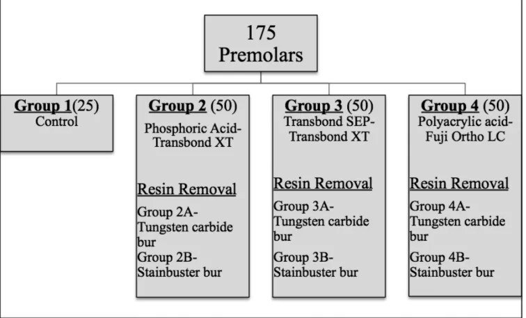
Of the 175 teeth, 25 served as control specimens. The remaining were randomly assigned to three experimental groups (n 5 50 each) with respect to the adhesive tested (Figure 1). The following adhesive protocols were employed:
Group 1 (control). The enamel surfaces were left untreated and were only subjected to color assess-ment before and after photoaging.
Group 2. Enamel was etched with 37% orthophos-phoric acid for 15 seconds, rinsed with air-water spray for 20 seconds, and air dried for 10 seconds. Transbond XT Adhesive Primer (3M Unitek, Monrovia, Calif) was used in conjunction with Transbond XT Adhesive Resin (3M Unitek) according to the manu-facturer’s instructions for bonding metal brackets with 0.018-inch slots (Ormco; Sybron Dental Specialties, Glendora, Calif). Following removal of excess adhe-sive from the margins, light curing was performed with an LED source (3M Elipar S10; 3M Unitek) for 10 seconds.
Group 3. A self-etch adhesive system (Transbond Self-Etching Primer [SEP]; 3M Unitek) was used in conjunction with Transbond XT Adhesive Resin as with group 2.
Group 4. Following pretreatment of enamel surfaces with 20% polyacrylic acid, the brackets were bonded with light-cured resin-modified glass ionomer cement (RMGIC; Fuji Ortho LC; GC Corp, Tokyo, Japan). The cement was light cured for 20 seconds from both the mesial and distal aspects.
Before photoaging, the bonded specimens were kept in distilled water at room temperature for 24 hours (Figure 2).
Color Assessment
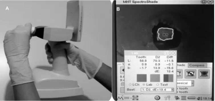
Color assessment was performed three times in the experimental groups and two times in the control group. Measurements were made from the rectangular area in the middle of the adhesive tags using a handheld spectrophotometer (SpectroShade Micro; MHT, Verona, Italy; Figure 3). Before each measure-ment, the spectrophotometer was calibrated. For each
specimen, three measurements were made in order to minimize the margin of error and calculate the average of three measurements. Color evaluation was made in accordance with the CIE (Commission Internationale de l’Eclairage) L*a*b* color system (lightness, red/ green, and blue/yellow). The following formula was used for color comparisons:
DE2{1~ ðDLÞ2zðDaÞ2zðDbÞ2 h i1 2 ~hðL2{L1Þ2zða2{a1Þ2zðb2{b1Þ2i 1 2
The D values calculated in this study were as follows:
Figure 2. Crown specimens embedded in acrylic blocks, with custom adhesive tags to standardize the area of color assessment. Figure 1. Schematic explanation of the four groups investigated.
DE 1: The difference between the values obtained at the beginning of treatment (before adhesive proce-dures) and after cleaning the surfaces (baseline-debonding). Clinically, this value indicates the color change throughout the orthodontic treatment.
DE 2: The difference between the values at the beginning of the treatment and the final values (baseline-ageing). Clinically, this value indicates the color change that occurred both during and after the orthodontic treatment.
DE 3: The difference between the values obtained after cleaning the surfaces and the final values (debonding-ageing), and clinically indicates the color-ing that occurs after the treatment.
In the control group, color measurement was conducted at the beginning and after photoaging. Thus, only the DE 1 value was calculated. In the experiment groups, tooth colors were measured before adhesive procedures, after cleaning of the enamel surfaces, and after the second photoaging. Thus, three D values were calculated.
Photoaging Procedure
Photoaging was performed in order to stimulate internal discoloration. This procedure induces aging equivalent to exposure to sun irradiation in Central Europe for 30 days.16For this purpose, the specimens
were placed in a photoaging device (Atlas Suntest CPS+; Atlas Inc, Geluhausen, Germany) with the
power configured at 50,000 kJ/m2
. Photoaging was achieved by continuous exposure of specimens to 135,000 Lux light at 400 nm for 24 hours. Photoaging
was applied once to the control group and twice to the experimental groups.
Debonding and Resin Removal
After photoaging, the brackets were removed with a debonding plier (Chifa; HPDR135.140 140; TMX Medical Instruments, Matick, Mass). In each experi-mental group, debonded specimens were divided into two subgroups (A and B; n 5 25 each) for testing the efficacy of the type of bur for cleaning residue.
In subgroups 2A, 3A, and 4A, residuals were cleaned with 12-blade tungsten carbide burs (Busch, Du¨sseldorf, Germany). In subgroups 2B, 3B, and 4B, cleaning was performed with Stainbuster composite burs (Abrasive Technology, Lewis Center, Ohio) reinforced with glass fiber and zirconium. The burs in both groups were mounted on a low-speed, water-cooled contra-angle, and cleaning was performed under loupe magnification (3.33) to simulate clinical conditions.
Statistical Analysis
The distribution of variables and the homogeneity of the variances were checked with Shapiro-Wilk and Levene tests. Descriptive statistics were indicated as the median and interquartile range. The reliability of the L, a, and b measurements at baseline (T0), after adhesive residue removal (T1), and after additional photoaging (T2) were determined with the intraclass correlation coefficient (ICC) at a 95% confidence interval.
The Kruskal-Wallis test and the Mann-Whitney U-test were used with Bonferroni correction to compare the effects of the adhesive systems and cleaning methods on DE 1, DE 2, and DE 3 median values. The differences between the three DE median values among the cleaning methods and the adhesive system subgroups were evaluated using the Wilcoxon signed-ranks test with Bonferroni correction.
RESULTS
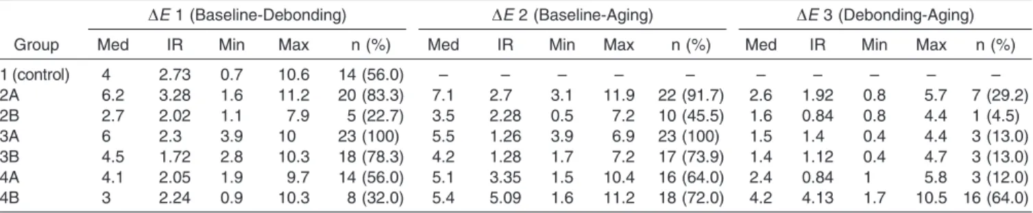
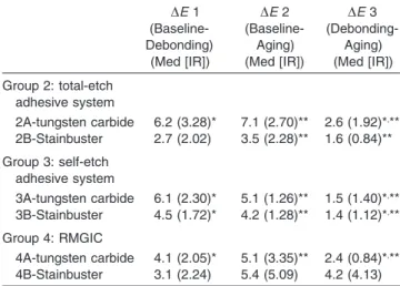
Eight teeth had enamel cracks after debonding and were excluded from the study. For each measurement, the ICC values indicating the reliability of the L, a, and b measurements repeated at T0, T1, and T2 were above 99% (Table 1). The median, interquartile range, and minimum-maximum DE values as well as the number and percentage of DE values that are over the clinical threshold (DE 5 3.7) are presented in Table 2. The color change was similar for the DE 1 and DE 2 values in groups 2A, 3A, 3B, and 4A, and both were significantly higher than that of DE 3 (Table 3). There was no significant difference between the DE 1 and DE 3 values in group 2B, whereas the DE 2 value was significantly higher than that of DE 3. No significant difference was observed between the D values in group 4B (Table 3).
In both the etch-and-rinse and the SEP groups, the color change observed with the tungsten carbide bur was significantly higher than that obtained with the Stainbuster bur (Table 4), with the former being significantly higher than that of the control. Within the RMGIC subgroups, the amount of change for the DE 1
value was similar for both the cleaning methods and the control group (Table 4).
Cross-comparisons of the DE 1, DE 2, and DE 3 levels with respect to the adhesive systems and cleaning methods are presented in Table 5. Within the etch-and-rinse subgroups, the color change observed at the DE 1, DE 2, and DE 3 levels in subgroup 2A were significantly higher than those of 2B. Within the SEP subgroups, the color change DE 1 and DE 2 levels in subgroup 3A were significantly higher than those of subgroup 3B. However, there was no significant difference between the subgroups at level DE 3. Within the RMGIC subgroups, the color change at level DE 3 in group 4B was higher than that of subgroup 4A. However, there was no significant difference between the subgroups at levels DE 1 and DE 2 (Table 5).
DISCUSSION
The CIE L*a*b* system is considered to be the standard color space, and the mathematical magni-tude of color changes is indicated as DE.17,18 The
human eye has restricted capability to see such differences and cannot perceive DE values below 1.19
The DE values between 2 and 3.7 represent the clinically perceivable but acceptable range of differ-ences. It has been reported that DE values of 3.7 and higher cannot be accepted under clinical conditions.20
Therefore, as with previous studies, the DE threshold value was accepted as 3.7 units herein.4,10–15
Enamel discoloration after orthodontic treatment is often overlooked in daily practice. According to
Table 1. Intraclass Correlation Coefficients and 95% Confidence Intervals for Repeatability of theL, a, and b Measurements at T0, T1, and T2a
Variables T0 (Mean [Min–Max]) T1 (Mean [Min–Max]) T2 (Mean [Min–Max])
L 0.9954 (0.9941–0.9965) 0.9988 (0.9984–0.9991) 0.9985 (0.9981–0.9989)
a 0.9930 (0.9909–0.9946) 0.9946 (0.9928–0.9960) 0.9962 (0.9951–0.9971)
b 0.9953 (0.9939–0.9964) 0.9986 (0.9982–0.9990) 0.9992 (0.9990–0.9994)
aL indicates lightness; a, red/green; b, blue/yellow; T0, baseline; T1, after adhesive residue removal; T2, after additional photoaging; Min,
minimum; Max, maximum.
Table 2. The Median, Interquartile Range, and the Minimum and Maximum DE Values of the Groupsa
Group
DE 1 (Baseline-Debonding) DE 2 (Baseline-Aging) DE 3 (Debonding-Aging)
Med IR Min Max n (%) Med IR Min Max n (%) Med IR Min Max n (%)
1 (control) 4 2.73 0.7 10.6 14 (56.0) – – – – – – – – – – 2A 6.2 3.28 1.6 11.2 20 (83.3) 7.1 2.7 3.1 11.9 22 (91.7) 2.6 1.92 0.8 5.7 7 (29.2) 2B 2.7 2.02 1.1 7.9 5 (22.7) 3.5 2.28 0.5 7.2 10 (45.5) 1.6 0.84 0.8 4.4 1 (4.5) 3A 6 2.3 3.9 10 23 (100) 5.5 1.26 3.9 6.9 23 (100) 1.5 1.4 0.4 4.4 3 (13.0) 3B 4.5 1.72 2.8 10.3 18 (78.3) 4.2 1.28 1.7 7.2 17 (73.9) 1.4 1.12 0.4 4.7 3 (13.0) 4A 4.1 2.05 1.9 9.7 14 (56.0) 5.1 3.35 1.5 10.4 16 (64.0) 2.4 0.84 1 5.8 3 (12.0) 4B 3 2.24 0.9 10.3 8 (32.0) 5.4 5.09 1.6 11.2 18 (72.0) 4.2 4.13 1.7 10.5 16 (64.0)
aMed indicates median; IR, interquartile range; Min, minimum; Max, maximum. 2A, and-rinse adhesive/tungsten carbide burs; 2B,
etch-and-rinse adhesive/Stainbuster burs; 3A, self-etch adhesive/tungsten carbide burs; 3B, self-etch adhesive/Stainbuster burs; 4A, resin-modified glass ionomer cement (RMGIC)/tungsten carbide burs; 4B, RMGIC/Stainbuster burs. For each DE value, the columns marked ‘‘n (%)’’ indicate the number and percentage of DE values of groups that are over the clinic threshold (DE 5 3.7).
Karamouzos et al.,10 the optical characteristics of
enamel are changed during orthodontic treatment, with the color change being affected by several factors. External coloring occurs as a result of superficial absorption of food pigments, while internal coloring occurs during aging. In the present study, photoaging was performed to stimulate internal color-ing. A color change above the threshold (ie, DE 5 3.7) observed in 56% of the control specimens confirms the efficacy of this method. As explained previously,14,15,21
the lack of saliva, the food coloring, and the inability to simulate the mechanic abrasion caused by brushing are the limitations of this methodology. In the present study, the magnitude of color change was greater during orthodontic treatment than after treatment, with the latter being below the threshold. These findings corroborate those of Eliades et al.4 and Jahanbin et
al.,15 who observed the highest color change after
debonding.
Tungsten carbide burs are known to produce minimal damage to the enamel during removal of adhesive residue.22–24Fiber-reinforced composite burs require a
longer time to remove adhesive remnants after debond-ing but may offer the advantage of providdebond-ing a smoother
surface compared to tungsten carbide.14,25 In the
present study, the total color changes observed in the etch-and-rinse and SEP groups were significantly lower in the Stainbuster subgroups than in the tungsten carbide group. This finding can be explained by the fact that Stainbuster burs provide a smoother enamel surface and increase light reflection.14,25
In the present study, the finding that the etch-and-rinse/tungsten carbide bur group exhibited the highest total color change can be explained by the occurrence of thicker and longer resin extensions formed after phosphoric acid etching.26
Accordingly, this combina-tion may not be suitable for clinical use. Compared to the etch-and-rinse adhesive, the lesser extent of total color change observed in the self-etch/tungsten carbide bur combination strongly suggests shorter resin extensions are produced by the SEP,27–29which
in turn causes less discoloration.30As for the RMGIC,
the changes observed after adhesive removal by both types of burs were low, confirming that the RMGIC did not produce resin extensions and that the bond between the enamel and the cement was predomi-nantly chemical rather than micromechanical.26
How-ever, with regard to the total change (ie, discoloration after the orthodontic treatment), it was interesting to observe that the RMGIC/Stainbuster group exhibited the only value among the test groups that was above the clinic threshold (DE 5 3.7). This unexpected result suggests that RMGIC residuals that are invisible to the eye at clinical magnification may still exist on the enamel surface after cleaning. Coupled with the unfavorable color stability of RMGIC,31these residuals
may lead to clinically unacceptable discoloration values as observed herein. Further studies using elemental analysis should be carried out to confirm this assumption. However, based on the present
Table 3. Comparisons Between the DE 1, DE 2, and DE 3 Levels Within the Adhesive Systems and Cleaning Methodsa
DE 1 (Baseline-Debonding) (Med [IR]) DE 2 (Baseline-Aging) (Med [IR]) DE 3 (Debonding-Aging) (Med [IR]) Group 2: total-etch adhesive system 2A-tungsten carbide 6.2 (3.28)* 7.1 (2.70)** 2.6 (1.92)*,** 2B-Stainbuster 2.7 (2.02) 3.5 (2.28)** 1.6 (0.84)** Group 3: self-etch adhesive system 3A-tungsten carbide 6.1 (2.30)* 5.1 (1.26)** 1.5 (1.40)*,** 3B-Stainbuster 4.5 (1.72)* 4.2 (1.28)** 1.4 (1.12)*,** Group 4: RMGIC 4A-tungsten carbide 4.1 (2.05)* 5.1 (3.35)** 2.4 (0.84)*,** 4B-Stainbuster 3.1 (2.24) 5.4 (5.09) 4.2 (4.13)
aMed, median; IR, interquartile range; RMGIC, resin-modified
glass ionomer cement.
* The difference between DE 1 and DE 2 is statistically significant (P , .001).
** The difference between DE 1 and DE 3 is statistically significant (P , .001).
Table 4. Comparisons Between the Adhesive System and the Cleaning Method With Respect to the DE 1 (Baseline-Debonding) Valuea
Control (Med [IR]) Total Etch (Med [IR]) Self Etch (Med [IR]) RMGIC (Med [IR]) P* 4.0 (2.73)ABEab
Tungsten carbide 6.2 (3.28)ACac 6.1 (2.30)BDac 4.1 (2.05)CD ,.001
Stainbuster 2.7 (2.02)EFbc 4.5 (1.72)DFc 3.1 (2.24)D ,.001
P** ,.001 ,.001 .064
aMed, median; IR, interquartile range; RMGIC, resin-modified glass ionomer cement.
* Comparisons made between the control group and the adhesive system groups among the cleaning methods are considered statistically significant forP , .025 according to the Kruskal-Wallis test with Bonferroni correction; ** comparisons made between the control group and the cleaning methods among the adhesive systems are considered statistically significant forP , .017 according to the Kruskal-Wallis test with Bonferroni correction. In each row, the difference between groups that are indicated with the same uppercase letter is statistically significant (P , .025); in each column, the difference between groups that are indicated with the same lowercase letter is statistically significant (P , .017).
results, the use of Stainbuster burs cannot be recom-mended for removal of RMGIC.
CONCLUSIONS
N Discoloration occurs on enamel during and after
orthodontic treatment. Both the orthodontic adhesive systems and the burs used to remove their residuals on tooth surfaces are responsible for this effect. N The highest color change was observed in the
etch-and-rinse/tungsten carbide bur group. This combi-nation is not recommended for clinical use in terms of enamel discoloration.
N When the brackets are bonded with the
etch-and-rinse or SEP systems, cleaning the adhesive residuals with Stainbuster burs is recommended for minimal color change. For the RMGIC, tungsten carbide burs may provide less enamel discoloration in the long run.
REFERENCES
1. Bishara SE, Trulove TS. Comparisons of different debond-ing techniques for ceramic brackets: an in vitro study. Part II. Findings and clinical implications.Am J Orthod Dentofacial Orthop. 1990;98:263–273.
2. Quirynen M, Bollen CML. The influence of surface rough-ness and surface-free energy on supra- and subgingival plaque formation in man. A review of the literature.J Clin Periodontol. 1995;22:1–14.
3. Van Waes H, Matter T, Krejci I. Three-dimensional measurement of enamel loss caused by bonding and debonding of orthodontic brackets.Am J Orthod Dentofacial Orthop. 1997;112:666–669.
4. Eliades T, Kakaboura A, Eliades G, Bradley TG. Compar-ison of enamel colour changes associated with orthodontic bonding using two different adhesives.Eur J Orthod. 2001; 23:85–90.
5. Kinch AP, Taylor H, Warltier R, Oliver RG, Newcombe RG. A clinical study of amount of adhesive remaining on enamel after debonding, comparing etch times of 15 and 60 seconds. Am J Orthod Dentofacial Orthop. 1989;95: 415–421.
6. Inokoshi S, Burrow MF, Kataumi M, Yamada T, Takatsu T. Opacity and color changes of tooth-colored restorative materials.Oper Dent. 1996;21:73–80.
7. Hodges SJ, Spencer RJ, Watkins SJ. Unusual indelible enamel staining following fixed appliance treatment. J Orthod. 2000;27:303–307.
8. Bulut T, Turkun M, Turkun LS, Isıksal E. Evaluation of the shear bond strength of 3 curing bracket bonding systems combined with an antibacterial adhesive. Am J Orthod Dentofacial Orthop. 2007;132:77–83.
9. Ireland AJ, Hosein I, Sherriff M. Enamel loss at bond-up, debond and clean-up following the use of a conventional light-cured composite and a resin-modified glass polyal-kenoate cement.Eur J Orthod. 2005;27:413–419.
10. Karamouzos A, Athanasiou AE, Papadopoulos MA, Kolo-kithas G. Tooth-color assessment after orthodontic treat-ment: a prospective clinical trial. Am J Orthod Dentofacial Orthop. 2010;138:537.e1–537.e8.
11. Eliades T, Gioka C, Heim M, Eliades G, Makou M. Color stability of orthodontic adhesive resins.Angle Orthod. 2004; 74:391–393.
12. Faltermeier A, Rosentritt M, Reicheneder C, Behr M. Discoloration of orthodontic adhesives caused by food dyes and ultraviolet light.Eur J Orthod. 2008;30:89–93.
13. Corekci B, Irgın C, Malkoc S, Ozturk B. Effects of staining solutions on the discoloration of orthodontic adhesives: an in-vitro study. Am J Orthod Dentofacial Orthop. 2010;138: 741–746.
14. Trakyalı G, Ozdemir FI, Arun T. Enamel colour changes at debonding and after finishing procedures using five different adhesives.Eur J Orthod. 2009;31:397–401.
15. Jahanbin A, Ameri H, Khaleghimoghaddam R. Effects of adhesive types on enamel discolouration around orthodontic brackets.Aust Orthod J. 2009;25:19–23.
16. Radlanski RJ. A new carbide finishing bur for bracket debonding.J Orofac Orthop. 2001;62:296–304.
Table 5. Cross-Comparisons of the DE 1, DE 2, and DE 3 Levels With Respect to the Adhesive Systems and Cleaning Methodsa
Total-Etch (Group 2) (Med [IR]) Self-Etch (Group 3) (Med [IR]) RMGIC (Group 4) (Med [IR]) P* DE 1 (baseline-debonding) Tungsten carbide 6.2 (3.28)*** 6.1 (2.30)**** 4.1 (2.05)***,**** ,.001 Stainbuster 2.7 (2.02)***** 4.5 (1.72)****,***** 3.1 (2.24)d ,.001 P** ,.001 ,.001 .023 DE 2 (baseline-aging) Tungsten carbide 7.1 (2.70)***,***** 5.1 (1.26)***** 5.1 (3.35)*** .002 Stainbuster 3.5 (2.28)*** 4.2 (1.28) 5.4 (5.09)*** .005 P** ,.001 ,.001 .347 DE 3 (debonding-aging) Tungsten carbide 2.6 (1.92) 1.5 (1.40) 2.4 (0.84) .009 Stainbuster 1.6 (0.84)*** 1.4 (1.12)**** 4.2 (4.13) ***,**** ,.001 P** ,.001 .668 ,.001
aMed, median; IR, interquartile range; RMGIC, resin-modified glass ionomer cement.
* Comparisons between the adhesive systems among the cleaning methods are considered statistically significant forP , .0083 according to Kruskal-Wallis test with Bonferroni correction; ** comparisons between the cleaning methods among the adhesive systems are considered statistically significant forP , .0056 according to Mann-Whitney U-test with Bonferroni correction; *** the difference between the total-etch group and the RMGIC group is statistically significant (P , .0083); **** the difference between the self-etch group and the RMGIC group is statistically significant (P , .001); ***** the difference between the total-etch group and the self-etch group is statistically significant (P , .0083).
17. Wee AG, Monaghan P, Johnston WM. Variation in color between intended and matched shade and fabricated shade of dental porcelain.J Prosthet Dent. 2002;87:657–666. 18. Hasegawa A, Ikeda I, Kawaguzhi S. Color and translucency
of in vivo natural central incisors.J Prosthet Dent. 2000;83: 418–423.
19. Johnston WM, Kao EC. Assessment of appearance matches by visual observation and clinical colorimetry. J Dent Res. 1989;68:819–822.
20. Ruyter IE, Nilner K, Moller B. Color stability of dental composite resin materials for crown and bridge veneers. Dent Mater. 1987;3:246–251.
21. Joo HJ, Lee YK, Lee GY, Kim TJ, Lim YK. Influence of orthodontic adhesives and clean-up procedures on the stain susceptibility of enamel after debonding. Angle Orthod. 2011;81:334–340.
22. Pus MD, Way DC. Enamel loss due to orthodontic bonding with filled and unfilled resins using various clean-up techniques.Am J Orthod. 1980;77:269–283.
23. Rouleau BD JR, Marshall GW JR, Cooley RO. Enamel surface evaluations after clinical treatment and removal of orthodontic brackets. Am J Orthod Dentofacial Orthop. 1982;81:423–426.
24. Zachrisson BU, Artun J. Enamel surface appearance after various debonding techniques. Am J Orthod. 1979;75: 121–137.
25. Karan S, Kırcelli BH, Tasdelen B. Enamel surface rough-ness after debonding. Angle Orthod. 2010;80:1081– 1088.
26. Fjeld M, Øgaard B. Scanning electron microscopic evalua-tion of enamel surfaces exposed to 3 orthodontic bonding systems. Am J Orthod Dentofacial Orthop. 2006;30: 575–581.
27. Hourichi S, Kaneko K, Mori H, et al. Enamel bonding of self-etching and phosphoric acid-self-etching orthodontic adhesives in simulated clinical conditions: debonding force and enamel surface.Dent Mater J. 2009;28:419–425.
28. Øgaard B, Fjeld M. The enamel surface and bonding in orthodontics.Semin Orthod. 2010;16:37–48.
29. Drummond JL, Roth JR, Al-Turki L, Evans CA, Montasser MA. Rebonding of orthodontic brackets. Part II, an XPS and SEM study.Angle Orthod. 2008;78:537–544.
30. Campbell PM. Enamel surfaces after bracket debonding. Angle Orthod. 1995;65:103–110.
31. Sidhu SK. Clinical evaluations of resin-modified glass ionomer restorations.Dent Mater. 2010;26:7–12.