T.C.
ALTINBAS UNIVERSITY
Institute of Graduate Studies
Department of Orthodontics
EVALUATION OF SHEAR BOND STRENGTH AND
ADHESIVE REMNANT INDEX OF TWO
ORTHODONTIC COLOR CHANGE ADHESIVES
SYSTEMS ON BONDING OF METAL BRACKETS
Muayad Muqdad Ghaidan ALBAYATI
Master of Science
Supervisor
Prof. Dr. S. İlhan RAMOĞLU
EVALUATION OF SHEAR BOND STRENGTH AND
ADHESIVE REMNANT INDEX OF TWO ORTHODONTIC
COLOR CHANGE ADHESIVES SYSTEMS ON BONDING
OF METAL BRACKETS
by
MUAYAD MUQDAD GHAIDAN ALBAYATI
Department of Orthodontics
Submitted to the Institute of Graduate Studies in partial fulfillment of the requirements for the degree of
Master of Science in Orthodontics
ALTINBAS UNIVERSITY 2021
The thesis titled “EVALUATION OF SHEAR BOND STRENGTH AND ADHESIVE
REMNANT INDEX OF TWO ORTHODONTIC COLOR CHANGE ADHESIVES SYSTEMS ON BONDING OF METAL BRACKETS” prepared and presented by
“Muayad Muqdad Ghaidan ALBAYATI” was accepted as a Master of Science Thesis in Orthodontics.
Prof. Dr. S. İlhan RAMOĞLU Supervisor Thesis Defense Jury Members:
Prof. Dr. Korkmaz SAYINSU
ALTINBAS UNIVERSITY
School of Dentistry
__________________
Prof. Dr. S. İlhan RAMOĞLU
ALTINBAS UNIVERSITY
School of Dentistry
__________________
Asst. Prof. Dr. Ufuk OK
GELISIM UNIVERSITY
School of Dentistry
__________________
I certify that this thesis satisfies all the requirements as a thesis for the degree of Master of Science.
Approval Date of Institute of Graduate Studies: ____/____/____
I hereby declare that all information in this document has been obtained and presented in accordance with academic rules and ethical conduct. I also declare that, as required by these rules and conduct, I have fully cited and referenced all material and results that are not original to this work
ACKNOWLEDGEMENTS
I would firstly like to thank my supervisor, Prof. Dr. S. İlhan RAMOĞLU, whose expertise was invaluable in formulating the research questions and methodology. His insightful feedback pushed me to sharpen my thinking and brought my work to a higher level.
I would also like to thank Prof. Dr. Korkmaz SAYINSU (Head of Orthodontic Department), who shared his knowledge and experience by guiding me during my post-graduation in Orthodontics.
I am also very grateful for the support of Dr. Seden AKAN BAYHAN, and Dr. Pelin ACAR ULUTAŞ, as well as all my other professors.
I am also very grateful for the support of my batchmates, They helped me a lot and were very supportive. This thesis would not be possible without the help of you all.
ABSTRACT
EVALUATION OF SHEAR BOND STRENGTH AND ADHESIVE REMNANT INDEX OF TWO ORTHODONTIC COLOR CHANGE ADHESIVES SYSTEMS ON BONDING OF METAL BRACKETS
ALBAYATI, Muayad Muqdad Ghaidan, M.Sc., Orthodontics, Altinbaş University, Supervisor: Prof. Dr. S. İlhan RAMOĞLU
Date: 02/2021 Pages: 55
The aim of this study was to evaluate the shear bond strengths (SBS) and adhesive remnant index (ARI) of two color change adhesives systems, new BracePaste adhesive paste (American Orthodontics, Sheboygan, WI) and, common Transbond Plus adhesive paste (3M Unitech, Monrovia, CA, U.S.A.), in bonding of metal brackets.
Materials and methods: 60 human upper premolar teeth were divided into 2 groups, For
bonding, with both the adhesive paste same steps were followed, according to the manufacturer’s instructions. The samples were thermocycled between 5° and 55° for 500 cycles. The specimens were stored at 37° in distilled water for 24 hours before SBS testing on a universal testing machine.
Results: Statistically significant difference in the shear bond strength between the two
groups. The highest SBS was observed in the Transbond Plus with a mean range of (11.03 ± 1.41) Mpa, and the mean range of the BracePaste was (7.79 ± 1.16) Mpa. However, BracePaste SBS value is considered clinically acceptable. ARI scoring showed that most of the debondings occurred in the adhesive layer. The ARI scores of Transbond Plus and BracePaste groups did not exhibit a statistically significant difference.
Conclusion: Our results were based on in-vitro laboratory conditions, and the clinical
relevance of our findings should also be confirmed with in-vivo studies. Further clinical studies are needed to substantiate the conclusions from this investigation. Both the adhesives had sufficient SBS values for a clinical use.
ÖZET
METAL BRAKETLERİN YAPIŞTIRILMASINDA İKİ ORTODONTİK RENK DEĞİŞTİREN ADEZİV SİSTEMİNİN KESME BAĞLANMA KUVVETİ VE ARTIK ADEZİV ENDEKSİNİN DEĞERLENDİRİLMESİ
ALBAYATI, Muayad Muqdad Ghaidan, M.Sc., Ortodonti, Altinbaş Üniversitesi, Danışman: Prof. Dr. S. İlhan RAMOĞLU
Tarih: 02/2021 Sayfalar: 55
Bu çalışmanın amacı, yeni BracePaste adeziv (American Orthodontics, Sheboygan, WI) ve yaygın Transbond Plus adeziv (3M Unitech, Monrovia, CA, ABD) olmak üzere metal braketlerin yapıştırılmasında iki renk değiştiren adeziv sisteminin kesme bağlanma kuvvetleri (SBS) ve artık adeziv endeksini (ARI) değerlendirmektir.
Materyaller ve yöntemler: 60 insan üst küçük azı dişi 2 gruba ayrılmıştır. Metal ortodontik
braketler için kullanılan yapıştırma için, imalatçının talimatlarına göre her iki adeziv için pastayla aynı adımlar takip edilmiştir. Numuneler, 500 döngüde 5° ile 55° arasındaki suda ısı döngüsüne alınmıştır. Örnekler, üniversal bir test makinesinde test edilmeden önce, 24 saat süresince distile suda bekletilmiştir.
Bulgular: İki grubun SBS’si arasında istatistiksel olarak anlamlı fark bulunmuştur.
Transbond Plus (11,03 ± 1,41) Mpa, BracePaste’e göre (7,79 ± 1,16) Mpa daha yüksek SBS değerine sahiptir. Bununla birlikte BracePaste SBS değeri klinik olarak kabul edilebilirdi. ARI skorları, en fazla ayrılmanın adeziv katmanında olduğunu göstermiştir. Transbond Plus ve BracePaste gruplarının ARI skorları, istatistiksel olarak anlamlı bir fark göstermemiştir.
Sonuç: Sonuçlarımız, in-vitro laboratuvar koşullarına dayalıdır ve bulgularımızın klinik
geçerliliği in-vivo çalışmalarda da teyit edilmelidir. Bu incelemenin sonuçlarını doğrulamak için başka klinik çalışmaların yapılması gerekmektedir. Her iki adeziv klinik kullanım için yeterli SBS değeri göstermiştir.
TABLE OF CONTENTS
ABSTRACT V
ÖZET VI
TABLE OF CONTENTS VII
LIST OF TABLES VIII
LIST OF FIGURES IX
ABBREVIATIONS AND SYMBOLS X
1. INTRODUCTION 1
1.1 FEATURE, STRUCTURE AND FORMATION OF ENAMEL 3
1.1.1 Features 3
1.1.2 Structures 3
1.1.3 Formations 4
1.2 COMPOSITE STRUCTURE AND CHARACTERISTICS 4
1.2.1 Self-Cured Materials 5
1.2.2 Light-Cured Materials 6
1.3 PREPARATION OF ENAMEL SURFACE 6
1.4 ENAMEL CONDITIONING METHODS 7
1.5 BOND STRENGTH 10
1.6 THERMOCYCLING 11
1.7 ADHESIVE REMNANT INDEX (ARI) 11
2. MATERIALS AND METHODS 13
3. STATISTICAL ANALYSIS 22
4. RESULTS 23
5. DISCUSSION 25
6. CONCLUSION 32
LIST OF TABLES
Table 1: Mean difference of shear bond strength values in (MPa)&(N) and descriptive
statistics. 23
Table 2: Correlation between the two groups. 23 Table 3: Distribution frequency and percentages of ARI scores. 24
LIST OF FIGURES
Figure 1: All the specimens, a total of 60 samples was included. 13 Figure 2: Teeth after cutting of the roots. 14 Figure 3: Teeth mounted in acrylic. 15 Figure 4: Pumice and dental brush rubber cup. 15 Figure 5: Acid etching with 37 % Phosphoric Acid. 16
Figure 6: Valo light device. 16
Figure 7: Mini-Master Brackets, (American Orthodontics, Sheboygan, WI). 17 Figure 8: BracePaste CCA and MTP Primer (American Orthodontics, Sheboygan, WI). 18 Figure 9: Transbond Plus CCA and XT primer (3M Unitech, Monrovia, U.S.A.). 18 Figure 10: Transbond Plus adhesive group. 19 Figure 11: BracePaste color Change adhesive group. 19 Figure 12: Specimens secured for debonding procedure, 20 A: Specimen undergoin SBS Test, B: Specimen after debonding. 20 Figure 13: Instron Universal Testing Machine, 21 (Model 3345, Instron Inc., Canton, Massachusetts, U.S.A). 21 Figure 14: Bar graph of SBS in MPa for BracePaste and Transbond plus CCAs. 24
ABBREVIATIONS AND SYMBOLS
SBS: Shear Bond Strength ARI: Adhesive Remnant Index MPa: Mega Pascal
UV: Ultraviolet N: Number Kg: Kilogram gr: Gram F: Force nm: Nanometer N: Newton mm: Millimeter cm: Centimeter μm: Micrometre
EVALUATION OF SHEAR BOND STRENGTH AND
ADHESIVE REMNANT INDEX OF TWO ORTHODONTIC
COLOR CHANGE ADHESIVES SYSTEMS ON BONDING
OF METAL BRACKETS
1. INTRODUCTION
Increasing knowledge related to bonding of adhesives to enamel surface in dentistry has caused developments in orthodontics as in other fields. With the advancement of new materials science of orthodontics is rapidly evolving. Mainly in the field of bonding, many new substances such as new adhesives, sophisticated base designs, and new bracket materials, faster and better efficient curing methods, self-etching primers, fluoride-releasing agents as well as sealants adhesive that were introduced for bonding the orthodontic brackets (1,2).
In fixed orthodontic treatments brackets and tubes used to transfer the applied forces to the teeth, previously performed by welding of orthodontic brackets to stainless steel bands which were cemented to all teeth. This procedure was impractical and took long chair time (1). In addition, it could harm and damage the periodontal, dental tissue and oral hygiene. Bonding of orthodontic attachments is widely used nowadays, not only in fixed appliances but also with removable appliance (3) and became universally applicable technique in orthodontics in the nineties of the last century (4).
Buonocore (5), in the year 1955, used 85% phosphoric acid and discovered the ability of acid conditioning of the enamel to increase adherence of acrylic resin which ultimately revolutionized the practice of restorative and preventive dentistry which greatly influenced the development of bonding in orthodontics.
In 1964, Newman (6), introduced the technique of bonding orthodontic attachments directly to tooth surfaces.
This development resulted in improving in orthodontic treatment, including greater comfort for the patient, elimination of pre-treatment separation, decreased gingival irritation and protecting periodontal health, easier oral hygiene, improved esthetics, shortening treatment period reduced chair side time and the risk of decalcification (7,8).
The proper removal of excess adhesive material from the tooth surface during placement of bracket, which has been considered an important role in orthodontics by bonding fixed appliances to tooth surfaces (9,10). Similarity in color and shade makes it difficult to clearly delineate the enamel adhesive interface, which could result in removal of the adhesive incompletely during flash clean-up (11-14). furthermore, still there are two major concerns about light cured adhesives,
I. The removal of excess adhesive material in an improper manner from the surface of the tooth surface while bracket placement, has been considered a potential risk for subsequent gingival irritation and white spot lesions on the surface of enamel, II. As closely as possible, restoring of the enamel, to a flawless and intact surface after
removal of the bracket (9,10).
The color change adhesives are a newly development in the orthodontic adhesive materials. They provide a distinct color and contrast and can easily be differentiated from enamel while bracket placement and adhesive removal, with the purpose of facilitating the discrimination between the adhesive material and enamel (10). Color change adhesives are becoming popular in orthodontics due to promising features and studies which prove they can be used in orthodontic practice efficiently (9,10,15-18).
The purpose of this work was to evaluate the shear bond strengths (SBS) and adhesive remnant index (ARI) of two color change adhesives (CCAs) systems, new BracePaste adhesive paste (American Orthodontics, Sheboygan, WI) and, common Transbond Plus adhesive paste (3M Unitech, Monrovia, CA, U.S.A.), on bonding of metal brackets.
1.1 FEATURE, STRUCTURE AND FORMATION OF ENAMEL 1.1.1 Features
The enamel is the hardest substance in the human body, consisting of 96% mineral mainly in the form of hydroxyapatite and 4% water and organic material one fully formed (3). Hydroxyapatite is the primary mineral, which is crystalline calcium phosphate. Enamel is formed on the tooth while the teeth develop inside the gums, before that it explodes in the mouth (4).
In humans, enamel difference in thickness on the surface of the teeth, often more thick on the cusp, up to 2.5 mm, and less thick on its border with cement the surface is not good shape at cement enamel junction (CEJ).The normal colour of enamel ranges from light yellow to Grey (bluish) white. On the edges of the teeth where there is no dentine underlying the enamel, the colour sometimes has white or slightly transparent tone, and can be easily seen on the upper incisors. As the enamel is semi-transparent, enamel strongly affects the appearance, the large amount of minerals in the enamel accounts do not only for its strength but also for its fragility. However, enamel does not contain collagen, as found in other hard tissues like dentine and bones, but the enamel contain two unique classes of proteins: Amelogenins and Enamelins (19).
1.1.2 Structures
The basic enamel unit is known as an enamel rod. Measuring 4-8 microns in diameter, the enamel rod, formally called the enamel prism, is a tightly packed block of hydroxyapatite crystals in a structured pattern. It is best to compare with a keyhole with a head or head oriented toward the crown of the tooth, or the tail, oriented toward the tooth root in the cross section (19).
The two enamel cells (cells that begin forming enamel) and Tomes processes (a cone-shaped process at the distal secretory end of the ameloblasts) affect the crystals. Enamel crystals in the head of the enamel rod are parallel oriented to the long axis of the rod. When found in the tail of the enamel rod, the crystal orientation diverges slightly (65 degrees) from the long axis (19).
The arrangement of enamel rods is better understood than its internal structure. Enamel rods are found in rows along the tooth and within each row, and the long axis of the enamel rod is generally perpendicular to the primary dentine.
Enamel rods near the cement enamel junction (CEJ) tilt slightly towards the root of the tooth in permanent teeth. Understanding the direction of enamel is very important in restorative dentistry, because unsupported enamel of dentine is vulnerable to break. The area around the enamel rod is known as interred enamel. Inter enamel rod has the same configuration as enamel rod (19).
1.1.3 Formations
Enamel formation also known as formation of amylogenesis, occurs after the creation of the first dentine by enamel cells which known as Ameloblasts, an amylogenesis is two phases (3).
The first phase produces partially mineralized (about 30%) enamel. Once the full width of this enamel is deposited, the second phase includes large flow of additional materials and water to achieve more than 96% mineral contain. This mineral flow makes the shape of the crystals, during the first phase grow wider and thicker. This complex process is under cellular control, and associated cells undergo major morphological changes throughout amylogenesis, reflecting its physiological evolution (19).
1.2 COMPOSITE STRUCTURE AND CHARACTERISTICS
Dental composites play a vital role in orthodontics. Dentists are able to choose from composites having a wide range of viscosities for different clinical applications, from the most fluid resin fissure sealants through flowable, microfill and hybrid composite formulations, to the composites having high and low viscosity. There are definite limitation and advantages of each category. The proprietary materials within each category differ significantly in handling, mechanical and physical properties (10).
During the past 15 years, a huge number of dental composite products have appeared, having common ans simple characteristics. They are combinations of silane-coated inorganic filler
particles with dimethacrylate resin, either urethane dimethacrylate (UDEMA) or bisphenol-A-glycidyl methacrylate (BISGMA).
In certain cases, to lower the viscosity an amount of lower molecular weight monomer such as triethylene glycol dimethacrylate (TEGDMA) is introduced. The filler particles used are either barium silicate glass, quartz or zirconium silicate, usually combined with 5-10% weight of minutely sized 0.04 μm particles of colloidal silica. Modern dental composite materials are hence a blend of glass or ceramic particles dispersed in a photo-polymerizable synthetic organic resin matrix. The polymer materials are blended together with the finely divided inorganic material such as a barium aluminosilicate glass or other glass compositions having an effective amount of radiopaque oxide that renders the resultant glass radiopaque to x-rays (20).
Filled dental restorative materials were also used as orthodontic adhesives (21). As mentioned above, these materials, consist of an organic diacrylate (BIS-GMA), a coupler (silane), and a higher percentage content of inorganic filler such as quartz and silica. The inorganic filler makes the material more abrasion resistant, also increasing the SBS values as well as decreasing the coefficient of thermal expansion to values closer to those of enamel in order to prevent long term microleakage (22).
These days, further additions of certain chromatic agents into composite resin adhesive are being done which results in the change of color during setting and turns in being translucent clear upon curing as it warms during treatment. This has an advantage of easy flash removal thus reducing the amount of plaque accumulation and decalcification and also helping patients to maintain better oral hygiene.
Polymerization type such as light-cured as well as self-cured materials form the distinctive groups of composites,
1.2.1 Self-Cured Materials
There is no mix adhesives set when one paste under light pressure is brought together with a primer fluid on the etched enamel and bracket backing or when another paste on the tooth is meant to be bonded. usually the primers are composites with minimal amount of fillings
(23). hence one adhesive component is applied to the bracket base while another is applied to the dried etched tooth and pressed tightly together in place for 30-60 sec (24).
1.2.2 Light-Cured Materials
The resins which are light cured have the advantage of extended, though not indefinite, working time. Light-cured resins used with metal brackets are usually dual-cure resins incorporating light initiators as well as a chemical catalyst. Ultraviolet light was for the first time used for curing the resins (24). Watts and Tavas, in 1979, were the first to use visible light to cure resins (13,22,25). For the orthodontist, the light cured material have an increased working time, maximum curing depth, less porosity and allows the clinician to clean the composite around the base of the bracket reducing the demineralization of the enamel through plaque accumulation (23,26,27).
1.3 PREPARATION OF ENAMEL SURFACE
Pumice prophylaxis before enamel etching is a procedure recommended by clinicians. Although acid applied to enamel surface shows effect of a thin organic layer, accumulations and deposits may reduce effectiveness of applied acid (28), therefore, providing an ideal surface for bonding, organic pellicle and debris on tooth surface should be cleaned.
The most common method is pumice and dental brush rubber cups that works at low speed, many other techniques, such as airflow and bicarbonate polishers could be used, but they can hurt tissue and contaminate surfaces (29-31). Cleaning the enamel surface with prophylaxis pastes containing fluoride or oil-based are not recommended by many clinicians, however some authors mentioed that prophylaxis pastes containing fluoride or oil-based could also be used (32). Y., Aboush et al, (33) indicated that the use of different prophylaxis pastes do not cause any changes. for cleaning the enamel surface, tooth surface needs to be washed thoroughly to remove debris from the enamel surface with compressed air/water spray after cleaning (33).
Barry et al, (34) in 1995, made a clinical investigation on the effects of pumice prophylaxis, he found no statistical difference on bond failure rates and general etched enamel surface characteristics.
Burgess et al, (35) in 2006, They reported that pumice prophylaxis has a significant effect on bond failure and noted that without prior pumice prophylaxis leads to increase in bond failure, but this material cannot be always recommended for a clinical use.
Lill et al, (36) studied the importance of pumice prophylaxis for bonding, bond strength of brackets that were bonded to teeth after surface preparation procedures, which either included or did not include prior pumice prophylaxis, was evaluated; scanning electron microscopy (SEM) was used to examine the surface characteristics of teeth that had been etched with and without prior pumice prophylaxis; and rate of bracket failure bonded with and without prior pumice prophylaxis was showed a little differences were noted in bond strength. Some specific differences, however, were noted on SEM in localized areas of the etched enamel surfaces, although these did not appear to affect the bond strength or bracket retention rates ultimately attained, In spite of that, they still suggested that the enamel surface should to be clean with pumice for achieving adequate enamel etching during orthodontic bonding procedures. Acid roughening itself is sufficient for removal of plaque and debris on the tooth surface prior to bonding according to Main et al, (37). Reisner et al, (38) reported in their studies that they had the most successful results in terms of bonding when the enamel surface was slightly abrased with carbide burs at the low speed prior to acid application.
1.4 ENAMEL CONDITIONING METHODS
Purpose of enamel roughening method is to increase surface area, obtain retentive areas, change the enamel surface from a low reactivity surface to a surface more susceptible to adhesion (39). Surface conditions can be changed with chemical, thermal and mechanical methods; as etching with acid on the enamel surface, spraying of aluminium oxide particles or laser application (40). The most common method is surface roughening or etching of enamel surface with 37% phosphoric acid, and this enamel bonding technique, known as the acid etching technique.
Acid-etch technique of the enamel surface which was developed by Buonocure, in 1955, gave a new meaning to dentistry in terms of adhesion. Acid-etching and bonding played an active role in the success of clinical results and has become usual and ordinary method of modern dentistry (40). With the use of acid-etching, approximately 10 μm of the enamel surface is resorbed and creates a microporous layer from 5 to 50 μm deep (41).
When the enamel surface is dried, a white appearance is formed and This appearance results from decalcification of the enamel and deterioration of crystal form (12,42-45). Areas of which this appearance is not occurred must be re-etched.
Three etching patterns of enamel introduced by Gwinnett, (46) in which there is predominant dissolution of the prism cores in first type, presence of predominant dissolution of the prism peripheries in the second pattern and last, in which no prism structures are evident. Two types of resin tags have been described by Bayne et al, (47). Macro-tags are formed circularly between enamel prism peripheries; micro-tags are formed at the cores of enamel prisms, where the monomer cures into a multitude of individual crypts formed where hydroxyapatite crystals have dissolved. Micro-tags probably contribute most to the strength because of their greater quantity and large surface area (48).
Some factors that influence acid etching of enamel for orthodontic bonding include the type and concentration of the acid and the duration of etching. Many studies have been published on the effect of phosphoric acid etching with the concentration of (5-65%) on SBS and found that concentration didn’t expressively affect on the shear bond strength (49-62). But most frequently used acid concentration ranges from 30% to 40% (63). A clear consensus could not be built among researchers in studies of etching duration. No drastic differences are seen in the bond strengths between 15 seconds and 60 seconds etching with phosphoric acid. If etching time is less than 10 seconds bond strength reduces significantly (59,61).
According Diedrich, (64) acid-etching with phosphoric acid causes approximately 10% reduction in the enamel layer. Due to some disadvantages, alternative acids have been tried to minimize the loss on the surface and to obtain clinically sufficient bond strength, these are maleic acid (65,66), nitric acid (67) and polyacrylic acid (68).
Etching of enamel surface with the use of 10 percent maleic acid for etching results in lower bond strength (69), some studies compared 37% phosphoric acid, they could not find a significant difference between them but they found it more appropriate to prefer maleic acid clinically (70-73).
Gardner et al, (67) compared phosphoric acid with nitric acid etching in their studies and reported that 37% phosphoric acid was much more effective on bond strength than 2.5% nitric acid on the enamel for 15, 30 and 60 seconds.
Polyacrylic acid-etching of enamel surface was first introduced as an alternative material for etching of enamel surface by Maijer and Smith, in 1979. The sulphate components in the polyacrylic acid creates activation in crystals on the enamel surface by reacting with calcium on the enamel surface (74). The bond strength values are lower compared to phosphoric acid-etching (75).
Farquhar’s et al, (76) compared polyacrylic acid and 43% phosphoric acid, and were found the bond strength values 3-4 times more in phosphoric acid compared to polyacrylic acid, that loss of polyacrylic acid on enamel surface was found 4.5 μm after removal of brackets and cleaning of tooth surface.
According to Zachrisson, (2) one of the main causes of bond failure is contamination. This contamination reduces the bond strength of adhesive materials by causing the precipitation of glycoproteins on the enamel surface during or after acid-etching (77). When contamination occurs, etching process must be repeated (78). The roughened surface must be absolutely avoided from contamination of saliva, mucous membranes, and etc for better bond strength.
Recently, lasers application offer practical advantages over traditional dental etching, it ensures the removal of tissue with high sensitivity (41). Laser etching of enamel surface causes thermal changes reaching 10 to 20 μm depth on the enamel surface depending on laser type and the energy applied to enamel surface. These changes are caused by micro explosions due to the continuous evaporation of water which is trapped in the hydroxyapatite matrix. The application of etching depends on the system used and the wavelength of the laser (70). Laser etching has been the subject to many researches whether retention of composite resins increases the same degree with conventional acid-etch techniques. While some studies suggested that laser etching caused poor bond strength (79-83), others reported that it showed bonding forces comparable to acid etching technique (84,85).
1.5 BOND STRENGTH
Direct bonded attachments were obtained to ensure that the orthodontic attachments remain attached to the teeth during treatment time without any failure (Millet and Gordon., 1994) (86). Ideally, orthodontic brackets should be able to show sufficient resistance to the forces generated by orthodontic treatment and forces by mastication during orthodontic treatment, and they can be removed without occurring any damage to the tooth surface at the end of treatment (87-89).
A maximum value of forces 60-80 kg/cm² (5.9-7.8 MPa) seems reasonable, adhesives giving tensile bond strength of approximately 30-50 kg/cm² are clinically accepted successfully in in-vitro studies (39). Ferguson et al, (90) have mentioned many factors that may influence the bond strength including the nature of the enamel surface, the conditioning procedure, the type of adhesive and the design of the bracket base itself. Orthodontic bond strength can be investigated using different test methods. Universal testing machine was used for measuring shear bond strength since it gives precise value and popularly used, this machine is capable of delivering a controlled and measured force to the bonded bracket via its moving crosshead (91).
Compressive fracture resistance test by universal testing machine is an important method used for measure the shear bond strengths of different types of orthodontic brackets bonded to extracted teeth, although it has a lot of advantages but the major disadvantage is that because in vitro shear bond strength test so does not accurately proliferate the clinical situation. While in the mouth there are multiple forces shear, tensile and torsion applied onto orthodontic brackets, whereas in vitro studies the universal testing machine is able to producing only pure debonding forces (shear, tensile or torsion) not the combination of them. Moreover, the rate of loading for the machine is stander while in vivo studies are not constant (91,92).
In spite of limitation of shear bond strength test to represent the real bond strength still continue as clinically relevant method used for comparison of bonding with different protocols and give significant awareness on bracket debonding clinically (93).
Bond strength tests can be classified as shear, tensile and torsion according to mode of load applied to break off the attachment. Since torsion and tensile is found less valid by many researchers, and shear experiments are preferred because it is easy to apply by simulating clinical situation more (94).
1.6 THERMOCYCLING
Thermocycling simulates the temperature dynamics in the oral environment, with bonding; thermocycling reduces the bond strength of orthodontic adhesives (95). Orthodontic adhesives are routinely exposed to temperature variations in oral cavity. Air temperature, humidity, and air velocity when breathing can also alter resting mouth temperature (96). Intraoral temperatures have been recorded as low as 0°C and as high as 67°C. Thermo cycling is believed to simulate oral conditions in vitro (61). While simulating the introduction of hot and cold extremes in the oral cavity it also shows the relationship of the linear coefficient of thermal expansion between tooth and restorative material (90).
1.7 ADHESIVE REMNANT INDEX (ARI)
The amount of adhesive which remains on the tooth surface or on the bracket base is an important factor for choosing an orthodontic adhesive. Although the changes in Adhesive Remnant Index (ARI) scores reflects the bond strength, bond failures within the adhesive layer or at the bracket-adhesive interface are preferred. The reason for that, need less chair time to remove and it’s easy to clean the tooth surface after debonding the brackets. Adhesives can reach 50 µm depth of the enamel surface (97) and may cause irreversible effects on the surface during removal of orthodontic attachments (98). Also, ARI scores are used to identify the site of bond failure between enamel, adhesive and bracket base. Attachment failure in two major sites, bracket base/adhesive interface and the enamel/adhesive interface have been used in most of in-vitro investigations of bond failure (99).
The bond failure can be adhesive and occur between bracket and adhesive or enamel and adhesive, cohesive failure in which it occurs in the main core of resin, or combination of both adhesive and cohesive failure (100,101). Knowing the failure mode in studies significantly makes bond strength data meaningful (102).
Powers et al, have stated that if the orthodontists know where the bond failure will occur, they should modify the debonding technique and advice patients to how to take care of their appliances (8).
Artun and Bergland et al, have introduced index system known as "Adhesive Remnant Index" (ARI) system (103), which is used for evaluating fracture sites and amount of adhesives left on the tooth after debonding.
The index score ranges from 0 to 3 as follows:
0= All of the adhesive remaining on the base of the bracket
1= more than half of the adhesive remaining on the base of the bracket 2= less than half of the adhesive remaining on the base of the bracket 3= no adhesive on the base of the bracket
In many studies, the ARI system scores have been modified or improved for a more precise evaluation (104).
2. MATERIALS AND METHODS
According to the power analysis result, which was performed to determine the number of samples, the minimum sample size was found to be 27 for each group. The analysis was performed with (G. Power 3) to test the difference between two independent group means using a one tailed test, a medium effect size (d=0.691), and alpha of 0.05. Results showed that a total sample of 54 samples with two equal sized groups of n=27 was required to achieve a power of 0.80. In this study, two groups were used, a total of 60 samples was included (Figure 1), and each group contained 30 samples.
All study was carried out in the Research Laboratory of Altinbas University, School of Dentistry, Department of Orthodontics and dentofacial Orthopedics, Istanbul, Turkey, with the permission of the Local Research Committee. Sixty premolar teeth, (first and second maxillary), were collected from patients whose teeth need to be extracted for orthodontic treatment purposes. The teeth were;
I. Intact without enamel cracks caused by forceps, II. Free of caries,
III. And have not tested with hydrogen peroxide, formalin, alcohol, or other chemical agents after extraction.
The extracted teeth were washed and immersed with distilled water in a sealed container for one to three months until testing. The distilled water which will keep the teeth were changed every week, to prevent the growth of bacteria and fungi. The roots of all premolars were then removed (Figure 2) with a high-speed diamond bur to aid in the subsequent steps.
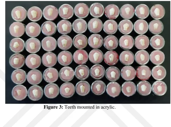
Each tooth was individually mounted in a cylinder of cold-cure acrylic resin (Vertex). The buccal surface of the crowns placed parallel to the metal edge of the rings, with at least 1mm of projection supra to the border of the cylinder (Figure 3).
Figure 3: Teeth mounted in acrylic.
In both the groups, same bracket bonding protocol was followed. Before bonding, prophylaxis was performed by using a handpiece along with rubber cup, pumice and water spray followed by rinsing with water (Figure 4).
For bonding, which is used to bond metal orthodontic brackets, with both the adhesive paste same steps were followed, according to the manufacturer’s instructions, i.e., etching with 37% phosphoric acid, rinsing, drying, priming (application of bonding agent), bracket placement with adhesive at the base of the bracket and curing. The tooth surface was etched using 37% phosphoric acid (Etching-Royale™ Pulpdent Corporatin, Watertown, USA) for 30 seconds (Figure 5), followed by rinsing and drying for 10 and 15 second; then respective bonding agents, were applied in thin layers and 3 mm away from the buccal surface of the tooth the primer cured by using Valo light cure device (Valo, Xtra power mode; 3200 mW/cm² Ultradent, SJ, UT), (Figure 6) for 3 seconds.
Figure 5: Acid etching with 37 % Phosphoric Acid.
A thin layer of adhesive was paste applied to the base of the metal bracket and the brackets were accurately pressed onto the buccal surface with a placement scaler. Once the bracket is in the correct position, the scaler was removed. The excess composite resin was removed from the margin of the bracket with a dental probe before setting. After removing excess resin, the adhesive paste was cured from the occlusal and gingival sides of the tooth for 3 seconds.
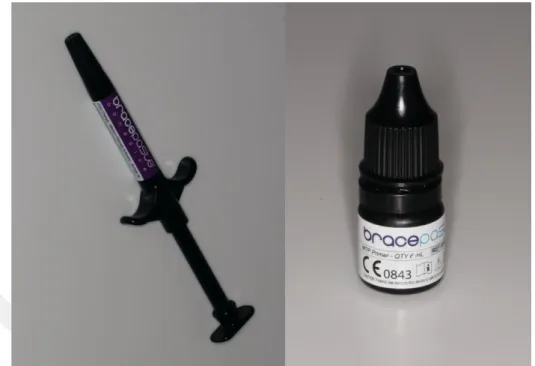
In our study, stainless steel brackets were placed to maxillary premolar teeth (0.22 slot) (Mini-master series, American Orthodontics, Sheboygan, WI), (Figure 7), Color change BracePaste adhesive material and MTP Primer (American Orthodontics, Sheboygan, WI), (Figure 8 A&B). Color change Transbond Plus adhesive material and Transbond XT primer (3M Unitech, Monrovia, U.S.A.) was used in this study (Figure 9 A&B).
Figure 8: BracePaste CCA and MTP Primer (American Orthodontics, Sheboygan, WI).
Figure 10: Transbond Plus adhesive group.
Figure 11: BracePaste color Change adhesive group.
Following bonding the brackets, all specimens of the teeth were stored in fresh distilled water for approximately 24 hours (±2 hours) before being debonded for shear bond testing SBS to ensure complete and uniform polymerization. All the samples were thermocycled between 5° and 55° for 500 cycles. During thermocycling, the dwelling time for the specimens in each well was 30 seconds. A Universal Testing Machine (Model 3345, Instron Inc., Canton, Massachusetts, USA), working at 0.5 mm/min speed, was used to deboned the
brackets from all the specimens, each mounted tooth was then placed in a universal testing machine with the bracket/tooth interface placed parallel to the blade motion. The brackets were debonded using a crosshead speed of 0.1 mm/min with the loading blade contacting between the tie wing and the bracket base as close to the base as possible (Figure 12). The maximum load was recorded and converted to shear bond strength in MPa. In order to convert force to debond brackets into stress to debond brackets, the following equation was used (105).
Stress (Megapascals N/m2) = Force (Newtons)/Area of bracket base mm2
The surface area of bracket base was calculated by taking the average sum of width and length of the bracket using a digital calipers, The bond force were recorded in Newton’s and then divided by the bracket base area, mm2, and converted to megapascals (MPa) (one MPa N/mm2).
A. B.
Figure 12: Specimens secured for debonding procedure,
Figure 13: Instron Universal Testing Machine,
(Model 3345, Instron Inc., Canton, Massachusetts, U.S.A).
After debonding, each tooth and debonded bracket was viewed under an optical stereomicroscope (SMZ 1000 Nikon; Tokyo, Japan) at 50x magnification. The Adhesive Remnant Index (ARI) score was recorded to determine where the bond failure occurred.
3. STATISTICAL ANALYSIS
Statistical analyses were realized with the help of SPSS version 17.0 software. Conformity of the variables with the normal distribution was analyzed with histogram graphs and Kolmogorov-Smirnov test. When descriptive analyses are presented, mean, standard deviation, medium values were used. Categorical variables were compared by using Pearson Chi-Square Test. While evaluating variables showing a normal distribution (parametric) among groups, Independent t-Test was used. In the analysis of measurable data with each other, Pearson Correlation Test was used. P-value below 0.05 (p<0.05) were considered as a statistically significant.
4. RESULTS
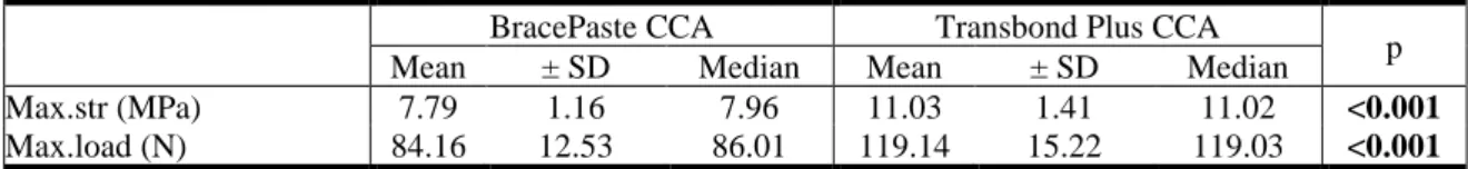
Shear bond strengths were measured for 60 samples in both experimental groups. The mean shear bond strength and standard deviation values for each group are shown in Table 1. BracePaste CCA group showed a mean SBS of 7.79 ±1.16 MPa and Transbond Plus CCA group showed a mean SBS of 11.03 ±1.41 MPa.
The t-test revealed that there was a statistically significant difference in between the SBS of the two groups Table 1. Pearson correlation showed that here was no significant correlation between the two groups Table 2.
Table 1: Mean difference of shear bond strength values in (MPa)&(N) and descriptive statistics.
BracePaste CCA Transbond Plus CCA
p Mean ± SD Median Mean ± SD Median
Max.str (MPa) 7.79 1.16 7.96 11.03 1.41 11.02 <0.001 Max.load (N) 84.16 12.53 86.01 119.14 15.22 119.03 <0.001
SD= Standard Deviation.
Table 2: Correlation between the two groups.
r p
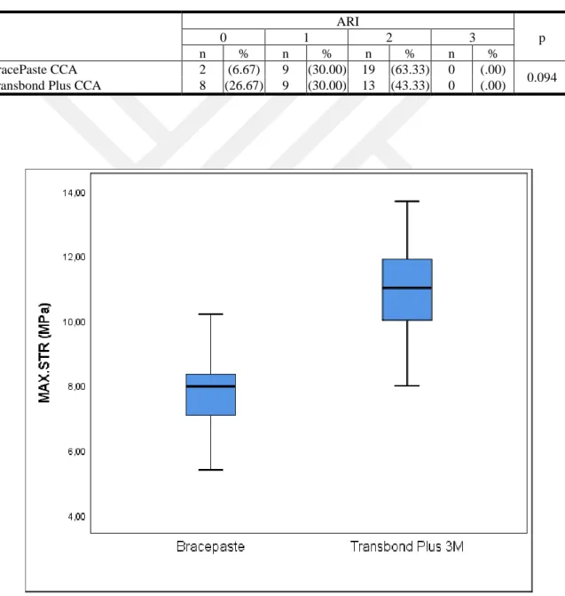
The ARI scores for adhesive remaining of the BracePaste and Transbond Plus CCAs groups are summarized in Table 3. The ARI score of 2 (i.e., more than half of the adhesive left on the tooth) of BracePaste CCA was found to be the most prevalent (63.33%), followed by Transbond Plus CCA (43.33%). Chi-square test showed that the ARI scores of Transbond Plus and BracePaste groups did not show a statistically significant difference.
Table 3: Distribution frequency and percentages of ARI scores.
ARI p 0 1 2 3 n % n % n % n % BracePaste CCA 2 (6.67) 9 (30.00) 19 (63.33) 0 (.00) 0.094 Transbond Plus CCA 8 (26.67) 9 (30.00) 13 (43.33) 0 (.00)
5. DISCUSSION
Today, there are different types of orthodontic bonding materials available like self-cure light cure, bonding material with primers, self-etch primers, color change adhesive, etc. These materials are introduced to clinics after certain initial test; one of the important test is shear bond strength to dental enamel. Bond strength of orthodontic adhesives has been the subject of study for many researchers.
The bond strength of bracket - adhesive - enamel system in orthodontic bonding varies and depends on factors such as the type of adhesive, bracket base design, enamel morphology, appliance force systems and the clinician's technique. In-vitro studies may not provide sufficient information regarding combination of forces and numerous factors involved in orthodontic treatment but they are useful as a guideline for the clinician in the selection of the bracket/adhesive system to be used in clinical settings (106).
Shorter curing times are always preferable to improve clinical outcomes and are especially advantageous for pediatric patients (107-109). In our study, The light sources used a high intensity LED. It has different curing modes, including standard mode with 1000 mW/cm2 of light intensity, highpower mode with 1400 mW/cm2 of light intensity and plasma mode, Valo Ortho with 3200 mW/cm2 of light intensity. The light sources were selected due to their high performances which were reported in previous studies (110-117). Which is an alternative to the conventional halogen lights, reduces the curing time for bracket bonding (118-120).
In most of the studies because of their similarities to the human teeth, their flat surfaces and for being easy to collect, bovine teeth have been used (121). More recently, Osterle et al, (121) found that the bond strength of bovine enamel was (21-44%) weaker than human enamel, however, the bond strength of deciduous bovine enamel was found to be significantly greater than permanent bovine enamel. The reason Oesterle et al, (121) found higher difference, between the SBS values of human and bovine teeth, is that human incisor teeth were used in their study while, in the current study human premolar teeth were used. Hobson et al, (122) and Mattick et al, (123) investigated the acid etch pattern and bond strength to etched enamel on different types of human teeth. They found that there were
significant differences in the acid etch pattern achieved on different tooth types and that in the upper arch bond strength was greater on the anterior teeth than posterior teeth. Weatherell, (124) suggested that the premolar teeth differed from the rest of the permanent dentition in the quality of enamel present. There is a higher percentage of aprismatic enamel, which causes the bond strengths of brackets in the premolar region to be among of the lowest. The present study was conducted on extracted human premolar teeth like some other in-vitro studies in the literature (111,112,116).
Ozbilgen et al, (23) showed that, the conditions the teeth were stored in, did not affect the bond strength. Other researchers have used thymol (72,125-127), 70% alcohol concentration (128,129), formaldehyde (27), and human serum (130-132). However in our study in order to conserve the organic structure of the enamel the teeth were stored at room temperature in distilled water like it has been suggested in the previously published literature (25,133,134). In the present study the extracted teeth were cleansed of soft tissue and then polished with no fluoridated pumice and rubber prophylactic cups at low speed and immersed in distilled water in a sealed container for one to three months until testing. The water was changed weekly, to prevent the growth of bacteria and fungi. Before the bonding procedure, the labial surface of each tooth was polished with a combination of a polishing agent and a brush at a low speed using a micro-motor as recommended in previous studies (135-137). In other studies, a combination of fluoride pumice and water which is not generally recommended because of fears that they might hinder the etching process (25,27,138), or silicon carbide discs at different thickness to flatten the surface of the tooth for better adaptation of the bracket base to the tooth, have been used (128,130).
Some acid-etching related factors that influence orthodontic bonding include the type and concentration of the acid and the duration of etching (139). A 37 % orthophosphoric acid gel was used for the acid etching of the teeth for 30 seconds in concordance with the results of other studies. Several studies have been published related with the effect of acid etching on SBS (52-62).
In the present study the brackets used for the both groups were stainless steel (upper premolar) Mini-Master Brackets, (American Orthodontics, Sheboygan, WI). Studies have shown that foil-mesh-based and integral based brackets bond better than perforated ones (140,141).
Wang et al, (141) reported that the size and design of a bracket base can affect bond strength. The Tomy bracket, with its circular concave base, produced greater bond strength than did the mesh-based brackets and that among the brackets with mesh-type bases, when the mesh spacing was larger; the bond strength was found to be greater.
After bonding the brackets all the samples were thermocycled, thermocycling of each group was carried out in accordance with the ISO 11405 standards i. e., 500 cycles at 5° and 55°, dwell time of 30 seconds and transfer time of 15 seconds (142). Thermocycling allows the bonded specimens to be subjected to extreme temperatures, to stimulate accelerated aging by thermally induced stresses (143-147). Bishara et al, (147) have suggested that thermal cycling should be part of the testing protocol of new adhesives. Thermocycling is believed to simulate oral conditions in vitro (61). While simulating the introduction of hot and cold extremes in the oral cavity it also shows the relationship of the linear coefficient of thermal expansion between tooth and restorative material (148). Godfrey et al, (146) found no difference between thermocycled samples and samples stored in 37 °C. Other studies in restorative dentistry have shown that thermocycling of samples can decrease the bond strength by 20-70% (149,150).
In most of the studies either shear bond strength (25,27,128,134,139,151,152) or tensile bond strength (131,153-155) was evaluated. Very few studies have evaluated both bonding strengths in the same experimental design (23,156). Shear bond strength of the samples was evaluated in our study. In our study through a special constructed device the teeth were mounted in a circle mould using chemically cured acrylic (71,133,134,157-159).
In this study, Adhesives used, new BracePaste color change Adhesive and conventional Transbond Plus color change adhesive to evaluate the shear bond strength values. Duers MW et al, (9) conducted in vitro studies on color change adhesive to evaluate the bond strength, and they concluded that color-change adhesive also demonstrated adequate bond strength to withstand orthodontic forces.
Adhesive used in orthodontic are constitute of two components which are resin matrix containing monomers/oligomers, polymerizing initiators, accelerators and inhibitors and inorganic filler particles. Transbond Plus adhesive has microhybridsilane treated glass and quartz with 75% wt (18). According to the manufacturer, the BracePaste Adhesive is a light curing adhesive used as an orthodontic bonding agent for metal brackets, ceramic brackets and buccal tubes.
The flow consistency (medium viscosity) of the adhesive allows for easy manipulation, yet does not allow bracket “drift” during placement. The flowable consistency also allows for precise dispensing and application of the adhesive. The adhesive contains Bis GMA and Bis EMA as resin fillers. A combination of silanized strontium aluminum boron silicate glass and silanized silica is used as fillers (Silane Treated Quartz; Silane Treated Silica). The inorganic loading is approximately 72% by weight. There is lack of in-vitro and in-vivo studies on new BracePaste color change adhesive. This study represents the first study evaluating new BracePaste color change adhesive in vitro the SBS amount and ARIs scores compared to the other adhesives.
In the BracePaste color change adhesive group, SBS was measured for 30 samples. The mean SBS and standard deviation values for the BracePaste CCA group was 7.79 ±1.16 MPa and for the 30 samples of Transbond Plus CCA group had a mean SBS of 11.03 ±1.4 MPa. The results of this research show that there was a statistically significant difference in the shear bond strength between these two groups. Reynolds, (39) stated that the bond strengths need to be in the range of 5.9-7.8 MPa to overcome normal intraoral forces and forces from orthodontic treatment. Although strong bond that adhesive can offer is desirable in orthodontic practice, bond strength values higher then 9.7 MPa can lead to enamel fractures (160). Both the adhesives had SBS values which is clinically acceptabile.
Turkkahraman et al, (18) In-vitro evaluation of shear bond strengths of colour change adhesives without thermocycling, measured the mean SBSs of colour change adhesives in the range of 16.0-22.1 MPa. In their study, the difference between the SBS of these CCAs was not statistically significant and higher amounts of SBS in their study compared to this study. Duers et al, (9) reported a mean SBS of 6.44 MPa for Greengloo and 7.69 MPa for Transbond Plus adhesives at 24 hours. Lower amounts of SBS in their study compared to
use of ground enamel surface instead of natural intact enamel surface (9). They used Transbond etching gel and Transbond primer for all the study groups and cured all the adhesives for 20 s. Ekhlassi et al, (10) used Transbond Plus self-etching primer and 20 s of light-curing time for all the groups and reported a mean SBS of 14.5 ±2.8 MPa for Transbond Plus at 24 hour, which is higher than the results of the present study.
Pickett et al, (160) and Arnold et al, (161) who reported SBSs of 11.2 and 9.7 MPa, for conventional acid-etch adhesive and Transbond XT, respectively. However, Scougall Vilchis et al, (88) compared Transbond XT (control group) with Transbond Plus and other three self-etching adhesives and found that the SBS of Transbond XT was highest (19.0 MPa), followed by Transbond Plus (16.6 MPa) and three other self-etching adhesives. Scougall Vilchis et al, (88) reported that etch-and-rinse primer systems that use acid etchants provided higher surface roughness and subsequently higher SBS, but in our study both the groups had a SBS with acceptable level, the authors suggested the use of etch-and-rinse systems as a step toward conservative treatment in an ideal clinical situation like perfect isolation and manipulation. In the present study, the range of SBS with BracePaste adhesive and MTP primer was lower than that with Transbond Plus adhesive and Transbond XT primer. The both adhesives showed acceptable mean bond strength values with no enamel fractures noticed. The probable reason for this wide range of shear bond quality in each group could be because of differences in morphology of extracted tooth which was selected for the study, might be due to the brackets and different primers applied, i.e.,(162).
After bracket failure, the enamel surface was examined under a SEM microscope at x50 magnification, and the amount of adhesive remaining on the tooth was recorded with the use of the adhesive remnant index (ARI) scores. The ARI scores in our study was the same as the one used by Artun and Bergland, (103). Some studies have used a modification of the ARI index used by Artun and Bergland (25,163), some others only evaluated if the bracket failure has taken place between adhesive and bracket or adhesive and enamel (129,143). After the bracket failure the enamel surface and the bracket base evaluation have been performed by a stereo light microscope (27,115,129,151,164-167), and by a SEM to be able to evaluate in more details (23,27,118,130,140,146,168).
Maintaining a sound and intact enamel surface is an important objective in orthodontic treatments. To this end, bond failures within the adhesive layer or at the bracket-adhesive interface are preferred to those at the enamel-adhesive interface. On the other hand, higher amounts of remnant adhesive need more chair time to remove (10). Thus, there is a tradeoff between conservancy of orthodontic adhesives and convenience of the procedure for clinicians.
CCAs are an impressive step forward toward faster and facilitated adhesive removal. In the present study, ARI scoring revealed that most of the debondings occurred in the adhesive layer. The ARI scores of Transbond Plus and BracePaste color change adhesive groups did not exhibit a statistically significant difference, most of the debondings occurred in the adhesive layer and no bond failure occurred at the bracket-adhesive interface.
Mavropoulos et al, (169) in their study have reported that ARI scores were not significantly different consistent with the results of the present study. In their study as well as some others, (17,88) most of the debondings occurred at the bracket-adhesive interface or in the adhesive layer. In the present study, no bond failure occurred at the bracket-adhesive interface. The majority of bond failures in Greengloo adhesive occurred within the adhesive layer, which is consistent with the results of a study by Turkkahramanet al, (18). Based on the results of the present study, regarding the averge amounts of SBS in BracePaste and high amount of SBS in Transbond Plus adhesive and the safe region of bond failure within the adhesive layer, the both CCAs may be a good candidate in cases where a high SBS is needed, such as rebonding of debonded brackets or bonding brackets to mutilated enamel.
In the present study as well as in the study by Turkkahraman et al, (18) the majority of debondings at enamel-adhesive interface occurred with Transbond Plus adhesive, suggesting a potential risk of enamel damage with the use of this adhesive. However, the question is to what extent bond failures at enamel-adhesive interface contribute to enamel damage. Although, most of the debondings at enamel-adhesive interface were observed with Transbond Plus adhesive. However, due to the limited number of debondings at enamel, drawing a conclusion based on the current data is not feasible. An ideal adhesive will show minimal amount of remnant adhesive and minimal change in enamel surface, while providing sufficient bond strength.
This is the first time that a research has been done in this particualr new material, BracePaste adhsive, According to the limitations of this study, precise comparison between studies in the literatures requires matching of factors such as brand of light cure device, composites, their manufacturer propositions, power, intensity, area, wavelength of light, distance and etc. These factors are very important that should be considered in conjuction with the time. In addition, the findings of this study were obtained under in-vitro conditions that may differ from intraoral conditions. Some factors such as enamel composition, saliva contamination and the difference between the universal testing machine forces and intraoral forces may affect the results in clinical situations (53,170-172).
6. CONCLUSION
This study evaluated the shear bond strength values of the new BracePaste color change adhesive and Transbond Plus color change adhesive when they are bonded directly to the enamel surface. Statistically significant difference in the shear bond strength between the two groups. Transbond Plus color change adhesive had a high SBS value, however, both adhesives had a sufficient SBS which is considered clinically acceptable.
ARI scoring showed that most of the debondings occurred in the adhesive layer. The ARI scores of Transbond Plus and BracePaste groups did not exhibit a statistically significant difference. There was no significant correlation between the two groups.
Our results were based on in-vitro laboratory conditions, and the clinical relevance of our findings should also be confirmed with in-vivo studies.
REFERENCES
1. Gange, P. (2015). The evolution of bonding in orthodontics. American Journal of
Orthodontics and Dentofacial Orthopedics, 147(4), 56–63.
2. Zachrisson, B. U. (1977). A posttreatment evaluation of direct bonding in orthodontics.
American Journal of Orthodontics, 71(2), 173–189.
3. Betteridge, M. A. (1979). Bonding of orthodontic attachments: its use and technique.
British Dental Journal, 147(6), 162–164.
4. Fox, N. A., McCabe, J. F., & Buckley, J. G. (1994). A Critique of Bond Strength Testing in Orthodontics. British Journal of Orthodontics, 21(1), 33–43.
5. Buonocore, M. G. (1955). A Simple Method of Increasing the Adhesion of Acrylic Filling Materials to Enamel Surfaces. Journal of Dental Research, 34(6), 849–853.
6. Newman, G.V., Snyder, W.H., & Wilson J. r. (1968) Acrylic Adhesives for Bonding Attachments to Tooth Surfaces. The Angle Orthodontist, 38(1), 12–18.
7. Bishara, S. E., Olsen, M. E., Damon c, P., & Jakobsen, J. R. (1998). Evaluation of a new light-cured orthodontic bonding adhesive. American Journal of Orthodontics and
Dentofacial Orthopedics, 114(1), 80–87.
8. Powers, J. M., Kim, H. B., & Turner, D. S. (1997). Orthodontic adhesives and bond strengthtesting. Seminars in Orthodontics, 3(3), 147–156.
9. Duers M. W., English J. D., Ontiveros J. C., Powers J. M., Bussa H. I., Frey G. N., Gallerano R. L., & Paige S. Z., 2011. Bond strength comparison of color change adhesives for orthodontic bonding. Texas Dental Journal. 128(3), 267–275.
10. Ekhlassi, S. (2011). Bond strength comparison of color-change adhesives for orthodontic bonding using a self-etching primer. Clinical, Cosmetic and Investigational
Dentistry, 29(3), 39–44.
11. Zachrisson, B. U., & Årthun, J. (1979). Enamel surface appearance after various debonding techniques. American Journal of Orthodontics, 75(2), 121–137.
12. Fitzpatrick, D. A., & Way, D. C. (1977). The effects of wear, acid etching, and bond removal on human enamel. American Journal of Orthodontics, 72(6), 671–681.
13. Pus, M. D., & Way, D. C. (1980). Enamel loss due to orthodontic bonding with filled and unfilled resins using various clean-up techniques. American Journal of Orthodontics,
14. Krell, K. V., Courey, J. M., & Bishara, S. E. (1993). Orthodontic bracket removal using conventional and ultrasonic debonding techniques, enamel loss, and time requirements. American Journal of Orthodontics and Dentofacial Orthopedics, 103(3), 258– 266.
15. Sunna, S., & Rock, W. P. (1999). AnEx VivoInvestigation into the Bond Strength of Orthodontic Brackets and Adhesive Systems. British Journal of Orthodontics, 26(1), 47–50. 16. Lowder, P. D., Foley, T., & Banting, D. W. (2008). Bond strength of 4 orthodontic adhesives used with a caries-protective resin sealant. American Journal of Orthodontics and
Dentofacial Orthopedics, 134(2), 291–295.
17. Chalgren, R., Combe, E. C., & Wahl, A. J. (2007). Effects of etchants and primers on shear bond strength of a self-ligating esthetic orthodontic bracket. American Journal of
Orthodontics and Dentofacial Orthopedics, 132(5), 1–5.
18. Turkkahraman, H., Adanir, N., Gungor, A. Y., & Alkis, H. (2010). In vitro evaluation of shear bond strengths of colour change adhesives. The European Journal of Orthodontics,
32(5), 571–574.
19. General Embryology. (2013). Ten Cate's Oral Histology, 14–25.
20. Jones, D. W. (1998). Dental Composite Biomaterials. Journal of the Canadian Dental
Association, 64, 732–734.
21. Eversoll, D. K., & Moore, R. N. (1988). Bonding orthodontic acrylic resin to enamel.
American Journal of Orthodontics and Dentofacial Orthopedics, 93(6), 477–485.
22. Uysal, T., Sari Z., & Demir A. (2004). Are the flowable composites suitable for orthodontic bracket bonding? The Angle Orthodontist, 74(5), 697–702.
23. Ozbilgen, E. O. (2000). Farklı yapıstırıcı ve braket sistemlerinin sıyırma ve cekme kuvvetlerine karsı tutuculuklarının in vitro olarak karsılastırılması. Doktora tezi Istanbul. 24. Zachrisson, B. U. (1994). Bonding in orthodontics. In Mosby. Current principles and techniques. Graber T.M. and Vanarsdall R. L. Jr. second edition, 542–626.
25. Bishara, S. E., Olsen, M., & Von Wald, L. (1997). Comparisons of shear bond strength of precoated and uncoated brackets. American Journal of Orthodontics and Dentofacial
Orthopedics, 112(6), 617–621.
26. Joseph, V. P., & Rossouw, E. (1990). The shear bond strengths of stainless steel and ceramic brackets used with chemically and light-activated composite resins. American
27. Evrenol B. I., Arun T., & Kucukkeles N. (1999). Comparison of shear bond strength of flouride releasing materials. Journal of Clinical Pediatric Dentistry, 23(4), 315–9. 28. Atik, E., & Ciğer, S. (2015). Ortodontik bonding işleminde self-etch primer ve konvansiyonel asit-etch tekniklerinin karşilaştirilmasi. Atatürk Üniversitesi Diş Hekimliği
Fakültesi Dergisi, 24(3), 3–12.
29. Rios, D., Honorio, H. M., Francisconi L. F., Magalhaes, A. C., Machado M. A., & Buzalaf M. A. (2008). In situ effect of an erosive challenge on different restorative materials and on enamel adjacent to these materials. Journal of Dentistry, 36(2), 152–7.
30. Boyde, A. (1984). Airpolishing effects on enamel, dentine, cement and bone. British
Dental Journal, 156(8), 287–291.
31. Honorio, H. M., Rios D., Abdo, R. C., & Machado M. A. (2008). Effect of different prophylaxis methods on sound and demineralized enamel. Journal of Applied Oral Science,
14(2), 117–23.
32. Alkış, H. (2008). Farklı adeziv sistemlerinin ortodontik braket bağlanma dayanımlarına etkilerinin karsılastırmalı olarak incelenmesi. SDÜ Sağlık Bilimleri Enstitüsü.
33. Aboush, Y. E., Tareen, A., & Elderton, R. J. (1991). Resin-to-enamel bonds: effect of cleaning the enamel surface with prophylaxis pastes containing fluoride or oil. British Dental
Journal, 171(7), 207–209.
34. Barry, G. R. (1995). A Clinical Investigation of the Effects of Omission of Pumice Prophylaxis on Band and Bond Failure. British Journal of Orthodontics, 22(3), 245–248. 35. Burgess, A. M., Sherriff, M., & Ireland, A. J. (2006). Self-etching primers: is prophylactic pumicing necessary? A randomized clinical trial. The Angle Orthodontist, 76(1), 114–118.
36. Lill, D. J., Lindauer, S. J., Tüfekçi, E., & Shroff, B. (2008). Importance of pumice prophylaxis for bonding with self-etch primer. American Journal of Orthodontics and
Dentofacial Orthopedics, 133(3), 423–426.
37. Main, C., Thomson, J. L., Cummings, A., Field, D., Stephen, K. W., & Gillespie, F. C. (1983). Surface treatment studies aimed at streamlining fissure sealant application.
38. Reisner, K. R., Levitt, H. L., & Mante, F. (1997). Enamel preparation for orthodontic bonding: A comparison between the use of a sandblaster and current techniques. American
Journal of Orthodontics and Dentofacial Orthopedics, 111(4), 366–373.
39. Reynolds, I. R. (1975). A Review of Direct Orthodontic Bonding. British Journal of
Orthodontics, 2(3), 171–178.
40. Dayangaç, B. (2000). Kompozit rezin restorasyonlar. Güneş Kitabevi.
41. Üşümez, S., Orhan, M., & Üşümez, A. (2002). Laser etching of enamel for direct bonding with an Er,Cr:YSGG hydrokinetic laser system. American Journal of Orthodontics
and Dentofacial Orthopedics, 122(6), 649–656.
42. Shey, Z., & Brandt, S., (1982). Enamel loss due to acid treatment for bonding. Journal
of clinical orthodontics, 16(5), 338–340.
43. Lehman, R., Davidson, C. L., & Duijsters, P. P. E. (1981). In vitro studies on susceptibility of enamel to caries attack after orthodontic bonding procedures. American
Journal of Orthodontics, 80(1), 61–72.
44. Brown, C. R. L., & Way, D. C. (1978). Enamel loss during orthodontic bonding and subsequent loss during removal of filled and unfilled adhesives. American Journal of
Orthodontics, 74(6), 663–671.
45. Kochavi, D., Gedalia, I., & Anaise, J. (1975). Effect of conditioning with fluoride and phosphoric acid on enamel surfaces as evaluated by scanning electron microscopy and fluoride incorporation". Journal of dental research, 54(2), 304–309.
46. Gwinnett, A. J. (1971). Histologic changes in human enamel following treatment with acidic adhesive conditioning agents. Archives of Oral Biology, 16(7).
47. Bayne, S. C., Flemming J. E., & Faison S. (1982). SEM-EDS analysis of marco and micro resin tags of laminates. Journal of Dental Research, 61, 304.
48. Summitt J., B. Robbins J., W. & Schwartz R. S. (2001). Fundamentals of operative
Dentistry a contemporary approach, 2, 191–194.
49. Silverstone, L. M., Saxton, C. A., Dogon, I. L., & Fejerskov, O. (1975). Variation in the Pattern of Acid Etching of Human Dental Enamel Examined by Scanning Electron Microscopy. Caries Research, 9(5), 373–387.
50. Moin, K., & Dogon, I. L. (1977). Indirect bonding of orthodontic attachments.