Contents lists available atScienceDirect
Animal Reproduction Science
journal homepage:www.elsevier.com/locate/anireprosciSperm cellular and nuclear dynamics associated with bull fertility
Naseer A. Kutchy
a, Erika S.B. Menezes
a, Muhammet R. Ugur
a, Asma Ul Husna
a,b,
Hazem ElDebaky
a,c, Holly C. Evans
a, Emily Beaty
a, Fagner C. Santos
a,d, Wei Tan
e,
Robert W. Wills
f, Einko Topper
g, Abdullah Kaya
h, Arlindo A. Moura
d,
Erdogan Memili
a,*
aDepartment of Animal and Dairy Sciences, Mississippi State University, Mississippi State, MS, USA bDepartment of Zoology, Pir Mehr Ali Shah Arid Agriculture University, Rawalpindi, Pakistan cNational Research Center, Cairo, Egypt
dDepartment of Animal Science, Federal University of Ceara, Fortaleza, Brazil
eFlow Cytometry Core Facility, College of Veterinary Medicine, Mississippi State University, Mississippi State, MS, USA fDepartment of Pathobiology and Population Medicine, Mississippi State University, Mississippi State, MS, USA gAlta Genetics, Inc., Watertown, WI, USA
hDepartment of Reproduction and Artificial Insemination, Faculty of Veterinary Medicine, Selcuk University, Konya, Turkey
A R T I C L E I N F O
Keywords:
Chromatin decondensation Sperm DNA integrity Male fertility Nuclear proteins Protamines
A B S T R A C T
The objective of this study was to ascertain cellular characteristics and the dynamics of the sperm chromatin proteins protamine 1 (PRM1) and protamine 2 (PRM2) in the sperm of Holstein bulls having a different fertility status. Important sperm variables were analyzed using computer-sisted sperm analysis (CASA). Sperm membrane, acrosome status, DNA integrity were also as-sessed using propidium iodide (PI),fluorescein isothiocyanate conjugated to Arachis hypogaea (FITC-PNA), and acridine orange (AO) followed byflow cytometry. In addition, abundances of PRM1 and PRM2 were analyzed usingflow cytometry experiments. Differences in sperm de-condensation capacity were assessed in bulls of varying fertility using a dede-condensation assay. As determined using CASA, average pathway velocity, amplitude of lateral head displacement and straightness were different (P < 0.05) for sperm from high and low fertility bulls. There, how-ever, were no differences between the high and low fertility bulls for characteristics of sperm plasma membrane, acrosome, and DNA integrity (P > 0.05). Relative abundances of PRM1 and PRM2 in sperm from the high and low fertility bulls were inversely related (P < 0.0001). Percentages of decondensed sperm were different between high and low fertility bulls (P < 0.0001) and total numbers of decondensed sperm were greater in low fertility bulls than high fertility bulls (R2= 0.72). Results of the present study are significant because molecular and morphological phenotypes of sperm that were detected affect fertility in livestock species.
1. Introduction
During spermiogenesis, spermatids differentiate into spermatozoa and most of the histones are replaced by protamines to protect the paternal genome. The chromosomal DNA of spermatozoa is compressed into a less than one nanometer space in the cell nucleus as
https://doi.org/10.1016/j.anireprosci.2019.106203
Received 26 May 2019; Received in revised form 18 September 2019; Accepted 9 October 2019
⁎Corresponding author at: Department of Animal and Dairy Sciences, Mississippi State University, Mississippi State, 4025 Wise Center, 120 Wise Center Road, MS, 39762, USA.
E-mail address:[email protected](E. Memili).
Available online 18 October 2019
0378-4320/ © 2019 Elsevier B.V. All rights reserved.
a result of the actions of protamines and histones. Histones are basic proteins, whereas protamines are arginine rich proteins, highly basic than histones and are abundantly present in sperm (Takeda et al., 2016). Nuclear remodeling and condensation during sper-miogenesis result in a controlled release of histonesfirst by transition nuclear proteins and possibly primarily as a result of protamine actions (Balhorn et al., 1984;Kistler et al., 1996). The histone and protamine transition is essential for efficient chromatin com-paction to inhibit sperm gene expression. In addition, for maintenance of sperm DNA integrity and for regulating the shape of the nucleus and thus the sperm head so these cells have greater hydrodynamic capacity (Braun, 2001).
Mammalian sperm chromatin is not entirely packed with protamines because in a 1%–15% portion of the sperm chromatin there is retention of the histones (Gatewood et al., 1987;Hammoud et al., 2009;Erkek et al., 2013;Samans et al., 2014). Among these retained histones few are specific to sperm such as TH2B, TH2A, H2AL1, H2AL2, H3.3A and H3.3B (Govin et al., 2007;Kutchy et al., 2017) and the linker histones H1T2 and HILS1 (Martianov et al., 2005). Protamines are important for efficient compression of sperm DNA into a 10-fold greater compacted state compared to other spermatogenic cell types such as spermatogonia until there is dif-ferentiation into round spermatids as a result of protamine toroid actions (Ward, 2010). Protamines are positively charged and induce increased condensation with the negatively charged paternal genomic DNA (Oliva, 1995;Queralt and Oliva, 1995; Lewis et al., 2003). Because of cysteine amino acids in protamines, tight disulphide bridges are created between the protamine molecules resulting in a stabilized nucleoprotamine complex (Lewis et al., 2003;Vilfan et al., 2004).
Most primates have a single copy of PRM1 and PRM2 genes (Nelson and Krawetz, 1993;Queralt and Oliva, 1993;Nelson and Krawetz, 1994;Schlueter et al., 1996). The PRM1 and PRM2 genes create a loop domain in conjunction with the transition protein 2 (TNP2) plus a sequence termed as gene 4 (Wykes and Krawetz, 2003;Martins et al., 2004). This three-dimensional spatial organi-zation may facilitate a coordinated expression of these genes during spermatogenesis. While protamines (PRM1 and PRM2) and transition proteins (TNP1 and TNP2) are present during specific spermatogenic stages the functions of these proteins having been extensively studied in humans and mice, but there is only limited information for cattle.
While protamines have important functions in bull sperm development and reproduction, the mechanisms regulating sperm physiology are unclear. Protamines function as a shield for the sperm genetic material and transport of the sperm DNA into the egg (Oliva and Dixon, 1991;Mengual et al., 2003). Compromised protamination leads to damage of sperm by endogenous or exogenous agents, such as nucleases, free radicals or mutagens (Irvine et al., 2000;Alvarez et al., 2002). As determined using a sperm chromatin structure assay (SCSA), abnormal protamination leads to DNA-fragmentation that in turn leads to less fertilization success when there is intracytoplasmic sperm injection (ICSI) and lesser in vitro fertilization (IVF) rates with use of these technologies (Evenson et al., 1980;Evenson and Wixon, 2005). Abundances of protamines in human sperm are directly related to sperm DNA integrity (Aoki et al., 2005) and developmental defects after fertilization have occurred as a result of decreased DNA integrity in mice (Tesarik et al., 2004; Lewis and Aitken, 2005; Suganuma et al., 2005). Studies of protamines could lead to an enhanced understanding of epigenetic programming, and development of novel approaches for early diagnosis and treatment of infertility (Bao and Bedford, 2016). The objectives of the present study were to determine the associations between sperm kinetics, semen characteristics and bull fertility, and to ascertain the differential abundances of PRM1 and PRM2 in spermatozoa from bulls with contrasting fertility phenotypes. 2. Material and methods
2.1. Semen procedures and determination of sperm kinetics using computer-assisted sperm analysis system (CASA)
Semen samples were collected from ten mature Holstein bulls using an artificial vagina, and sperm were cryopreserved using standard procedures at Alta Genetics (Table 1; Alta Genetics, Inc., Watertown, WI, USA). Cryopreserved sperm stored in semen straws, from each bull, were used to analyze sperm motion characteristics. Methods for determination of bull fertility have been previously described (Peddinti et al., 2008). The calculation of bull fertility was similar to those used in previous studies (Killian et al., 1993; Moura et al., 2006, 2007). The conception rates were confirmed by either uterine palpation by a veterinarian or ultrasonic assessments in thefield, and factors affecting fertility of sires such as environment and herd management were adjusted Table 1
Fertility data of bulls with sperm samples used in study.
Bulls Fertility group Number of breeding’s In vivo fertility scores Std. dev. High fertility bulls
1 HF-1 748 3.0 1.406
2 HF-2 965 3.0 1.371
3 HF-3 682 2.9 1.339
4 HF-4 3647 2.8 1.311
5 HF-5 6051 2.6 1.198
Low fertility bulls
6 LF-1 1020 −3.6 −1.658
7 LF-2 927 −3.6 −1.664
8 LF-3 901 −3.6 −1.649
9 LF-4 1105 −3.7 −1.5979
(Zwald et al., 2004a,2004b). The average conception based on breeding records and conception rates were determined using the Probit F90 software (Chang et al., 2004). There was selection of bulls for the study based on conception rates when there was use of their semen for AI. This occurred by selecting bulls that were two standard deviations greater and lesser than the conception rate average for bulls in the population for which there were available records. For this study, the fertility phenotypes were grouped into high fertility (HF; n = 5) when bulls had percent differences in conception rates two standard deviations greater than the average and low fertility (LF; n = 5) when sires had percent differences that were two standard deviations less than the average for conception rates.
For determination of cellular characteristics of the sperm, semen in straws (250μl) were thawed in a water bath at 37 °C for 30 s. Semen was then transferred to 2.0 ml centrifuge tubes and diluted to 5 × 10⁶sperm/ml in 0.0067 M phosphate buffered saline (HyClone, GE Healthcare Life Sciences, Logan Utah). Motility and kinetic variables for bull sperm were assessed using HTM-IVOS (Hamilton-Thorne Biosciences; Beverly, MA, USA), adjusted for bull (SETUP“bovine species”). An aliquot of diluted semen (5 μl) was placed on a pre-warmed standard count four chamber slide (depth 10μm) (Leja® Products B.V. Luzernestraat, Nieuw Vennep, Netherland), and at least 200 sperm were assessed using a phase contrast microscope at 100X magnification. Three aliquots from each bull semen sample were placed in a standard chamber and evaluated in randomly selectedfields of view for each sample. The following variables were subsequently evaluated: total motility (%), progressive motility (%), average path velocity (VAP,μm/s), straight line velocity (VSL,μm/s), curvilinear velocity (VCL, μm/s), amplitude of lateral head displacement (ALH, μm), beat cross frequency (BCF, Hz), straightness (STR, %, VSL/VAP × 100), and linearity (LIN, %, VSL/VCL × 100). The spermatozoa were also classified as rapid (VAP > 80 μm/s), medium (80 μm/s > VAP > 25 μm/s), slow (VAP > 25 μm/s), and static (VAP = 0). Progressive motility cutoff values were VAP ≥ 80 μm/s and STR ≥ 75%.
2.2. Evaluation of sperm membrane integrity and acrosome status usingflow cytometry
The acrosome status of bull sperm was assessed usingfluorescein isothiocyanate conjugated to Arachis hypogaea (peanut; L7381 FITC-PNA, Sigma-Aldrich Co., St. Louis, MO, USA) and using Propidium Iodide (PI) staining, as described previously (Nagy et al., 2003). Three semen straws (30 × 106) were thawed in a water bath at 37 °C for 30 s and washed with PBS three times using centrifugation at 700 g at 4 °C for 10 min. The supernatant was carefully discarded, and the pellet was re-suspended in 1 ml of PBS and werefiltered (20 μm syringe filter) through a flow cytometric tube using a cell strainer cap (Becton Dickinson Labware, Franklin Lakes, NJ, USA). Sperm concentration was determined using a hemocytometer. The FITC-PNA working solution (Flesch et al., 1998) at afinal concentration of 1 μg/ml, 1 mg/ml of stock solution with DMSO (working solution further diluted five-fold with PBS) and 12μM PI solution (P4864, Aldrich Sigma-Aldrich Co., St. Louis, MO, USA) were added to 1 ml of diluted semen. Samples were mixed and incubated at 37 °C for 5 min. and remixed before analysis.
The processed semen samples were screened using a FACSCaliburflow cytometry (BD Bioscience San Jose, CA, USA), an auto-mated benchtopflow cytometry system, and CellQuest Pro software (BD Biosciences, San Jose, CA, USA) were used to record a total of 10,000 events. The cytometer was used at the lowflow rate of 12 μl/min. The instrument was calibrated using BD Calibrite beads. Thefluorescence from the sperm samples stained with FITC-PNA and PI was excited with a 488 nm laser and recorded with the use of FL1 (530/30 nmfilter) and FL2 (584/42 nm filter). Compensation was used to minimize the spillover of fluorescence. Fluorescence parameters were displayed in log representation. The unstained sample was used as the negative control. The subpopulations of sperm were divided by quadrants, and the frequency of each subpopulation was quantified. Three replicates were performed for each bull sample.
2.3. Flow cytometric evaluation of DNA integrity in bull spermatozoa
The DNA integrity assay was used to measure the DNA fragmentation index and was conducted using the method previously described by Evenson et al. (Evenson et al., 2002). Briefly, frozen/thawed semen samples (10 × 106) from each bull were thawed at 37 °C for 30 s, washed three times with PBS (700 g; 4 °C; 10 min), andfiltered through a flow cytometric tube. Following the last centrifugation, the supernatant was discarded, and the pellet was diluted with TNE buffer (0.15 M NaCl, 0.01 M Tris HCl, 1 mM disodium EDTA; pH 7.4) to a concentration of 2 × 10⁶cells/ml and immediately mixed with 400μl of the acid-detergent solution (0.08 M HCl, 0.15 M NaCl, 0.1% Triton X-100, pH 1.2, 4℃). Then, cells were incubated for 30 s in 1.2 ml acridine orange solution (0.037 M citric acid, 0.126 M Na2HPO4, 0.0011 M disodium EDTA, 0.15 M NaCl, pH 6.0) containing 6μg/ml electrophoretically purified AO (24603-10 2% Acridine Orange, Polysciences, Inc, Warrington, PA) in a phosphate citrate buffer (0.037 M citric acid; 0.126 M Na2HPO4; 0.0011 M EDTA (disodium), 0.15 M NaCl, pH 6.0).
Sperm samples were analyzed using the BD-FACSCaliburflow cytometer (BD Bioscience, USA) as previously described (Section 2.2of this manuscript). After calibration, 10,000 events were collected per sample. Values for green and redfluorescence intensities were collected with use of 530/30 nm and 584/42 nmfilters, respectively utilizing BD CellQuest software. With these conditions, AO intercalated with double‐stranded DNA emits a green fluorescence, and AO associated with single‐stranded DNA emits a red fluor-escence. Measurements were determined in triplicate, and unstained samples were used as negative controls. The DNA denaturation represents the ratio of red-to-red plus greenfluorescence intensity.
2.4. Flow cytometric evaluation of PRM1 and PRM2 abundance in bull spermatozoa
flow cytometry. Semen straws from five HF and five LF Holstein bulls were removed from liquid nitrogen and thawed at 37 °C for 30 s. Samples were washed twice in PBS with 0.1% Bovine Serum Albumin (BSA) and centrifuged at 1,000 g at 4 °C for 5 min. Sperm were counted using a Neubauer-chamber and approximately 2–3 million cells were distributed in a separate centrifuge tube and then fixed in 1 ml of 4% formaldehyde at room temperature for 1 h. The samples were then centrifuged at 5000 g and 4 °C for 5 min. and the pellets were re-suspended in 250μl of PBS and immediately permeabilized in 250 μl of 0.1% Triton X-100 in 0.1% of sodium citrate in PBS on ice for 2 min. The pellets were re-suspended in 500μl of PBS, filtered through a flow cytometric tube using a cell strainer cap (Becton Dickinson Labware; catalogue no. 352235), and then incubated with the primary antibodies at 4 °C overnight. The primary antibodies used were rabbit polyclonal PRM1 (M-51) monoclonal (Santa Cruz Biotechnology, Dallas, TX, USA; catalog # sc30174; 1/250 dilution) and mouse monoclonal antibody PRM2 (Briar Patch Biosciences, Livermore, CA, USA; catalog # Hup 2B; 1/ 250 dilution). On the following day, samples were centrifuged at 3,000 g at 4 °C for 5 min, washed once in 500μl washing buffer, centrifuged at 3,000 g and 4 °C for 5 min. and incubated with secondary antibodies at room temperature for 1.5 h. The secondary antibodies were Donkey anti rabbit IgG (FITC; Santa Cruz, Dallas, Texas, USA; catalog # sc2090; 1/250 dilution) and goat anti-mouse IgG H&L (DyLight® 650; Abcam, Cambridge, MA, USA; catalog # 96874; 1/250 dilution) Following the incubation, the samples were washed twice in PBS with 0.1% BSA (3,000 g at 4 °C for 5 min.). A total of 6 × 10⁶sperm was used from each bull. The experiment was repeatedfive times, and sperm samples were then analyzed using the BD-FACSCalibur flow cytometer (BD Bioscience, USA) and data were collected as FL1 (530/30 nmfilter) compared with FL4 (661/16 nm filter). There were 150,000 spermatozoa evaluated per sample in each replicate.
2.5. Sperm decondensation assay
The nuclear chromatin decondensation assay (NCD) was performed using a modified protocol as described by Gopalkrishnan (Gopalkrishnan et al., 1991) and Sreenivasa et al (Sreenivasa et al., 2012). Semen samples were thawed at 37℃ for 30 s, and centrifuged (700 g; 4℃) for 10 min. Supernatant was then removed, and the pellet was washed thrice using 1 ml of TNE buffer. A sperm sample from each bull was collected with treatment which served as a control. The remaining sperm pellet from each bull was re-suspended with 1 ml of 5 mM of DTT and 40μM heparin in double distilled water followed by incubation (37 ℃ for 60 min.) and centrifugation (10 min.). Washing was performed three times with 500μl of TNE buffer at 700 g at 4 ℃ for 5 min. After removing the supernatant, 500μl of 3% glutaraldehyde was added and mixed by vortexing. There was 500 μl of sample placed on clean glass slides and allowed to settle for 10 min., then 1 ml of DAPI in PBS was added for 10 min. Washing of the slide with 1 ml of PBS was performed twice. The slide was covered with a coverslip and sealed with vecta-shield.
2.6. Statistical analyses
Originalflow cytometry data were analyzed using FlowJo (FlowJo LLC). Data collected using the CASA were analyzed using the GraphPad Prism software, version 5 (GraphPad, San Diego, CA, USA). Data were assessed for normality using the Shapiro-Wilk test and variables not normally distributed were arcsine transformed prior to the statistical analyses. The Levene’s test was used to evaluate the homogeneity of variances. Statistical differences between the two groups (HF compared with LF bulls) were calculated using the t-test, and a P-value of P < 0.05 was considered significant. Data were reported as the mean and standard deviation (SD). The relationship between PRM1 and PRM2 was determined using SAS for Windows 9.4 (SAS Institute, Inc., Cary, NC). To meet the assumptions of the statistical tests, values for PRM1 and PRM2 were natural log transformed (LPRM1 and LPRM2). The linear mixed models were individuallyfit with PROC MIXED for LPRM1 and LPRM2 as outcomes. Grouping of bulls based on fertility score was included as afixed effect with bull included as a random effect in each model, and individual fertility score was the intra-subject factor of the mixed model. In addition, separate linear mixed models werefit with LPRM1 and LPRM2 as response variables with LPRM2 and LPRM1 asfixed effects, respectively. Group as a fixed effect and bull as a random effect were also included in each of the models. Separate models with fertility score as thefixed effect were fit for both the HF and LF groups to improve the understanding of the association between fertility score and both LPRM1 and LPRM2. Regression line plots were developed using PROC SGPLOT. The regression line was determined using the model-predicted values of LPRM1 and LPRM2 for each value of fertility score, LPRM2, or LPRM1 as indicated using the mixed effects model. An alpha level of 0.05 was used to determine statistical significance. Differences between HF and LF bulls related to sperm decondensation were analyzed using the Kruskal-Wallis Test (SAS Institute, Inc., Cary, NC; alpha level = 0.05).
3. Results
3.1. Sperm kinetics, sperm plasma membrane, acrosome status and DNA chromatin damage
Based on analyses of semen samples using CASA, HF bulls had sperm with a greater average pathway velocity, lateral head displacement and straightness of sperm motility as indicated by its amplitude (P < 0.05). Both the percentage of motile and rapidly progressive motile sperm, however, were similar (P > 0.05) for the two groups of bulls (Table 2). Values obtained for sperm plasma membrane integrity, acrosome status, and DNA fragmentation assessments usingflow cytometry were not different (P > 0.05) between HF and LF bulls (Fig. 1).
3.2. PRM1 and PRM2 abundance in sperm from HF and LF bulls
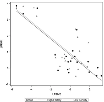
Flow cytometric measurements of PRM1 and PRM2 sperm indicated there were different histogram profiles for sperm from the HF and LF bulls (Figs. 2A–C;3A&B). The abundance of LPRM1 and LPRM2 was not different between HF and LF bulls (Figs. 4A&B;5A& B). Considering all bulls (both HF and LF bulls), the relative abundances of LPRM1 and LPRM2 were inversely related to each other (P < 0.0001;Fig. 6).
3.3. Sperm decondensation and bull fertility
The nuclear chromatin decondensation assay was performed to compare sperm nuclear decondensation between sperm from HF and LF bulls. The percentages of decondensed sperm cells were different between HF and LF bulls (P < 0.0001;Fig. 7A–C). The percentage of decondensed sperm was greater in sperm from LF compared with HF bulls. The numbers of decondensed sperm were greater in sperm from LF than HF bulls (R2= 0.72;Fig. 8).
4. Discussion
Optimal rates of reproduction are important for efficient, profitable and sustainable production of cattle. The effects of sperm nuclear proteins on sperm head shape, size, motility and fertilizing capacity aid in determining male fertility of mammals. Although even with the production of ample sperm numbers having optimal motility and morphology, some bulls are infertile due to molecular defects in their sperm (Blaschek et al., 2011;Memili et al., 2012). The CASA system andflow cytometry analyses were used to test the hypothesis that sperm cellular phenotypes as well as PRM1 and PRM2 abundances are associated with bull fertility. There was a focus in this pioneering study on improving the knowledge base and bio-techniques that could be used for cattle reproduction.
The average pathway velocity, amplitude of lateral head displacement, and path straightness were different in sperm from HF and LF bulls. In the present study, proportions of rapidly progressive motility and lateral head displacement spermatozoa were less in sperm from LF compared with HF bulls which is consistent withfindings in previous studies (Hoflack et al., 2007;Birck et al., 2010; Singh et al., 2016). Surprisingly, in the present study sperm plasma membrane integrity and acrosome status, as well as sperm DNA fragmentation status, were not different between semen samples from high and LF bulls. A functional plasma membrane is dependent on membrane ion channels (Petrunkina et al., 2001,2004) and directly related to sperm fertilizing potential (Tartaglione and Ritta, 2004). The mechanisms underlying the alterations of membrane functionality and its effect on fertility are not fully understood. Results from previous studies also indicate there is a non-significant association between sperm characteristics and plasma membrane integrity among bulls (Mishra et al., 2013).
Sperm DNA is tightly packaged when replacement of histones by protamines occurs, which makes sperm chromatin highly condensed. This highly condensed state allows for protection of the integrity of paternal genetic material during the period of transiting through the female reproductive tract. Following fertilization, disulfide bonds between protamines are decreased, and highly condensed and transcriptionally inactive sperm chromatin is converted into the decondensed state by replacing protamines by maternal histones. Results from previous studies indicate incomplete protamine deposition during spermiogenesis is the major de-terminant for chromatin packaging (Sakkas et al., 1999). Furthermore, results from previous studies indicate abnormal sperm chromatin decondensation during pronucleus formation can cause fertility problems (Queralt and Oliva, 1995), and that variability in the PRM1 abundances within sperm are associated with bull fertility (Dogan et al., 2015). In the present study, LF bulls produced larger numbers of decondensed sperm compared with HF bulls (Figs. 7&8). It is known that PRM1 and PRM2 are important for sperm molecular morphology and chromatin condensation. Insufficient condensation of sperm chromatin limits fertility (Balhorn et al., 1988) by causing DNA denaturation, DNA strand breaks (Belokopytova et al., 1993;De Yebra et al., 1993), and is associated with an increase in the retention of histones by sperm chromatin (Foresta et al., 1992). As such, abnormal sperm decondensation makes the male genome more susceptible to external factors that lead to increases in sperm damage (Auger et al., 1990).
Relative abundances of PRM1 and 2 were inversely associated in the bulls in the present study. Results of the dynamic changes of PRM2 in sperm indicated that there is no change in abundance of this protein, thus, the functions of this protein remain unexplained. Possible reasons could be that both PRM1 and PRM2 protamines, if present together in sperm, have a synergistic effect, where PRM2 functions in the incorporation of PRM1. Another explanation would be that the absence of one protamine allows for augmentation of
Table 2
Sperm variables (Mean values ± S) of frozen−thawed bull spermatozoa with different fertility phenotypes; Different su-perscript letters in the same row indicate differences (P < 0.05).
Evaluations Fertility phenotypes
Semen variables High fertility Low Fertility Motile spermatozoa (%) 55.6 ± 2.4 52.4 ± 3.6 Rapidly progressive motile spermatozoa (%) 40.5 ± 2.4 38.5 ± 3.0 Average path velocity (μm/s) 91.5 ± 1.7a 82.61 ± 3.4b
Amplitude of lateral head displacement (μm/s) 8.8 ± 0.4a 7.69 ± 0.4b
the other protamine. Alternatively, the presence of PRM2 in minute amounts could be an indicator of a viable sperm population in bulls. This assumption is well-supported by results from a previous study (Cho et al., 2001), where results indicated PRM1 requires PRM2 to for incorporation into sperm chromatin. Also, the lesser abundances of PRM2 in the majority of infertile bulls occurred because of an abnormal PRM1/PRM2 ratio (Aoki et al., 2005,2006). This abnormal ratio is associated with lesser fertility, DNA fragmentation, and morphological changes in bull sperm (Carrell and Liu, 2001;Razavi et al., 2003;Nasr‐Esfahani et al., 2004;Aoki et al., 2005,2006). In other studies with different mammals, results indicate that abnormal quantities of protamines lead to con-formational changes in sperm formation during spermatogenesis and hence a decrease in male fertility (Carrell and Liu, 2001; Mengual et al., 2003;Aoki et al., 2005).
In conclusion, average pathway velocity, amplitude of lateral head displacement and path straightness of frozen-thawed sperm evaluated using CASA, but not sperm plasma membrane, acrosome status or DNA integrity, were distinct between bulls with Fig. 1. Assessment of plasma membrane integrity, acrosome status, and DNA integrity of bull spermatozoa; Fluorescence cytograms for samples stained with (A) Propidium iodide, (B) FITC-PNA, and (C) acridine orange stain followed byflow cytometry evaluation.
contrasting in vivo fertility. Results from analysis of PRM1 and PRM2 abundances usingflow cytometry indicated there were different histogram profiles in sperm from HF and LF bulls. Thus, flow cytometry and CASA analysis are valid approaches for evaluation of potential bull fertility. Furthermore, sperm nuclear proteins (PRM1 and PRM2) can also be important biomolecular markers of fertility. Results of the present study will advance the fundamental understanding of mammalian male gametes and provide in-formation for reliable evaluation of semen quality and a more accurate prediction of bull fertility.
Funding
The research was partially supported in part by Mississippi Agricultural and Forestry Experiment Station, Alta Genetics. Inc., Conselho Nacional de Desenvolvimento Científico e Tecnológico (CNPq) of Brazil, Foundation for Research and Scientific Development of the State of Ceara, Brazil (FUNCAP), and by Selcuk University, Konya, Turkey. NAK was funded by the Netaji Subhas-Indian Council of Agricultural Research for International Fellowships 2015–2017, India (NS-ICAR). ESB Menezes was supported by a Fig. 2. Flow cytometry negative controls. There was an absence offluorescence and unstained sperm in negative control samples (Q4;Fig. 2A); Negative control FITC as secondary antibody (Q4;Fig. 2B) to target PRM1; Negative control for DyLight 650 as secondary antibody (Q4;Fig. 2C) to target PRM2.
Fig. 3. Differential expression of PRM1 and PRM2 in bull spermatozoa; Percentage of spermatozoa with PRM2 (Q1), double positive (PRM1 and PRM2 together) (Q2), PRM1 (Q3), and unstained cells (Q4) in high fertility (Fig. 3A) and low fertility bulls (Fig. 3B).
competitive fellowship from Coordenação de Aperfeiçoamento de Pessoal de Nível Superior (CAPES) of Brazil. MRU was funded by the Turkish Ministry of National Education. Hazem ElDebaky, was funded by Arab Republic of Egypt. Asma Ul Husna was funded by Higher Education Commission of Pakistan under International Research Support Program. MR Ugur was founded by a competitive fellowship from Ministry of National Education of Turkey.
Author contributions
This study was conceptualized by NAK, ESBM, EM, AK and AAM. Data were curated by NAK, MRU, RWW. The bull sperm samples and the fertility phenotypic data were provided by AK and ET. Investigations were conducted by NAK, ESBM, WT, MRU, AUH, HED, EB, HCE, FCS, and EM. The original manuscript draft was written by NAK, ESBM, MRU, EM. Reviews and editing were completed by Fig. 4. Differential abundance of PRM1 in HF and LF bull sperm; Regression plot depicts the relationship of PRM1 with high (Fig. 4A) and low (Fig. 4B) bull fertility score.
Fig. 5. Differential abundance of PRM2 in HF and LF bull sperm; Regression plot depicts the relationship of PRM2 with high (Fig. 5A) and low (Fig. 5B) bull fertility score.
NAK, ESBM, HED, EB, FS, AUH, HED, WT, RWW, ET, AK, AAM and EM. All authors read and approved thefinal version Data statement
The data analyzed during the current study are available from the corresponding author on reasonable request. Declaration of Competing Interest
The authors declare that there is no conflict of interest that could affect our objectivity in this study. Acknowledgement
Authors thank Graham Wells for critical editing of the manuscript.
Fig. 6. Relationship between the abundances of PRM1 and PRM2 in bull sperm; Regression plot depicts the inverse relationship of log transformed PRM1 and PRM2 (P < 0.0001).
Fig. 7. Fluorescent microscopy of decondensed sperm cells from high and low fertility bulls; Sperm samples from high fertility and low fertility bulls were treated with heparin glutaraldehyde, DTT, and then stained with DAPI; A) Negative Control; Sperm incubated for 1 h at room temperature in TNE buffer; B) Sperm from high fertility bull treated at 37 °C in heparin and glutaraldehyde, and DTT for 1 h; C) Sperm from low fertility bull treated at 37 °C in heparin and glutaraldehyde and DTT for 1 h; Arrows indicate de-condensed sperm.
References
Alvarez, J.G., Sharma, R.K., Ollero, M., Saleh, R.A., Lopez, M.C., Thomas, A.J., Evenson, D.P., Agarwal, A., 2002. Increased DNA damage in sperm from leukocy-tospermic semen samples as determined by the sperm chromatin structure assay. Fertil. Steril. 78, 319–329.
Aoki, V.W., Christensen, G.L., Atkins, J.F., Carrell, D.T., 2006. Identification of novel polymorphisms in the nuclear protein genes and their relationship with human sperm protamine deficiency and severe male infertility. Fertil. Steril. 86, 1416–1422.
Aoki, V.W., Liu, L., Carrell, D.T., 2005. Identification and evaluation of a novel sperm protamine abnormality in a population of infertile males. Hum. Reprod. 20, 1298–1306.
Auger, J., Mesbah, M., Huber, C., Dadoune, J., 1990. Aniline blue staining as a marker of sperm chromatin defects associated with different semen characteristics discriminates between proven fertile and suspected infertile men. Intern. J. Androl. 13, 452–462.
Balhorn, R., Reed, S., Tanphaichitr, N., 1988. Aberrant protamine 1/protamine 2 ratios in sperm of infertile human males. Experientia 44, 52–55.
Balhorn, R., Weston, S., Thomas, C., Wyrobek, A.J., 1984. DNA packaging in mouse spermatids: synthesis of protamine variants and four transition proteins. Exp. Cell Res. 150, 298–308.
Bao, J., Bedford, M.T., 2016. Epigenetic regulation of the histone-to-protamine transition during spermiogenesis. Reproduction 151, R55–R70.
Belokopytova, I.A., Kostyleva, E.I., Tomilin, A.N., Vorob’ev, V.I., 1993. Human male infertility may be due to a decrease of the protamine P2 content in sperm chromatin. Mol. Reprod. Dev. 34, 53–57.
Birck, A., Christensen, P., Labouriau, R., Pedersen, J., Borchersen, S., 2010. In vitro induction of the acrosome reaction in bull sperm and the relationship tofield fertility using low-dose inseminations. Theriogenology 73, 1180–1191.
Blaschek, M., Kaya, A., Zwald, N., Memili, E., Kirkpatrick, B., 2011. A whole-genome association analysis of noncompensatory fertility in Holstein bulls. J. Dairy Sci. 94, 4695–4699.
Braun, R.E., 2001. Packaging Paternal Chromosomes with Protamine [comment].
Carrell, D.T., Liu, L., 2001. Altered protamine 2 expression is uncommon in donors of known fertility, but common among men with poor fertilizing capacity, and may reflect other abnormalities of spermiogenesis. J. Androl. 22, 604–610.
Chang, Y.M., Gianola, D., Heringstad, B., Klemetsdal, G., 2004. Effects of trait definition on genetic parameter estimates and sire evaluation for clinical mastitis with threshold models. Anim. Sci. 79, 355–363.
Cho, C., Willis, W.D., Goulding, E.H., Jung-Ha, H., Choi, Y.-C., Hecht, N.B., Eddy, E.M., 2001. Haploinsufficiency of protamine-1 or-2 causes infertility in mice. Nat. Gen. 28, 82–86.
De Yebra, L., Ballesca, J., Vanrell, J., Bassas, L., Oliva, R., 1993. Complete selective absence of protamine P2 in humans. J. Biol. Chem. 268, 10553–10557.
Dogan, S., Vargovic, P., Oliveira, R., Belser, L.E., Kaya, A., Moura, A., Sutovsky, P., Parrish, J., Topper, E., Memili, E., 2015. Sperm protamine-status correlates to the fertility of breeding bulls. Biol. Reprod. 92 (92), 91–99.
Erkek, S., Hisano, M., Liang, C.-Y., Gill, M., Murr, R., Dieker, J., Schübeler, D., Van Der Vlag, J., Stadler, M.B., Peters, A.H., 2013. Molecular determinants of nucleosome retention at CpG-rich sequences in mouse spermatozoa. Nat. Struct. Mol. Biol. 20, 868–875.
Evenson, D., Darzynkiewicz, Z., Melamed, M., 1980. Relation of mammalian sperm chromatin heterogeneity to fertility. Science 210, 1131–1133.
Evenson, D.P., Larson, K.L., Jost, L.K., 2002. Sperm chromatin structure assay: its clinical use for detecting sperm DNA fragmentation in male infertility and com-parisons with other techniques. J. Androl. 23, 25–43.
Evenson, D.P., Wixon, R., 2005. Comparison of the Halosperm® test kit with the Sperm Chromatin Structure Assay (SCSA®) infertility test in relation to patient diagnosis and prognosis. Fertil. Steril. 84, 846–849.
Flesch, F.M., Voorhout, W.F., Colenbrander, B., van Golde, L.M., Gadella, B.M., 1998. Use of lectins to characterize plasma membrane preparations from boar spermatozoa: a novel technique for monitoring membrane purity and quantity. Biol. Reprod. 59, 1530–1539.
Foresta, C., Zorzi, M., Rossato, M., Varotto, A., 1992. Sperm nuclear instability and staining with aniline blue: abnormal persistance of histones in spermatozoa in infertile men. Int. J. Androl. 15, 330–337.
Gatewood, J., Cook, G., Balhorn, R., Bradbury, E., Schmid, C., 1987. Sequence-specific packaging of DNA in human sperm chromatin. Science 236, 962–965.
Fig. 8. Differential decondensation rates in HF and LF bull sperm; Regression line represents percentage of decondensed sperm in high and low fertility bull sperm; 100 sperm cells/bull were evaluated usingflorescent microscopy to compare percentage of decondensed sperm cells in high and low fertility bulls; Difference between these two groups in terms of de-condensation tendencies (P < 0.0001).
Gopalkrishnan, K., Hinduja, I., Anand Kumar, T., 1991. In vitro decondensation of nuclear chromatin of human spermatozoa: assessing fertilizing potential. Arch. Androl. 27, 43–50.
Govin, J., Escoffier, E., Rousseaux, S., Kuhn, L., Ferro, M., Thévenon, J., Catena, R., Davidson, I., Garin, J., Khochbin, S., 2007. Pericentric heterochromatin repro-gramming by new histone variants during mouse spermiogenesis. J. Cell Biol. 176, 283–294.
Hammoud, S.S., Nix, D.A., Zhang, H., Purwar, J., Carrell, D.T., Cairns, B.R., 2009. Distinctive chromatin in human sperm packages genes for embryo development. Nature 460, 473–478.
Hoflack, G., Opsomer, G., Rijsselaere, T., Van Soom, A., Maes, D., De Kruif, A., Duchateau, L., 2007. Comparison of computer‐assisted sperm motility analysis parameters in semen from belgian blue and Holstein–Friesian bulls. Reprod. Domest. Anim. 42, 153–161.
Irvine, D.S., Twigg, J.P., Gordon, E.L., Fulton, N., Milne, P.A., Aitken, R., 2000. DNA integrity in human spermatozoa: relationships with semen quality. J. Androl. 21, 33–44.
Killian, G.J., Chapman, D.A., Rogowski, L.A., 1993. Fertility-associated proteins in Holstein bull seminal plasma. Biol. Reprod. 49, 1202–1207.
Kistler, W.S., Henriksén, K., Mali, P., Parvinen, M., 1996. Sequential expression of nucleoproteins during rat spermiogenesis. Exp. Cell Res. 225, 374–381.
Kutchy, N.A., Velho, A., Menezes, E.S., Jacobsen, M., Thibaudeau, G., Wills, R.W., Moura, A., Kaya, A., Perkins, A., Memili, E., 2017. Testis specific histone 2B is associated with sperm chromatin dynamics and bull fertility-a pilot study. Reprod. Biol. Endocrinol. 15, 59.
Lewis, J.D., Song, Y., de Jong, M.E., Bagha, S.M., Ausió, J., 2003. A walk though vertebrate and invertebrate protamines. Chromosoma 111, 473–482.
Lewis, S.E., Aitken, R., 2005. DNA damage to spermatozoa has impacts on fertilization and pregnancy. Cell Tissue Res. 322, 33–41.
Martianov, I., Brancorsini, S., Catena, R., Gansmuller, A., Kotaja, N., Parvinen, M., Sassone-Corsi, P., Davidson, I., 2005. Polar nuclear localization of H1T2, a histone H1 variant, required for spermatid elongation and DNA condensation during spermiogenesis. Proc. Nat. Acad. Sci. 102, 2808–2813.
Martins, R.P., Ostermeier, G.C., Krawetz, S.A., 2004. Nuclear matrix interactions at the human protamine domain a working model of potentiation. J. Biol. Chem. 279, 51862–51868.
Memili, E., Dogan, S., Rodriguez-Osorio, N., Wang, X., de Oliveira, R.V., Mason, M.C., Govindaraju, A., Grant, K.E., Belser, L.E., Crate, E., 2012. Makings of the Best Spermatozoa: Molecular Determinants of High Fertility, Male Infertility. InTech.
Mengual, L., Ballescá, J.L., Ascaso, C., Oliva, R., 2003. Marked differences in protamine content and P1/P2 ratios in sperm cells from percoll fractions between patients and controls. J. Androl. 24, 438–447.
Mishra, S., Kundu, A., Mahapatra, A., 2013. Effect of ambient temperature on membrane integrity of spermatozoa in different breeds of bulls. Bioscan 8, 181–183.
Moura, A.A., Chapman, D.A., Killian, G.J., 2007. Proteins of the accessory sex glands associated with the oocyte-penetrating capacity of cauda epididymal sperm from holstein bulls of documented fertility. Mol. Reprod. Dev. 74, 214–222.
Moura, A.A., Chapman, D.A., Koc, H., Killian, G.J., 2006. Proteins of the cauda epididymalfluid associated with fertility of mature dairy bulls. J. Androl. 27, 534–541.
Nagy, S., Jansen, J., Topper, E.K., Gadella, B.M., 2003. A triple-stainflow cytometric method to assess plasma-and acrosome-membrane integrity of cryopreserved bovine sperm immediately after thawing in presence of egg-yolk particles. Biol. Reprod. 68, 1828–1835.
Nasr‐Esfahani, M.H., Razavi, S., Mozdarani, H., Mardani, M., Azvagi, H., 2004. Relationship between protamine deficiency with fertilization rate and incidence of sperm premature chromosomal condensation post‐ICSI. Andrologia 36, 95–100.
Nelson, J.E., Krawetz, S., 1993. Linkage of human spermatid-specific basic nuclear protein genes. Definition and evolution of the P1–& P2–& TP2 locus. J. Biol. Chem. 268, 2932–2936.
Nelson, J.E., Krawetz, S.A., 1994. Characterization of a human locus in transition. J. Biol. Chem. 269, 31067–31073.
Oliva, R., 1995. Sequence, Evolution and Transcriptional Regulation of Avian-Mammalian P1 Type Protamines. Advances in Spermatozoal Phylogeny and Taxonomy. Editions du Museum National d’Histoire Naturelle, Paris, pp. 537–548.
Oliva, R., Dixon, G.H., 1991. Vertebrate protamine genes and the histone-to-protamine replacement reaction. Prog. Nucleic Acid Res. Mol. Biol. 40, 25–94.
Peddinti, D., Nanduri, B., Kaya, A., Feugang, J.M., Burgess, S.C., Memili, E., 2008. Comprehensive proteomic analysis of bovine spermatozoa of varying fertility rates and identification of biomarkers associated with fertility. BMC Syst. Biol. 2, 1.
Petrunkina, A., Harrison, R., Ekhlasi-Hundrieser, M., Töpfer-Petersen, E., 2004. Role of volume-stimulated osmolyte and anion channels in volume regulation by mammalian sperm. Mol. Hum. Reprod. 10, 815–823.
Petrunkina, A., Harrison, R., Hebel, M., Weitze, K., Topfer-Petersen, E., 2001. Role of quinine-sensitive ion channels in volume regulation in boar and bull sperma-tozoa. Reproduction 122, 327–336.
Queralt, R., Oliva, R., 1993. Identification of conserved potential regulatory sequences of the protamine-encoding P1 genes from ten different mammals. Gene 133, 197–204.
Queralt, R., Oliva, R., 1995. Demonstration of trans-acting factors binding to the promoter region of the testis-specific rat protamine P1 gene. Biochem. Biophy. Res. Commun. 208, 802–812.
Razavi, S., Nasr‐Esfahani, M.H., Mardani, M., Mafi, A., Moghdam, A., 2003. Effect of human sperm chromatin anomalies on fertilization outcome post‐ICSI. Andrologia 35, 238–243.
Sakkas, D., Mariethoz, E., Manicardi, G., Bizzaro, D., Bianchi, P.G., Bianchi, U., 1999. Origin of DNA damage in ejaculated human spermatozoa. Rev. Reprod. 4, 31–37.
Samans, B., Yang, Y., Krebs, S., Sarode, G.V., Blum, H., Reichenbach, M., Wolf, E., Steger, K., Dansranjavin, T., Schagdarsurengin, U., 2014. Uniformity of nucleosome preservation pattern in Mammalian sperm and its connection to repetitive DNA elements. Dev. Cell 30, 23–35.
Schlueter, G., Celik, A., Obata, R., Schlicker, M., Hofferbert, S., Schlung, A., Adham, I.M., Engel, W., 1996. Sequence analysis of the conserved protamine gene cluster shows that it contains a fourth expressed gene. Mol. Reprod. Dev. 43, 1–6.
Singh, R.K., Kumaresan, A., Chhillar, S., Rajak, S.K., Tripathi, U.K., Nayak, S., Datta, T., Mohanty, T., Malhotra, R., 2016. Identification of suitable combinations of in vitro sperm-function test for the prediction of fertility in buffalo bull. Theriogenology 86, 2263–2271 e2261.
Sreenivasa, G., Kavitha, P., Vineeth, V.S., Channappa, S.K., Malini, S.S.N., 2012. Evaluation of in vitro sperm nuclear chromatin decondensation among different subgroups of infertile males in Mysore, India. J. Res. Med. Sci. 17, 456.
Suganuma, R., Yanagimachi, R., Meistrich, M.L., 2005. Decline in fertility of mouse sperm with abnormal chromatin during epididymal passage as revealed by ICSI. Hum. Reprod. 20, 3101–3108.
Takeda, N., Yoshinaga, K., Furushima, K., Takamune, K., Li, Z., Abe, S.-i., Aizawa, S.-i., Yamamura, K.-i., 2016. Viable offspring obtained from Prm1-deficient sperm in mice. Sci. Rep. 6, 27409.
Tartaglione, C., Ritta, M., 2004. Prognostic value of spermatological parameters as predictors of in vitro fertility of frozen-thawed bull semen. Theriogenology 62, 1245–1252.
Tesarik, J., Greco, E., Mendoza, C., 2004. Late, but not early, paternal effect on human embryo development is related to sperm DNA fragmentation. Hum. Reprod. 19, 611–615.
Vilfan, I.D., Conwell, C.C., Hud, N.V., 2004. Formation of native-like mammalian sperm cell chromatin with folded bull protamine. J. Biol. Chem. 279, 20088–20095.
Ward, W.S., 2010. Function of sperm chromatin structural elements in fertilization and development. Mol. Hum. Reprod. 16, 30–36.
Wykes, S.M., Krawetz, S.A., 2003. The structural organization of sperm chromatin. J. Biol. Chem. 278, 29471–29477.
Zwald, N.R., Weigel, K.A., Chang, Y.M., Welper, R.D., Clay, J.S., 2004a. Genetic selection for health traits using producer-recorded data. I. Incidence rates, heritability estimates, and sire breeding values. J. Dairy Sci. 87, 4287–4294.
Zwald, N.R., Weigel, K.A., Chang, Y.M., Welper, R.D., Clay, J.S., 2004b. Genetic selection for health traits using producer-recorded data. II. Genetic correlations, disease probabilities, and relationships with existing traits. J. Dairy Sci. 87, 4295–4302.