Volume 20 Number 11
November 2014
Unusual case of frontal mucocele presenting with forehead ulcer
Altıntaş Kakşi S
1, Kakşi M
2, Balevi A
1,
Özdemir M
1, Çakır A
3Dermatology Online Journal 20 (11): 7
1
Medipol University Hospital- İstanbul, Department of dermatology
2Eyüp Government Hospital-
İstanbul, Department of neurosurgery
3Medipol University Hospital-
İstanbul, Department of pathology
Correspondence:
Sümeyye Altıntaş Kakşi,
Medipol Üniversitesi Hastanesi
TEM Avrupa Otoyo
lu Göztepe çıkışı, no:1 bağcılar 34214 İstanbul, Türkiye
Telephone number: 0505 2481940
[email protected]
Abstract
Paranasal mucoceles are benign slow-growing paranasal sinus lesions, which usually develop following the obstruction of the sinus ostiu. They most frequently occur in the frontal sinus. Frontal mucoceles are expansive lesions usually causing visual clinical signs and symptoms such asdiminution of vision, visual field defects, diplopia, orbital swelling, retroorbital pain, displacement of eye globe, ptosis, and proptosis. When the frontal mucocele extends intracranially, it can manifest with meningitis, meningoencephalitis, intracranial abscess, seizures, or cerebrospinal fluid fistula. Very rarely it can cause forehead swelling. We report an 80-year-old woman presenting with a forehead skin ulcer and painless subcutaneous forehead
induration. Histopathologic examination revealed mucin deposition and inflammation. Computerized tomography (CT) and magnetic resonance imaging (MRI) scans showed a mass originating from the frontal sinus with frontal bony defect and frontocutaneous fistula. Surgical excision of the mass confirmed the mucocele diagnosis. In this article, we present a case of frontocutaneous fistula and skin ulcer, which is an unexpected complication of frontal mucocele. We propose that in the case of a localized non-healing ulcerated forehead skin lesions, mucocele should be considered in the differential diagnosis.
Keywords: frontal mucocele, frontocutaneous fistula, frontal ulcer
Introduction
Paranasal mucocelesare mucus-containing benign cysts, which usually develop when the ostium of a paranasal sinus becomes obstructed by chronic sinusitis, polyps, bone tumors, surgical procedures, or,rarely, tumors. They mostly involve the frontal and ethmoid sinuses [1-8]. They can erode through the surrounding bone and spread both intraorbitally and intracranially [1,11,12,13]. Frontal sinus mucoceles can also very rarely extend into the subcutaneous region and present as a forehead mass [1,11,14,15].
We report an unusual case of frontal mucocele with subcutaneous extension presenting as only a forehead ulcer localized on the glabella without any neurologic or ophthalmologic involvement.
Case synopsis
At our clinic, an 80-year-old woman presented with a non-healing ulcer on her forehead. She reported that this initially began as a small painless subcutaneous indurated area. Subsequently, an ulcer formed spontaneously at the center within a few months. Despite various medical treatments, including topical and systemic antibiotics, no response was seen. There was neither history of surgery or trauma at that site nor any symptoms of an ocular or neurological condition.
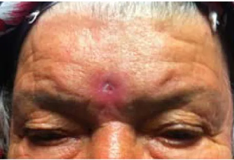
Dermatological examination revealed non-tender erythema and firm induration at the glabellar region of the forehead with a 0.3 cm diameter ulcer at the center of the lesion (Figure 1). There was a small amount of discharge from the ulcer. Further history indicated that the discharge had been continuous during the past year.
Figure 1. Non-tender erythema, induration and ulcer on the forehead
Physical examination of the patient upon admission revealed no abnormalities. Her visual and neurological examinations were unremarkable. The complete blood count, erythrocyte sedimentation rate, liver function tests, fasting blood glucose, urinalysis, serum creatinine, urea, calcium, electrolytes, IgA, IgG, IgM, and C-Reactive Protein were within normal limits. Bacterial culture of the drainage material was negative. Punch biopsy was performed on a segment of the indurated skin.
Histopathologic examination revealed chronic inflammation and mucin deposition. Waters’ view X-ray showed opacity of the frontal sinus. Otolaryngology consultation was obtained and the patient underwent cranial computerized tomography (CT), which revealed frontal sinus expansion and a defect of the frontal bone. Neurosurgical consultation suggested a diagnosis of a frontal mucocele. Therefore, a cranial magnetic resonance imaging (MRI) study was performed. It demonstrated the presence of high-density material, causing obliteration and expansion of the frontal sinus, with a 3-4 mm defect on the anterior wall of the frontal sinus and a 3 mm sinus tract opening to the skin of the frontal region (Figure 2). Based on clinical and radiological findings, the most likely diagnosis was considered to be a frontocutaneous fistula secondary to a frontal mucocele.
Figure 2. Axial magnetic resonance imaging showing frontocutaneous fistula tract from mucocele to frontal bone
Figure 3. Lesion partially lined by stratified squamous epithelium including mucus glands, with chronic inflamation, on histopathological examination of surgical biopsy (HE, X 40)
Following aright ciliary skin incision by the neurosurgeon, a small erosion of the frontal bone was observed below the frontotemporal fascia. Mucocele diagnosis was confirmed at surgery. The mucocele was well isolated and completely excised, the diseased sinus mucosa was removed, the sinus was packed with absorbable hemostatic gelatin sponge (Spongostan™, Ethicon, Inc. Somerville, NJ, USA) and the ostium was closed. Histopathology showed a fibrous connective-tissue cyst wall, partially lined by stratified squamous epithelium, with chronic inflammation (Figure 3). These findings were consistent with mucocele diagnosis. Following surgery, the localized ulcer healed. No fistula or ulcer developed during the twenty-four month follow-up, and a repeat MRI scan showed no recurrence of the mucocele.
Discussion
We report an unusual patient with frontal sinus mucocele whose only symptom was a non-healing forehead ulcer. Paranasal sinus mucocelesare benign lesions that result from continuous or intermittent obstruction of the paranasal sinus ostium. They are most commonly found in the frontal sinus, with less frequent involvement of the other sinuses [1-8]. Despite their benign character, paranasal sinus mucoceles can be serious owing to their ability to destroy the bone structure of the sinus wall. When located in the frontal sinus, they may cause serious pathological lesions in the neighboring structures such as the orbit and cranium. Obstruction of the frontonasal duct can lead to the development of a frontal mucocele, the cause of which could be the abnormal density of mucus (as in cystic fibrosis), a mechanical obstruction of the channel owing to previous trauma, nasal polyposis, post-operative scarring, or neoplasm [9,10]. Another important factor in the pathogenesis of mucocele is mucosal inflammation.
Gradual distension, thinning, and the eventual erosion of the bony wall of the sinus are caused by progressive accumulation of mucoid material. This usually results in pressure necrosis of the posterior and inferior walls. Mucoceles can extend into the orbital cavity or intracranial compartment by destroying bony structures. Because a mucocele usually expands in the direction of least resistance, frontal mucoceles tend to erode the thin bone of the orbit and displace the globe inferiorly[1,11,12,13]. They can present with diminution of vision, visual field defects, diplopia, orbital swelling, retroorbital pain, displacement of eye globe, ptosis, or proptosis [4,6]. Intracranial extension of a frontal mucocele can present with meningitis,
meningoencephalitis, intracranial abscess, seizures, or cerebrospinal fluid fistula. Because the anterior wall of the sinus is composed of relatively thick bone, it is resistant to erosion [16]. In our patient, there was an erosion of the anterior wall of the frontal sinus and a fistula formation from the mucocele to the frontal skin.
In the literature, a subcutaneous mass was very rarely reported as the sole presenting complaint of a frontal mucocele with intracranial or intraorbital extension [1,14,15]. Borkar S et al reported a 53-year-old female with a frontal mucocele with cranio-orbital extension, presenting with orbital swelling with forehead extension [15]. Tan CSH et al reported a 33-year-old female with blurring of the inferior visual field in the left eye, associated with periorbital swelling and a painless subcutaneous forehead mass [14].Akiyama M et al reported a similar case in a 57-year-old female who presented with a three-month history of an asymptomatic subcutaneous tumor on the forehead. CT scanning and magnetic resonance images revealed a sharply-demarcated cystic mass from the subcutaneous area on the forehead expanding into the frontal sinus and intracranial space [1]. In our patient, there was initially a forehead swelling that spontaneously formed a skin ulcer. All other cases in the literature with subcutaneous swelling had ocular or intracranial involvement. It may be that in our patient the frontocutaneous fistula formation and drainage from the skin stopped the mucocele from expanding toward the ophthalmic and cranial structures. CT and MRI are both considered the gold standard for the diagnosis of these lesions [17]. CT scans are used to evaluate the amount of expansion and bone erosion and characterize typical space-occupying lesions in the paranasal sinus with
surrounding bone erosion. MRI is used mainly to identify the relationship between the mucocele and the brain, orbit, and affected soft tissue. CT and MRI are complementary in complicated cases [14]. In our patient, CT and MRI findings revealed dense material that caused obliteration and expansion of the frontal sinus, a 3-4 mm bony defect of the anterior wall of the frontal sinus, and a 3 mm sinus tract opening to the frontal region.
The definitive treatment of mucocele is surgery. Surgical treatment of mucoceles can be accomplished with a minimally-invasive endoscopic procedure, craniotomy with craniofacial surgery, or both. In our patient, frontal bony defect, a fistula tract, and a frontal mucocele were detected. Complete obliteration of the sinus with exenteration of all its mucosa and packing with absorbable hemostatic gelatin sponge was successfully performed through a transciliar incision. The wound healed without any complications and no ulcer developed during the twenty-four month follow-up. A repeat MRI scan showed no recurrence of the mucocele.
Delayed diagnosis of a frontal mucocele can cause paranasal sinus expansion and invasion of vital structures such as the skull base and the orbit. Although sinus pathology is not a common source of localized forehead skin diseases, it is important to consider frontal mucocele in the differential diagnosis during the clinical workup of a patient presenting with only a forehead skin lesion without neurological or ophthalmological signs. Diagnosis and treatment of frontal mucocele requires careful clinical examination, radiological evaluation, and cooperation between related clinical medical specialists such as ophthalmologists, dermatologists, neurosurgeons, and otolaryngologists.
References
1. Akiyama M, Inamato N, Hashigucci K. Frontal mucocele presenting as a subcutaneous tumour on the forehead. Dermatology 199(9): 263-264, 1999. [PMID: 10592411].
2. Arrue P, Kanny MT, Serrano E et al. Mucoceles of the paranasal sinuses: uncommon location. J Laryngol Otol 112(9): 840-844, 1998. [PMID: 9876373].
3. Har –EL G: Endoscopic management of 108 sinus mucoceles. Laryngoscope 111(12): 2131-2134, 2001. [PMID: 11802010].
4. Hayasaka S, Shibasaki H, Sekimato M et al. Opthalmic complications in patients with paranasal sinus mucopyoceles. Opthalmologia 203(2): 57-63, 1991. [PMID: 1762720].
5. Lai PC, Lia o [?] SL, Jou JR et al. Transcaruncular approach for the management of frontoethmoid mucoceles. Br J Opthalmol 87(6): 699-703, 2003. [PMID: 12770964].
6. Leventer DB, Linberg JV, Ellis B. Frontoethmoidal mucoceles causing bilateral chorioretinal folds. Arch Opthalmol 119(6): 922-923, 2001. [PMID: 11405853].
7. Natvig K, Larsen TE. Mucocel of the paranasal sinuses. A retrospective clinical and histological study. J Laryngol. Otol. 92(12): 1075-1082, 1978. [PMID: 739180].
8. Ormerod LD, Weber AL, Rauch SD, Feldon SE. Opthalmic manifestations of maxillary sinus mucoceles. Opthalmology. 94(8): 1013-1019, 1987. [PMID: 3658361].
9. Rinna C, Cassoni A, Ungari C et al. Fronto-Orbital mucoceles: our experience. J Craniofac Surg 15: 885-9, 2004. [PMID: 15346041].
10. Koudstaal MJ, Van der Wal GH, Bijvoet HVC et al. Post-trauma mucocele formation in the frontal sinus; a rationale of follow-up. Int J Oral Maxillofac Surg 33: 751-4, 2004. [PMID: 15556321].
11. Kawaguchi S, Sakati T, Okuna S et al. Giant frontal mucocele extending into the anterior cranial fossa. J Clin Neurosci 9: 86-9, 2002. [PMID: 11749028].
12. Woegels RL, Balbani AP, Santos Junior RC et al. Frontoethmoidal mucocele with intracranial extension: a case report. Ear Nose Throat J 77: 117-20, 1998. [PMID: 9509725]
13. Tasman W, Jaeger EA. Duane’s clinical Opthalmalogy. Vol 2 New York: JB Lippincott Co, 1994: 3-7.
14. Tan CSH, Yong VK, Yip LW et al. An unusual presentation of a giant frontal mucoceles manifesting with a subcutaneous forehead mass. Ann Acad Med Singapore 34: 397-398, 2005. [PMID: 16021233].
15. Borkar S, Tripathi AK, Satyarthee G et al. Frontal mucocele presenting with forehead subcutaneous mass: An unusual presentation. Turkish Neurosurgery 2008 18(2): 200-203, 2008. [PMID: 18597239].
16. Marfatia HK, Muranjan SN, Navalakhe MM et al. Persistent frontal fistula. J Postgrad Med 43: 102, 1997. [PMID: 10740736].
17. Rao VM, Sharma D, Madan A. Imaging of frontal sinus disease: concepts, interpretation, and technology. Otolaryngol Clin North Am. 34(1): 23-39, 2001. [PMID: 11344059].