SIGNAL DETECTION THEORY ANALYSIS
OF CATEGORY-BASED VISUAL SEARCH
IN NATURAL MOVIES
a thesis submitted to
the graduate school of engineering and science
of bilkent university
in partial fulfillment of the requirements for
the degree of
master of science
in
electrical - electronical engineering
By
Osman Tutaysalgır
August 2016
Signal Detection Theory Analysis of Category-Based Visual Search in Natural Movies
By Osman Tutaysalgır August 2016
We certify that we have read this thesis and that in our opinion it is fully adequate, in scope and in quality, as a thesis for the degree of Master of Science.
Tolga C¸ ukur(Advisor)
Emine ¨Ulk¨u Sarıta¸s
Didem G¨ok¸cay
Approved for the Graduate School of Engineering and Science:
Levent Onural
ABSTRACT
SIGNAL DETECTION THEORY ANALYSIS OF
CATEGORY-BASED VISUAL SEARCH IN NATURAL
MOVIES
Osman Tutaysalgır
M.S. in Electrical - Electronical Engineering Advisor: Tolga C¸ ukur
August 2016
The human brain changes its inner hierarchy and connection strength between the neurons in order to apprehend the real world. In visual search, It is thought that the human brain changes the sensitivity of neurons in the favor of the attended object. Here, we investigate these tuning shifts of the voxels in signal detection theory perspective. Brain activities of human subjects were recorded while they were watching a natural movie. To assess the attentional effect on the human brain, the decoding procedure was employed on the BOLD responses and the natural movie stimuli. Decoding procedure tries to predict the stimuli that form the BOLD responses. In order to bridge the gap between the stimuli and BOLD responses, logistic regression which is a classification algorithm is applied to form models of the subjects’ brain. The model performances were assessed with d-prime, ROC and AUC parameters.
Our results suggest that category-selective regions in the human brain boost their detection performances further for the objects that they are not inherently selective.
Keywords: Visual Search, Logistic Regression, D-prime, Receiver Operating char-acteristic (ROC), Area Under Curve (AUC) .
¨
OZET
KATEGOR˙ISEL G ¨
ORSEL D˙IKKAT˙IN S˙INYAL
ALGILAMA TEOR˙IS˙I ILE ˙INCELENMESI
Osman Tutaysalgır
Elektrik - Elektronik M¨uhendisli˘gi, Y¨uksek Lisans Tez Danı¸smanı: Tolga C¸ ukur
A˘gustos 2016
˙Insan beyni, ger¸cek d¨unyayı anlamlandırabilme i¸cin kendi i¸c hiyerar¸sisini ve n¨oronlar arasındaki ba˘glantı g¨uc¨un¨u de˘gi¸stiren bir yapıya sahiptir. Bu do˘grultuda, insan beyninin dikkat edilen nesneye g¨ore n¨oronların hassasiyetini de˘gi¸stirdi˘gi d¨u¸s¨un¨ulmektedir. Bu ¸calı¸smada voxellerde meydana gelen bu du-yarlılık de˘gi¸simleri sinyal algılama teorisi ¸cer¸cevesinde ele alınacaktır. Deneklerin beyin aktiviteleri, onlara bir video izletilirken kayıt altına alınmı¸stır. Dikkatin beyin ¨uzerindeki etkisini anlayabilmek i¸cin hem beyin h¨ucrelerinin aktivite sinyal-leri (BOLD) hem de videodaki objelere ¸c¨oz¨umleme (decoding) prosed¨ur¨u uygu-lanmı¸stır. Bahsedilen ¸c¨oz¨umleme (decoding) prosed¨ur¨u beyin aktivitelerine neden olan uyarıcıyı (stimuli) tahmin etmeye ¸calı¸smaktadır. Bu ba˘glamda, etki ve tep-kiler arasındaki ba˘gıntının kurulması amacıyla bir sınıflandırma algoritması olan ”logistic regression” ile deneklerin beyin modelleri olu¸sturulmaya ¸calı¸sılmı¸stır. Bu modellerin performansları d-prime, ROC ve AUC parametreleriyle hesaplanmı¸stır ve de˘gerlendirilmi¸stir.
Bu ara¸stırma sonucu ortaya ¸cıkan sonu¸clara g¨ore, insan beyninin kategori-se¸cici b¨olgeleri g¨orsel dikkatle birlikte se¸cici olmadıkları nesneleri algılayabilmek i¸cin o nesnelerin se¸cicili˘gini se¸cici oldukları nesnelere g¨ore daha fazla arttırmaktadır.
Acknowledgement
Firstly, I would like to express my kindest gratitude to my advisor Dr. Tolga Cukur for his immense support, patience, motivation and sharing his knowledge during my MS study. His guidance helped me in all the time of research and writing of this thesis. Besides my advisor, I would like to thank Dr. Emine Ulku Saritas and Dr. Didem Gokcay for their insightful comments and encouragement to widen my research from various perspectives. I also want to mention about my friends: Salman, Yonus, Gokhan, Mahmut and Mehmet. I would like to thank them one by one for their encouragement and support during the years we are together. I also would like to thank TUBITAK for TUBITAK BIDEB and TUBITAK 3501 114E546 supports throughout my MS study.
Last but not the least, I would like to thank my family especially my granddad, Osman, for supporting me spiritually throughout writing this thesis and my my life in general.
Contents
1 Introduction 1
2 Functional Magnetic Resonance Imaging 4
2.1 Statistical Analysis of FMRI Data . . . 6
2.2 Data Acquisition . . . 8
2.3 FMRI Noise Sources and Characteristics . . . 9
2.3.1 Thermal and System Noise . . . 9
2.3.2 Physiological Noises . . . 10
2.4 Components of FMRI Preprocessing . . . 10
2.4.1 Quality Assurance . . . 10
2.4.2 Slice Time Correction . . . 11
2.4.3 Head Motion Correction . . . 11
2.4.4 Spatial Normalization . . . 12
CONTENTS vii
3 Human Brain and Vision Related Parts 14
3.1 Brain and Its Structure . . . 14
3.2 Cerebrum and Its Subparts . . . 15
3.2.1 Vision Related Regions . . . 17
4 Methods 20 4.1 Subject . . . 20
4.2 Stimuli . . . 20
4.3 Experiment . . . 21
4.4 MRI Data Acquisition . . . 22
4.5 Data Pre-Processing . . . 22
4.6 Category Model . . . 23
4.7 Model Fitting . . . 23
4.7.1 fMRI Models . . . 24
4.7.2 Signal Detection Theory and D-prime . . . 27
4.7.3 Logistic Regression . . . 32
4.7.4 Searchlight Analysis . . . 34
4.7.5 Procedures . . . 34
CONTENTS viii
5.1 Conclusion . . . 57
A Appendix 63 A.1 MRI: Magnetic Resonance Imaging . . . 63
A.2 MRI Physics . . . 64
A.3 Image Formation . . . 68
List of Figures
2.1 A typical hemodynamic response. Measured BOLD activity starts to increase two seconds after from beginning of the neural activity. It reaches its maximum about 5s after the onset. After the neu-ral stimulus ends, hemodynamic response amplitude falls below a baseline and recovers its initial state. The figure is reinterpreted from [1]. . . 5
4.1 Signal and noise trial. The threshold value is selected to minimize false alarm rate and maximize true positive rate for a given experi-ment. D-prime value represents how far means of these two curves are located between each other. This figure is adapted from [2]. . 30 4.2 ROC curve with different d-prime values. Discrimination between
the trials increases with the d-prime value. Different d-prime values yield various ROC curves. This figure is reinterpreted from [3]. . . 31 4.3 Sigmoid function. Our modeling function is changed to estimate
LIST OF FIGURES x
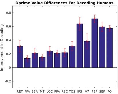
5.1 Decoding performance difference of the ‘Human’ object between attended and unattended stimuli. This illustration was obtained by subtracting the d-prime values of the ‘Human’ when subjects were attending and not attending to the object of interest. All of the regions of the brain, whether they are inherently selective to the ‘Human’ object or not, enhance their detection performances with visual search. Error bars show standard deviation that was calculated by bootstrapping d-prime values across subjects. . . 38 5.2 Decoding performance difference of ‘Vehicle’ object between
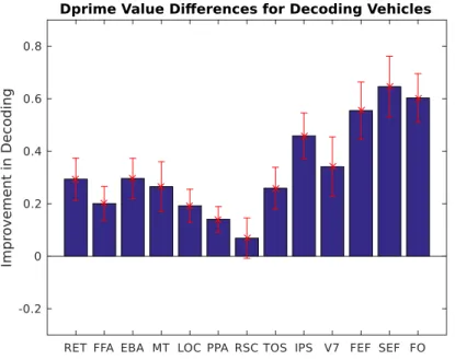
at-tended and unatat-tended stimuli. This graph is calculated by sub-tracting obtained d-prime values from the subjects’ two conditions (attending or not attending the category of interest). All of the regions benefit from the visual search and increases their detection performances. . . 39 5.3 Decoding differences between two object categories. This graph
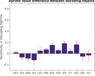
illustrates the decoding sensitivity changes over attention in dif-ferent ROIs. This plot is acquired by subtracting the Fig:5.1 from Fig:5.2. ‘Human’ and ‘Scene’ selective areas shows different char-acteristics. Human selective areas take advantage of the visual search of ‘Vehicle’ more than the visual search of ‘Human’. Scene selective areas tend to show the opposite relation. . . 41 5.4 Area under the curve graph in group analysis, horizontal axis
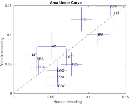
repre-sents the decoding improvement in ‘Human’ object with attention, vertical axis represents the increase in ‘Vehicle’ object decoding with attention. Scene selective areas enhance their areas under the ROC curves to a greater extent on visual search of ‘Human’ objects. Human selective regions tends to show the opposite relation. 44
LIST OF FIGURES xi
5.5 ROC curve for subject ‘TC’ with selected ROIs. FFA, EBA and MT selected as human selective ROIs and PPA, RSC, TOS selected as object selective ROIs. Horizontal axis represents the false posi-tive rate and vertical axis represents the true posiposi-tive rate. . . 45 5.6 ROC curve for subject ‘SN’ with selected ROIs. FFA, EBA and
MT selected as human selective ROIs and PPA, RSC, TOS selected as object selective ROIs. Horizontal axis represents the false posi-tive rate and vertical axis represents the true posiposi-tive rate. . . 46 5.7 ROC curve for subject ‘JG’ with selected ROIs. FFA, EBA and
MT selected as human selective ROIs and PPA, RSC, TOS selected as object selective ROIs. Horizontal axis represents the false posi-tive rate and vertical axis represents the true posiposi-tive rate. . . 47 5.8 ROC curve for subject ‘AH’ with selected ROIs. FFA, EBA and
MT selected as human selective ROIs and PPA, RSC, TOS selected as object selective ROIs. Horizontal axis represents the false posi-tive rate and vertical axis represents the true posiposi-tive rate. . . 48 5.9 ROC curve for subject ‘AV’ with selected ROIs. FFA, EBA and
MT selected as human selective ROIs and PPA, RSC, TOS selected as object selective ROIs. Horizontal axis represents the false posi-tive rate and vertical axis represents the true posiposi-tive rate. . . 49 5.10 Flatmap representation of subject ‘TC’, red areas represent
in-crease in human detection performance with attention, blue areas represent sensitivity enhancement in vehicle detection with attention. 52 5.11 Flatmap representation of subject ‘SN’, red areas represent
in-crease in human detection performance with attention, blue areas represent sensitivity enhancement in vehicle detection with attention. 53
LIST OF FIGURES xii
5.12 Flatmap representation of subject ‘JG’, red areas represent in-crease in human detection performance with attention, blue areas represent sensitivity enhancement in vehicle detection with attention. 54 5.13 Flatmap representation of subject ‘AH’, red areas represent
in-crease in human detection performance with attention, blue areas represent sensitivity enhancement in vehicle detection with attention. 55 5.14 Flatmap representation of subject ‘AV’, red areas represent
in-crease in human detection performance with attention, blue areas represent sensitivity enhancement in vehicle detection with attention. 56
A.1 Slice selection: Hydrogen atoms are tipped with slice selection gra-dient. Hydrogen atoms within the selected slice are tipped right after the RF pulse. In the left pane, the magnetization vector points toward a discrete set of angles. In the real world, how-ever, particles are exposed to a slightly different magnetic field and therefore form continuous bands of tipped angles. This figure is adapted from [1]. . . 69 A.2 A sample MR sequence. The RF and the slice selection gradient
excite a 2-D plane in a 3-D volume. After the excitation phase, phase and frequency encoding gradients are applied. Frequency and phase encoding gradients constitute a sufficient k-space cover-age. Sampling occurs at the same time as the frequency encoding gradient. This illustration is adapted from [5]. . . 71 A.3 An MR sequence and its k-space coverage. If we use number 1
of the phase encoding gradient, we will cover the line 1 on the k-space. By changing the Gx gradient, we shift from the middle to
the left and from the left to the right of the k-space and sample as we use the Gx gradient. The figure is adapted from [6]. . . 74
Chapter 1
Introduction
The brain is a highly flexible organ that evaluates, transforms and modifies the data from the sensors of the human body. It also governs many different tasks related to cognition and perception. It is a network interconnection system that consists of over 100 billion nerve cells. Although an individual nerve cell has lim-ited capacity, the interconnection of this system controls not only motor muscle movements such as walking but also fulfills feature specific, computationally ex-pensive tasks such as categorization of seen objects. The human brain’s process of understanding starts with the signaling pathways of several sensory organs, the communication between the neural cells and their formation of the intercon-nection system.
Vision is one of the most advanced features of a living organism. Most of our perception of the real world is obtained from our eyes. Vision starts in the retina. The retina converts analog real world signals into electrical ones [7]. Signals from the retina are transmitted to the cerebral cortex (that is the outermost layer of the brain) via neurons. This data first arrives at the primary visual cortex where the basic shape and orientation of the object are identified. In the higher levels of the processing, the brain combines information from several subregions to build the perception. It also selects information about specific attributes to construct short and long term memory [7].
The human visual system carries out various tasks in order to apprehend the surrounding world. Recognition is one of the most computationally expensive and challenging task. To recognize an object, the brain needs to compare the perceived object with countless possibilities and find the right match. Palmer et al. [8] described the perception process such that the brain converts an object into a reference plane. Size and orientation of the object seen in the reference plane are compared with the objects stored in the memory. This model creates an advantage on computational intensity in that only a few models need to be stored in the memory and processed in order to recognize a particular object.
Olshausen et al [9] presented a mechanism to explain how objects are repre-sented in visual areas. They tried to explain how attention and pattern recogni-tion occur in the brain. According to their model, input and output relarecogni-tionship between different brain regions are dynamical, changed with neural connections. Also, the strength of these neural connections are modified without loss of spatial and temporal resolution.
In earlier studies, researchers tried to evaluate the relationship between iso-lated individual voxel responses and the the stimuli represented. These analyses are not sensitive enough to decode information in certain aspects. These classical methods select the voxels that show significant response to the experiment stim-uli and take the average of their responses. Despite spatial averaging smoothing the data, it blurs the dataset and might eliminate unique information. Further-more non-significant voxels might convey stimuli-specific information, and this information can’t be taken into account for processing [35, 36].
In another recent study [10], researchers investigated the attentional effect on extracting categorical information. They proposed that visual search of a particular object biases the processing in the favor of that object so that only the attended object is represented in high-level cortical areas. In another study [11], researchers tested whether task-relevant features of the attended category are selected. According to their results, when subjects paid attention motion, they found out that the acquired signal is bigger as subjects see moving faces instead of moving houses in face-selective areas. Reddy et al [12] also showed that attention
removes the clutter effects and increases visual search performances under natural viewing conditions.
In this thesis, we tested how attention changes tuning shifts of the category-selective regions of the human brain. We used a signal detection theory perspec-tive and modeled different areas of the brain as detectors of the target category. Our results suggest that the detection performances of the category-selective re-gions are the highest for objects for which they are not inherently selective.
Outline of the thesis is as follows: In Chapter 2, building blocks of the MRI (magnetic resonance imaging), fMRI (functional magnetic resonance imaging) and fMRI signal processing are briefly explained. In Chapter 3, we talk about the human brain and its category-selective regions. In Chapter 4, methods used in this study are elaborated. In Chapter 5, obtained results are presented and explained.
Chapter 2
Functional Magnetic Resonance
Imaging
As the information processing takes places in the brain, the energy required is provided by the vascular system in the form of glucose and oxygen. Oxygen is transmitted to the brain via blood. It appends to the hemoglobin molecules in the blood. It changes the magnetic property of the hemoglobin. Functional magnetic resonance imaging (fMRI) uses these alterations in the magnetic field to form images of neural activity correlated with physiological changes. Contrast based on neural activities is also known as the blood-oxygenation-level-dependent (BOLD) contrast.
When oxygen molecules attach themselves to the hemoglobin, hemoglobin molecules show the diamagnetic property. Diamagnetic materials have no mag-netic moments when they are placed inside a magmag-netic field, thus they do not effect the MR signals. Deoxygenated hemoglobin, on the other hand, shows paramagnetic property, and it can be used as a contrast material for MR. This phenomenon was first discovered by Seiji Ogawa, who was a research scientist at Bell Laboratories [1].
on the transverse plane to decay faster and reduce the MR signal (underlying physical phenomena are described in the appendix part). Although oxygenated hemoglobin has no effects on the MR signal, neural activity related MR images are collected with the help of the oxygen molecules. Whenever a neural activation occurs in a brain region, the concentration of oxygenated hemoglobin molecules increase more than that of the deoxygenated hemoglobin molecules. Therefore, signal decrease due to the T2 effect is relatively reduced.
The MR signal triggered by neural activity is known as the hemodynamic response. Neurons usually fire within milliseconds after the stimulus. But hemo-dynamic responses start 1-2 seconds following the neural event and usually reach their maximum within the following 5 seconds. After a few seconds, hemody-namic responses fall below their baseline and refine to their initial state within 10 seconds.
Figure 2.1: A typical hemodynamic response. Measured BOLD activity starts to increase two seconds after from beginning of the neural activity. It reaches its maximum about 5s after the onset. After the neural stimulus ends, hemodynamic response amplitude falls below a baseline and recovers its initial state. The figure is reinterpreted from [1].
The spatial resolution of an MR image is defined as the separability of a voxel from the nearby spatial location. It is dependent on several different factors re-lating to the MR sequence and the machine. On the MR device, the field of view parameter describes the total imaged size in 2 dimensions, and it is usually expressed in centimeter. On the MR sequence, the total number of imaged voxels is defined as sampling period on each frequency and phase encoding gradient. So,
for example, a field of view of 25 by 25 cm and 125 samples on each encoding gradient results in a 2mm x 2mm in 2-D. The third dimension that is needed to construct the voxel is provided by the thickness of the slice that is formed with the slice selection gradient [13].
Increasing the voxel size is beneficial for fMRI studies. SNR of fMRI signals are dependent on the BOLD and if we decrease the spatial resolution by a factor of 2, BOLD signals reduce by half so does the SNR value. The second effect of increasing the voxel size is that time of acquisition decreases as we increase the voxel size. This increase in the acquisition time causes images to suffer from T2∗ effects and reduces the BOLD signal. On the contrary, increasing the voxel size too much causes a decrease in discrimination performances [13].
Temporal resolution can be described as the ability to discriminate different consecutive stimuli from the BOLD signals. fMRI can ensure a temporal reso-lution that is on the order of a few seconds. Like spatial resoreso-lution, temporal resolution is dependent on both the MR sequence and BOLD signal characteris-tic. Repetition time of the MR sequence is the first parameter in constructing the temporal resolution. Repetition time might change from 500ms to 3000 ms in a typical MR experiment [1]. The second factor that effects the temporal resolution is the hemodynamic response characteristic. As mentioned in previous sections, BOLD responses last around 10 seconds to recover their original state. It is bet-ter to sample the BOLD signals in a small inbet-terval because the smaller repetition intervals help us better identify the BOLD response of a neural activity.
2.1
Statistical Analysis of FMRI Data
In a typical fMRI study, researchers need to find out how to deal with the inher-ently noisy data coming from a complex spatiotemporal structure. To elucidate the underlying reasons behind the fMRI data, scientists utilize statistics as a valuable tool.
fMRI is a noninvasive imaging modality that is used to unveil brain functions. In a typical fMRI experiment, the researchers acquire a set of images (directly measuring the blood oxygenation level in different parts of the brain) that are related to the current task of the brain. This novel technique uncloaks the brain actions and inner hierarchies which previously were very difficult to gather infor-mation about. Stages of the systematics of neural connections and task-related responses of the brain can now be understood with a simple noninvasive brain scan.
In an fMRI study, researchers try to achieve information about several features and actions of the brain. These comprise of task-related activation, hierarchies, the cooperation of different parts of the brain and psychological state of the subject [13]. However, the images that are acquired by a standard MRI machine are inherently noisy and contain complex spatiotemporal structure. Scientists try to clear data from unwanted noise and simplify the complicated data set to achieve more meaningful results with the use of statistical properties.
Several components make an analysis of an fMRI study prone to errors: These include head movement during the scan and inconsistency in data, including the variability between or within the subjects during the time course of fMRI. The analysis of the fMRI data is to deal with all these inherent problems, and remedies for these problems can be grouped into several components. Correction of the fMRI data set may be accomplished by [14]
• Fixing the spatial distortion on the data set
• Alignment of the images over the time course to get rid of the relative head movement
• Alignment of the time sequence of slices and the subject to create a frame-work so that the data can be used in group analysis
• Smoothing the data temporally and spatially to reduce the noise over the relatively weak signals
After these steps, the data might be ready for the further analysis.
2.2
Data Acquisition
In this section, an overview of data collection is presented in a concise manner. The data collected in an fMRI experiment is an aggregation of several MRI im-ages. These images are obtained while the subject is carrying out an individual task. As mentioned earlier, a subject is located inside an MRI machine. The hydrogen atoms (H) in the body align with the magnetic field. A radio frequency pulse is used to tip over the aligned H atoms from their initial position. Im-mediately after the RF pulse, atoms that have been tipped precess from their aligned point in a way that they induce a current in the receiver coils of the MRI machine. The fully aligned slices in the brain can be altered via gradient coils throughout the experiment to complete the full brain images as they change the magnetic field over the brain.
The raw MRI images represent the spatial frequency of the proton densities of the brain tissue. Different tissues in the body create distinct spatial frequencies. Thus, they form a different contrasts on the reconstructed image. To acquire meaningful images, k-space representation of the imaged tissue needs to be sam-pled adequately. Different sampling schemes can be applied (uniform and non-uniform) over the k-space. In the uniform sampling, uniformly distributed points on the k-space are sampled, while in the non-uniform sampling, the k-space is sampled with different trajectories. Each sampling scheme has different advan-tages and disadvanadvan-tages regarding resolution, SNR, and speed. Once raw data are collected, the Fourier transform is applied to it. Using the Fourier transform on the raw data creates actual images of the imaged tissue, since the raw data only represents the spatial frequencies of the tissues.
Researchers scan subject’s brain while the subjects are performing certain tasks. Extracting the BOLD (blood-oxygen-level dependent) responses from the MRI images is quite challenging and the results are often indeterminable due to
spatial and temporal changes in the imaged brain tissue, the variability of the MR machine during the scan and physiological effects like breathing, head motion and heart beats [1]. Preprocessing algorithms are applied to readily acquired images in order to overcome these issues.
2.3
FMRI Noise Sources and Characteristics
2.3.1
Thermal and System Noise
MR imaging studies suffer from thermal noise related to the free motion of the electrons inside the subject and the machine. During the slice selection and excitation stages, the temperature inside the MR hardware increases, which in turn causes the electrons to collide with the atoms more frequently and results in unwanted current distortion inside the imaging machine. The same incident happens to the receiver hardware of the device. As induced currents pass through the receiver device, temperature increases, which results in more collision inside the hardware and causes more distortion on the image. Besides the thermal noise, another major type of noise is the MR system noise that is caused by the unsteadiness and variability in the imaging hardware. Known causes for this type of noises are [1]
• Static magnetic field inhomogeneities which in turn deteriorate the quality of image,which can also disrupt the contrast and geometry of the image because of the varying resonant frequency of the hydrogen atoms
• Instability of the gradient coils and their characteristics similar to the mag-netic field inhomogeneities, which changes the shape and location of parts of the image
• Off-resonance effects in the RF transmitter and receiver circuitries, and drift in the magnetic field of the primary magnets, which result in inefficient excitation and causes enormous decrease in image quality.
2.3.2
Physiological Noises
The human body is not a stationary, inert object. Muscles interact with each other, the respiratory system changes the position of the body and heartbeats make small variations in the status of the body or subject may swallow or move his/her head unintentionally. These minor changes create motion related artifacts in MR signals. Even if we don’t take into consideration the intrinsic noise sources of MRI hardware, these subject related movements create excessive degradation in the fMRI signals. BOLD related signals in MR are relatively small compared to the non-BOLD (anatomically) related signals. Since these variations are digitized, artifacts due to problems mentioned above can be modeled and removed from the digitized data if sampling is sufficient according to the Nyquist sampling rate.
2.4
Components of FMRI Preprocessing
The success and reliability of statistical processing algorithms depend on the consistency of the raw fMRI data sets. The primary objective of the preprocessing stage of the fMRI study is to ensure that the data is free from the artifacts causing the problems as mentioned above. Preprocessing has several steps.
2.4.1
Quality Assurance
Quality assurance is the starting point of the data analysis. The first, most important and easiest method in quality assurance is eye examination of the raw data because many artifacts in the raw data can be seen with the naked eye. There are several programs that can show time series images as an animation and this way any scanner dependent distortion can be detected quickly. Furthermore, researchers perform tests to ensure the statistical consistency of the raw data. No further processing is done on the data without passing this stage in order to save researchers from unnecessary work [13].
2.4.2
Slice Time Correction
In most standard MR scanners, data acquisition occurs in an interleaved manner, which means that the collected data belongs to the different time instants. The BOLD responses of adjacent slices would not be identical in consecutive slices even if they are related to the same event. These gaps between acquisition times form problems in data analysis. One remedy for this artifact is to interpolate time during preprocessing. Through interpolation the MR signals are corrected as if they are collected in the same instant so that the differences are minimized.
2.4.3
Head Motion Correction
Physical and mental status of the subject cannot be completely stable during 1-2 hour long sessions. Tasks that involve muscle movements, swallowing, and respiration, are the underlying causes of head motion during experiments. Sev-eral precautions (small breaks during a session to relieve subject, use of head stabilizer) can be done during and before the scan to prevent head motion. Pre-venting head movement is relatively easy compared to correcting it [1]. Although cautions are made, head movements still occur during the sessions. Some types of head movement only change the location of the voxel in the resulting images and can be corrected with software tools while others transform the data into meaningless throughputs.
Aligning consecutive images to a reference image is called co-registration. Since the shape and the volume of the brain don’t change, the rigid body transform can be used to align the images. In this transformation, a cost function is applied to maximize the similarities between images. Six variables (x-axis, y-axis, z-axis, roll, pitch, yaw) are formulated to shift the input images onto the reference images as close as possible [13]. Once the alignment is successfully implemented, co-registered images are spatially interpolated to reduce the noise that comes with the co-registration and this procedure is applied to all acquired images.
Co-registration is also used between functional and structural images. fMRI images are relatively low-resolution images that only show a silhouette of the brain. Structural brain images, on the other hand, show more anatomical details. With structural images, the regions of interests in the brain can easily be located, and activation patterns can easily be seen.
2.4.4
Spatial Normalization
Morphology of the brain across subjects is different from each other. These varia-tions prevent us from an inter-subject analysis. Spatial normalization techniques are types of co-registration that scale images to a normalized framework. Spa-tial normalization algorithms determine the sizes of individuals brains and try to compress or enlarge them to fit a known space. With this powerful tool, fMRI signals of different subjects can be aggregated for group analysis and let us test our hypothesis across subjects. A well-known and widely used method for nor-malization framework is the Talairach space. This scheme was created by Jean Talairach and imitated from the brain of an elderly woman [1].
2.4.5
Temporal and spatial smoothing
Temporal and spatial filtering are applied to the collected fMRI signals to increase the functional SNR [1]. Functional SNR is a term that expresses the differences between BOLD activities of ROI in two different conditions. In a nutshell, a func-tional SNR value enables us to detect and differentiate states of the experimental conditions.
Sampled data might be corrupted by physiological effects such as breathing and heartbeats. These effects can be diagnosed by applying a physiological exam-ination on the subject before the experiment (typical respiration rate for humans
is around 0.2-0.3 Hz, and heart rate is 1-1.5 Hz). If we sample the data suffi-ciently, we can eliminate these physiological variables through the use of appro-priate filters without effecting the task-dependent BOLD signals. Rectification of corruptions is also dependent on the experiment itself. If the experiment is fast-event based this means that conditions change every 1-2 second. In this case, the BOLD signals can be coupled with these unwanted effects, which makes re-moving them much more challenging. Researcher need to take precautions before the experiment. Another problem with the BOLD signals is the temporal corre-lation between the time points of the experiment. These irregularities cause data to become non-eligible to be used in statistical analysis and cause results to be more erroneous [13]. The remedy for this is whitening (intentionally adding white noise in) the data so that the temporal correlation between pairs is corrupted.
Spatial smoothing is applied in order to reduce the high-frequency component in the fMRI data sets. There are several reasons to reduce the spatial frequency in the data. Firstly, spatial smoothing blurs the images and increases the functional SNR. Generally, activation in the human brain occurs in multiple voxels. Smooth-ing data across voxels reduces susceptibility effects and minimizes unwanted noise in the data set. Secondly, it decreases the variability and the mismatch that is not adjusted with the spatial normalization. Most common method for spatial smoothing is convolution with a Gaussian kernel [14]. The degree of smoothing on the data set depends on the variance of the Gaussian kernel in that more voxels are involved in the smoothing with a large width kernel. There are different mea-sures to the required Gaussian kernel but in general starting from a twice-voxel sized kernel and increasing from that start point is recommended because there is a trade-off between the increase in the functional SNR and the attenuation of the meaningful information. A Gaussian kernel with a smaller width has no positive effects on SNR and a wider one diminishes the significant information on the brain.
Chapter 3
Human Brain and Vision Related
Parts
3.1
Brain and Its Structure
The brain is the main organ that is responsible for decision-making processes and sensory information. It is located inside the skull. The human brain shares the same structure as other mammals but, it has a more advanced cerebral cortex. It reacts real world stimuli and interacts with the environment. It contains millions of neuron cells, and each cell contributes to the transfer and the processing of specific information gathered from the other organs in the body [7].
The brain is the central organ of the central nervous system and interacts with the rest of the body via the spinal cord. It constitutes a centralized control mechanism over the body. The way to control the body is forming muscles’ ac-tivities through the spinal cord and releasing feature specific hormones. It also processes sophisticated sensory inputs and integrates them to collect informa-tion from the environment. From a neurologist’s point of view, the brain is the biological computer that collects and processes information from the world.
Brain consists of three main parts of which each one is responsible for a different type of processing. Basic descriptions of the brain parts are as follows [15]:
• Cerebrum: Cerebrum is the most advanced and largest part of the brain. It is located at the top of the brain. It comprises of two hemispheres (right and left) and their cortices. Cerebrum carries out functions such as triggering and coordination of movement, visual and audial processing, learning and interpretation, sense of touch and emotion.
• Brainstem: The brainstem is adjacent to the spinal cord and can be thought as the continuation of it. It structurally connects the cerebrum and cerebellum to the spinal cord. The brainstem controls all the auto-matic life dependent functions such as cardiac and respiratory systems. • Cerebellum: Cerebellum is located under the cerebrum at the back of
the head. It plays important roles in the motor movements. The cerebel-lum also coordinates some intentional actions such as speech, balance, and coordination.
The first aim of this study is to understand the attentional effects of the human visual system. Different subjects’ brains were scanned and stimuli related data were collected while they were watching a movie. Evaluation of the attentional effects was made on the category-selective and attention-related regions of interest in the human brain. These category-selective and attention-related regions were localized with the MR scans for different subjects, , the data from which were used in this study. These regions of interest will be further explained in the following sections.
3.2
Cerebrum and Its Subparts
The cerebrum is the largest and the most developed part of the human brain. It includes two hemispheres each controlling opposite sides of the human body. The
hemisphere on the right side is considered to control the creativity and artistic skills. The other hemisphere is thought to relate to the arithmetic, comprehension and writing abilities.
The surface of the cerebrum is called the cerebral cortex. Most of the compu-tational processing takes place in this area of the brain. It has a structure with folds that are called gyri and sulci which enhance its surface, therefore, increasing its computational power. It is the most superior part of the brain and encom-passes more than half of it. Nearly all of the interesting calculations (such as thinking, perception, language processing, sensory information processing) occur in this area. Cerebral cortex is directly involved with the sensory informations and perception of the surrounding world. The cerebral cortex is subdivided into four parts [15]:
• Frontal lobe: This part is found at the front of the brain. Its functions are correlated with attention, concentration, short term memory, motivation, and judgment. It also controls the daily activities like walking and plays a role in speaking and writing skills.
• Parietal Lobe: The parietal lobe is adjacent to the frontal lobe , stretch-ing towards to the end of the brain. The parietal lobe interprets sensory information and integrates information coming from different locations in the body. It is also responsible for the information about spatial and visual perception. It plays important roles in language processing.
• Occipital Lobe: Occipital lobe is located at the rear of the brain behind the parietal lobe. It is the primary visual processing center in the human brain. It comprises of most of the visual processing regions.
• Temporal Lobe: Temporal Lobe is located beneath the other three lobes towards to the brainstem. It interprets informations to reveal and organize the long term memory. It is also responsible for language and comprehen-sion.
sensory information and maintains cognitive functions of the human brain. Re-gions associated with the visual system and attention are further explained in the next sections.
3.2.1
Vision Related Regions
Discovery of the noninvasive imaging modality, specifically of the fMRI, facilitates the study of the human visual cortex and therefore enhances the understanding of the hierarchies of it. Two principles are employed to explain the visual cortex. In the first model (called the hierarchical processing), an image is first expressed in a local and conventional form. Through this stage of the processing, the image is turned into a more complete and complex representation [16]. In the second model, different properties of the visual scene are handled with parallel hierarchical streams [17]. Both these themes are approved by several studies in the fMRI researches.
In this thesis, not the whole human visual cortex, but only the human and scene selective regions are described and assessed. The recent finding about these areas for these two objects of interest are clarified. Attention-related regions in the brain are also explained. List of the areas of interests and their role in the higher visual system is as follows:
• FFA (Fusiform Face Area): This section, as its name indicates, is ac-tivated with perception of faces. Kanwisher, et al. found that activation pattern of this area six times greater when subjects passively watch human faces [18].
• EBA (Extrastriate Body Area): This region is named to indicate that it shows significantly higher responses for body parts than for inanimate objects [19]. Kanwisher et al. also showed that this area shows relatively higher responses to human body parts except for faces.
• MT (Middle Temporal Visual Area): This region responds more sig-nificantly when the subject sees non-stationary objects. This area performs
a significant role in the recognition of the motion. MT is also selective for biological motion, meaning the region is activated when a subject views walking people, hand and mouth movements of a person [20].
• PPA (Parahippocampal Place Area): Results in [21] indicates that this region is processing the information about the spatial domain. They reported that the PPA is responsible for scene recognition and also gives more marked responses to familiar places.
• RSC (Retrosplenial Cortex): RSC is involved with the spatial naviga-tion, memory related actions and scene processing in the brain [22]. Dam-ages that occur on the RSC might result in significant memory loss and navigation deficiency. In another study [23], It is stated that RSC is more selective when the number of stable items in the scene is increased.
• TOS (Transverse Occipital Sulcus): This area is located on the dorsal occipital-temporal cortex and is activated for construction and scene related stimuli [24].
• RET (Retinotopic (Early Visual Areas)): Early visual areas are rep-resented under the category of RET. In this area, a 2-D projection of the 3-D objects is demonstrated. Retinotopic mapping was introduced by the research of Tootell et al. [25].
• LOC (Lateral Occipital Complex): This area is associated with the recognition of an objects. LOC is considered the general purpose system for object analysis and categorization [26].
• V7 This area responds strongly to the spatial attention. V7 also shows robust activation in response to several different categories of stimuli[27]. • IPS (Intraparietal Sulcus): IPS serves as one of the visual attention
centers in the brain. This area is activated in directed attention regardless of the stimuli presented [28].
• FEF (Frontal Eye Field): This section is responsible for the generation and control of the eye movements and adjusting the visual attention [29].
• SEF (Supplementary Eye Field): According to the work of Purcell et all [30], SEF is the main region responsible for auditing the visual search performance. This study also states that it has a minor effect on continuing search behavior.
• FO (Frontal opperculum): This region is responsible for controlling the activity of other brain areas to fulfill cognitive tasks such as attention [31].
Chapter 4
Methods
In this work, we investigate the effects of attention on the responses of the hu-man brain. Our experiment, preprocessing and methods to analyze the data are explained in the following parts.
4.1
Subject
Five healthy male subjects took part in the experiment. Age of subjects were between 25 − 30. All subjects had normal or corrected to normal vision, and they all signed a consent form. The procedure of the experiment was approved by The Institutional Review Board at the University of California Berkeley.
4.2
Stimuli
To understand the effects of attention, a natural movie clip was created using 10 − 20s long short clips without repetition. For each attention condition (human or vehicle) 1800s of a natural film was created. Visual angle for the video was 24◦ x 24◦ and its resolution was 512x512. Each movie was created in a similar
manner where only one of the attended objects appeared in the movie for 450s, they co-appeared for 450s, and none of them appeared for 450s. Attended objects appeared in immensely different sizes, shapes, visual angles, and positions. A fixation point for the subjects (0.16◦ square) was located on the film and its color was switched at 1 Hz to ensure its visibility. The resultant video was shown to the subjects with a mirror and projector system during the scan.
4.3
Experiment
All experimental data were acquired at the University of California Berkeley, as part of a previous study on visual attention [33]. This thesis reanalyzes that dataset to address different neuroscientific questions than the previous study. Each subject was scanned seven times to collect not only functional data but also anatomical, functional localization and retinotopic mapping data in the original experiment. The functional data was collected in one session. During this session, six attention conditions each lasting 600s were performed by the subjects (three runs for the human and three runs for the vehicle category objects). Subjects were fixated to a point while they were seeking for a category of interests in the movie. To stay alert during sessions, they pressed a button whenever the attended object group was seen in the film. A cue word was shown before each run to inform the subjects about the attended category. The attended category was switched after each run and same with all the other runs. To create mutually exclusive stimuli clusters, each type of stimulus (human, vehicle, both of them, none of them) was randomly picked and evenly distributed within and between the runs. The hemodynamic offsets between the voxel responses and the movie stimuli were compensated by adding the last 10s of the film to the start of the film of that run. The offset data were extracted from further processing. Subjects passively watched 7200s of natural video in the other three sessions. These extra sessions were not used in this study.
4.4
MRI Data Acquisition
The MRI data acquisition took place in the University of Berkeley with a 3T Siemens scanner with a 32-channel head coil. The MRI sequence for the functional data was a T2 weighted gradient-echo echo planar imaging that was modified with
water-excitation RF pulse. Head motion was prevented by using a foam padding. Sequence parameters was as follows: Slice number = 32, TR = 2s, echo time = 34 ms, flip angle = 74◦, voxel size = 2.24 x 2.24 x 3.5mm3, FOV = 224 x 224 mm2.
Anatomical data was collected with a T1 - weighted MP-RAGE sequence. The
parameters for anatomical scan were voxel size = 1 x 1 x 1 mm3 and FOV = 256
x 212 x 256 mm3. The anatomical and the retinotopic data for two subjects were
collected with a 1.5T MRI scanner. Retinotopic mapping data were collected in four different sessions. Each session lasted nine minutes with various stimuli (rotating polar wedges and widening and shrinking rings). Motor localizer was a ten-minute scan. Each subject performed six different motor tasks (hand, mouth, foot, speech, rest and saccade blocks). Middle temporal visual area (MT) was localized with using four 90 seconds natural movie as stimuli. Category-selective regions were extracted with six 4.5 minutes scans. Stimuli used in the visual localization consisted of places, faces, animals, objects and human body parts. Auditory cortex was localized with various types of sound stimuli [32, 33].
4.5
Data Pre-Processing
Non-Brain tissue was extracted from the brain tissue using the Brain Extraction Tool (BET). Functional data between and within the runs was aligned to the first functional image acquired from the subject. The alignment was done with the Statistical Parameter Mapping Toolbox (SPM8). The BOLD responses arising from button press task were identified and removed from further processing. The cortical surface of the subjects was constructed with Caret5 software. Voxels of the cortical surface were determined such that the voxels which is located
within the 4mm radius of the cortical surface were selected. To remove the low-frequency drift (baseline wander) from the datasets, the Savitzky-Golay filter (which uses the low-degree polynomial to smooth the data without distorting the signals) was applied. No extra spatial and temporal smoothing was applied. The datasets for each subject were normalized to become zero mean and unit variance (z-score) in order to enable us to compare the results from different subjects easily. No subjects’ data were transformed into known brain space. Instead, two-dimensional flat-map representations were used. The data from the functional localizer and the retinotopic mapping was used to identify the particular region of interest (ROI) for each subject.
4.6
Category Model
WordNet Lexicon [34] database was used to label the object categories within the natural movies. In this database, English words are not organized based on their spellings but they are arranged with respect to their meanings and semantic features. Conceptually related words are linked to each other within hierarchical orders. The natural movie was labeled by three raters. Since the words are connected in a hierarchical manner, the existence of any category in the movie also denotes that its higher order classes also exists. It means that the presence of a ‘lorry’ also indicates the existence of following: ‘vehicle’, ‘conveyance’. The raters labeled 604 different object categories in the film. 331 higher order categories of these objects were also included. The stimulus matrix (category × time) for further processing is composed of 1s and 0s (former indicates the presence, and the latter represents the absence of the category of interest).
4.7
Model Fitting
In this work, we used the BOLD responses and the natural movie as inputs to our system and tried to create a model that reveals the effects of attention on the
human brain. As a starting point, we chose decoding routines as our modeling procedure between the stimuli and the response. In other words, we first tried to predict the stimuli that formed the obtained responses based on constructed models. Secondly, some metrics were computed to evaluate the performance of the model and clarify how attention effects decoding performance. Besides, these procedures were applied to different regions of the brain to gather a complete set of results on the visual attention.
Some alterations were done on both stimuli and the response data sets to apply a decoding procedure successfully. Firstly, stimulus matrix (category × time course) was down-sampled by 2 to balance time points of the BOLD sig-nals and the stimuli. Additionally, some changes were applied to the response matrix to compensate for the hemodynamic shift. Originally, the response ma-trix contained the responses of each voxel over the period of the experiment (voxels × time course). We added shifted versions of the each voxel response as a new column to the response matrix. The new matrix (4 × voxels × time course) contains the time slided version of each voxel response from 2s to 6s.
We modeled voxel responses as input and stimuli as output in order to create a transformation model. We divided our datasets into training, cross-validation and evaluation chunks to form and evaluate a model. Logistic regression algorithm was applied on the training datasets to obtain the models of the brain. The per-formance metrics were calculated on the cross-validation and the evaluation sets. We repeated this procedure 1000 times with different training, cross-validation and evaluation sets to create aggregations of the results and evaluate their sta-tistical consistency.
4.7.1
fMRI Models
Decoding models use voxel responses to estimate information about the real world (stimuli). Their complimentary operation is called encoding models that uses the opposite relationship between stimuli and response. If we formulate the response and the stimuli as probabilistic events, we can express our decoding and encoding
models with these prior probabilities. Let
• P(r) be the probability of the voxel responses
• P(s) be the probability of stimuli that form the responses.
• P(r,s) be joint probability that r is the response, s is the stimulus
• P(r|s) be conditional probability of a formed response r given that stimuli is present.
• P(s|r) be the conditional probability of stimuli was present given that re-sponse is occurred.
variables represent the probabilistic events. Joint probability can be described as the conditional probability of evoked response given stimuli times stimuli or the conditional probability of stimuli given formed response multiplied by the probability of response.
P (r, s) = P (r|s)P (s) = P (s|r)P (r) (4.1) A decoding model can be modeled as the likelihood of stimuli given response P(s|r) on the other hand an encoding model can be modeled as the likelihood of BOLD response patterns given the stimuli present P(r|s). Using Bayes Theorem these probabilities can be expressed with each others.
P (r|s) = P (s|r)P (r)
P (s) (4.2)
P (s|r) = P (r|s)P (s)
P (r) (4.3)
From these equation sets, we formulated our decoding procedure and tried to model human brain with prior stimuli and response probabilities.
In our work, we used decoding models to assess attentional effects on the human brain. We collected BOLD responses from each subject while they were viewing the movie sets. These datasets have low SNR value and massive size.
Because of these inherent characteristics of the fMRI datasets, tremendous efforts needs to be exerted on them to reveal any meaningful data.
Multivariate analysis leverages pattern recognition and, unlike the single voxel analysis, uses a population of voxels as a whole to increase its sensitivity [37]. It also includes the responses of non-significantly sensitive voxels in the calcu-lation and extracts substantial information. Multivariate analysis are used by a majority of the decoding studies in this area [36]. This procedure can disclose the information hidden in a set of data which is its advantage over conventional spatial averaging type analysis [38, 39].
Decoding studies might also be used to decode mental and cognitive states of the subject’s brain. Previously studies can only show which stimuli or task activate which region of the brain entirely because of resolution constraints. By increasing sensitivity using the multivariate analysis, decoding studies shows sig-nificant improvements and can even detect sudden (2s−4s) changes in the mental states.
The main advantage of multivariate analysis comes from representing the re-sponses and the stimuli as high dimensional spaces [37]. Rere-sponses of the brain and the stimuli are represented in these high-dimensional vector spaces, and every component and property of stimuli or activity and voxel of interest are described as new dimensions. If we measure the 1000 voxel over 900 s of time course, we represent the response matrix with 900x1000 dimensional space. Similarly, the stimuli pattern over the time course is formed and described with 1 × 900 di-mensional space. Once these representation spaces are created, they may be ma-nipulated with various mathematical tools to unveil substantial information and differentiate the several mental states required for one task. Multivariate analysis use machine learning algorithms and try to constitute a decision threshold that discriminates differences between the cognitive states and the high dimensional patterns [37].
In terms of decoding and encoding, multivariate analysis may be considered a manipulation and evaluation of the response and stimuli vectors on the hy-perspace. Decoding can be evaluated in order to form a transformation matrix that converts response patterns to stimuli patterns. Although cortical topogra-phy and anatomical structure are the essential characteristics of brain responses, these features are discarded. They can be evaluated with other analysis methods such as searchlight [40].
In our work, we performed our analysis on different regions of the brain. Voxels on each region were treated as a whole, and logistic regression algorithms were applied to each of them separately. We began our analysis by splitting response and stimuli matrices as training, test and evaluation subsets. These datasets were entirely independent of each other. The decision threshold that determines the presence of stimuli on a given response pattern was computed on the training sets with different regression parameters for each subject. Best regularization parameter was selected on the test set. Validation of the decision threshold and regularization parameter was done on the evaluation set. Test and evaluation data had no effect on the classifier to have a viable testing. We perform this procedure on each subject for both of the attention conditions. The performance of the classifiers was calculated with the d-prime parameter.
4.7.2
Signal Detection Theory and D-prime
Signal detection theory helps us to detect information and elucidate how deci-sion is made under ambiguity. This method provides an invaluable mathematical framework to probe the relationship between stimuli and response in psychologi-cal studies. Consider the following example to come to a deeper understanding of detection theory in psychological studies. A person is passively viewing the natu-ral video while their BOLD responses are recorded. BOLD responses for different objects are distinct from each other for a particular ROI. Because of the loss of attention the subject could not recognize the object of interest and this creates noise on the BOLD responses. If we try to determine the object using the data
from the BOLD responses, the signal detection theory accounts those noises into its calculation and try to formulate an optimal decision threshold. The threshold might be used to resolve the presence of the object.
To explain detection framework, some basic terminology needs to be explained. A complete picture can be described with 4 different metrics in detection theory. The first metric for performance of a detector is the hit rate. Hit rate can be explained as the proportion of the correct YES responses when stimuli is present during the basic YES/NO trials.
Hit Rate = P correct Yes P target (4.4) It is obvious that a good detector performs with a high hit rate. But this met-ric unfortunately is not enough to completely evaluate the performance of the detector. This is because the hit rate disregards the information when there is no stimuli presented during the trial. The hit rate depends solely on the correct answers and does not account for the mistake. The second metric for the detector is known as false alarm. The false alarm rate is the number of deceptive YES answers when there is no stimuli present on the trial. False alarm rate can be explained as follows:
False Alarm Rate =
P
f alse
Yes
P
no target (4.5) The third metric in detection theory stands for misses in the signal trials. It evaluates the performance of a trial with its wrong answers. It calculates the ratio of the wrong responses on the signal trials (when stimuli are present on the trial). Miss Rate = P f alse No P target (4.6) The last metric of the detection theory calculates the correct differentiation of the absence of the stimuli on the noise trials.
Correct Rejection Rate =
P
correct
No
P
no target (4.7) These 4 metrics can be tabulated as follows:
Yes Response No Response Stimuli Present Hit Miss Stimuli Absent False Alarm Correct Rejection
In detection theory, the metrics that are most commonly used are hit and false alarm rate. Others might be though of a complement of the first two. Although, these two metrics together might explain the behaviour of the detectors under cer-tain conditions, one single quantity that stands for the sensitivity might be more desirable. Signal detection theory uses both hit and false alarm rates to create a concrete mathematical formulation for the detection process. It also provides a metric for detectability useful for ambiguous decision-making processes. In the signal detection framework, hit and false alarm rate is turned into significant quantities.
Modeling trials as probabilistic events led us to utilize signal detection frame-work. Simplest form of the model is the Gaussian model in which both distribu-tions are the same but one of them is shifted to one side. We can model noise and signal trials as follows:
Xn∼ N (0, 1) (4.8)
Xs ∼ N (d0, 1) (4.9)
where N represents the Gaussian distribution
D-prime value explains the difference in the means the two identical Gaussian curves. When the d-prime value is large, the two curves are well separated from each other which is an indication of an acute detector of the conditions. Since we model the trials as equal variance Gaussian models as above, the d-prime value can be calculated from the hit and false alarm rates as follows [2].
d-prime = z(Hit Rate) − z(False Alarm Rate) (4.10) Where z is inverse cumulative distribution of the normal Gaussian distribution.
Weak signals can be hidden because of the bias effects [41]. Here in our study we directly measure the attention effect on the category selectivity of particular
Figure 4.1: Signal and noise trial. The threshold value is selected to minimize false alarm rate and maximize true positive rate for a given experiment. D-prime value represents how far means of these two curves are located between each other. This figure is adapted from [2].
brain regions. The d-prime metric enables us to measure bias-free statistical tests on our datasets.
In our work, the signal detection theory connects response of the subjects to the natural movie stimuli. Responses for both cases (when stimuli are present and absent) can be considered as two separated (with different means) Gaussian curves. We trained our model on the training sets for several regularization parameters using logistic regression. D-prime value is calculated to assess model performances. The best regularization parameter was selected in the trial. The final d-prime value was found on evaluation sets. D-prime value on our experiment shows us how particular ROI responds to a specific category and how its responses change with the directed attention.
Another metric used to evaluate the performance of a classifier is the receiver operating characteristic (ROC) plots. ROC curves illustrate the classifier perfor-mance while the decision threshold is varied. ROC curves are plotted with the false alarm rate located on the horizontal axis and the hit rate on the vertical axis and show trade-off between these two metrics [42]. With this two-sided mapping, the performance of the detector may easily be evaluated based on a single curve. If the probability distributions of the signal and the noise trial are known, the ROC curve can be plotted as the cumulative distribution function of both of the distributions that are calculated from various decision thresholds to inf.
false alarm = Z inf T f0(x)dx (4.11) hit rate = Z inf f1(x)dx (4.12)
On the ROC curve, the best possible outcome is the one in upper left corner that indicates %100 hit rate and %0 false alarm rate. On a given trial if the two Gaussian-shaped curves are well separated from each other, we might easily choose an appropriate decision criterion to increase the hit alarm rate. A suitable threshold changes ROC to take a shape of an upward bow like curve. On the other hand, an entirely random classification might be located on the diagonal line [42].
Figure 4.2: ROC curve with different d-prime values. Discrimination between the trials increases with the d-prime value. Different d-prime values yield various ROC curves. This figure is reinterpreted from [3].
ROC curves are two-dimensional plots of the classifier’s performance on a given trial. The area under the ROC curve is calculated to evaluate the performance of classifier with a single value. The area under the curve (AUC) always takes a value between 0 and 1 since probability distribution is used to calculate the curve. Better classifiers constitute higher AUC values for a given trial. On the other hand, a random classifier forms the diagonal line on the ROC curves and its area is 0.5, so no practical classifier should have a value below 0.5.
4.7.3
Logistic Regression
In our experiment, we are interested in the relations of input and output between real-world stimuli and BOLD responses of the subjects. We were trying to find a model of the subjects’ brains that enables us to classify information. Because we modeled the natural movie as a binary variable, ‘1’ is the indication of the presence of the object category. Classification is the name for this procedure and its one of the cornerstones of statistics and machine learning algorithms.
In the previous parts, we modeled response and stimuli datasets as conditional probabilities of one another. Since we are using decoding procedure we are using P(s|r) as a starting point and any unknown parameter in the probability distri-bution can be estimated with suitable estimators. In case of linear regression we can approximately model stimuli as a function of response patterns.
hω(x) = ω0+ ω1x1+ ω2x2+ ... (4.13) h(x) = n X i=0 ωixi = ωTx (4.14)
Above formula ωi are weights that maps input to output variables, n is the
number of inputs, x represents input patterns and y represent the output. We first assume that our stimuli datasets contains values of 1 and 0 so that we can change our modeling function for logistic regression as follows:
hω(x) = g(yωTx) =
1
1 + e−yωTx (4.15)
g(−yωTx) is known as the logistic function and take a value between 0 and 1 [4]. If we formulate our decoding assumption with a logistic function, formulation for the decoding procedure becomes as follows:
P (s = 1, 0|r; ω) = hω(r) =
1
1 + e−sωTr (4.16)
where ω represent the model parameters, s represents the stimuli, r represents the response patterns
There are several ways to estimate the model parameters on the above equa-tion. A common method is to use the maximum-posteriori (MAP) estimator [43].
Figure 4.3: Sigmoid function. Our modeling function is changed to estimate the binary classification [4].
The starting point in the MAP estimator is defining a prior probability. Let us define a gaussian probability with zero mean and parametric deviation. Variance parameter in the equation below is known as the regularization parameter and accelerates the algorithm in high dimensional spaces [44].
P (ω) ∼ N (0, λ−1I) (4.17) On a given response and stimuli dataset, we want to find model parameters ω that maximize the likelihood function. Let’s model likelihood of m different training samples l(ω) = − m X i=1 log(1 + exp(−siωTri)) + λ 2ω T ω (4.18) For maximizing the above equation, we need to take the partial derivative with respect to ω and find the point that maximizes the likelihood. There are sev-eral iterative algorithms to find the parameter spaces but explanation for those algorithms is beyond scope of this thesis.
4.7.4
Searchlight Analysis
Searchlight analysis technique is easily applied to the multivariate analysis in fMRI researches. Its aim is to identify and characterize informative areas on the human brain. The important assumption underlying the searchlight analysis is that adjacent voxels in the brain show similar brain activation patterns over time courses of the trials. Searchlight analysis takes advantage of this assumption and applies the multivariate analysis technique to the brain responses. With the searchlight analysis we might answer the question of where the localization occurs in the brain for specific stimuli and therefore how the spatial structure of the human brain is connected [45].
Searchlight technique creates synthetic spheres, composed of several adjacent voxels, for each voxel of the brain. Those groups of voxels might indicate the pattern of interest far better than the voxelwise analysis [40].
In our procedure, we created circular spheres for each voxel on the cortical sur-face of the subject’s brain. We again divided our data into training, evaluation and test samples. We trained our model for each sphere with the logistic re-gression and found the best regularization parameter that yielded better d-prime values on the evaluation set. We repeated this procedure for two different atten-tion condiatten-tions. In each attenatten-tion condiatten-tion, we decoded two separate categories of object and found the differences of d-prime for each attention condition. The difference in the d-prime value is visualized on the flat maps of the subject’s brain which are the projection of a human brain on a flat surface.
4.7.5
Procedures
In this work, readily available data of responses from 5 different subjects and stimuli were used. Data was collected at the University of Berkeley and used in several studies [33, 32]. In this study, we applied preprocessing algorithms on the


![Figure 4.3: Sigmoid function. Our modeling function is changed to estimate the binary classification [4].](https://thumb-eu.123doks.com/thumbv2/9libnet/6016537.126922/45.918.290.674.188.495/figure-sigmoid-function-modeling-function-changed-estimate-classification.webp)


