Published online 2017 October 10. Case Report
Ultrasound Elastography and Magnetic Resonance Imaging Findings
of Breast Angiosarcoma Mimicking a Benign Lesion by Elastography:
A Case Report
Hulya Aslan,
1,*Aysin Pourbagher,
1Ali Ezer,
2Filiz Aka Bolat,
3and Hakan Yabanoglu
21Baskent University Faculty of Medicine, Department of Radiology, 01250 Adana, Turkey 2Baskent University Faculty of Medicine, Department of General Surgery, 01250 Adana, Turkey 3Baskent University Faculty of Medicine, Department of Pathology, 01250 Adana, Turkey
*Corresponding author: Hulya Aslan, Department of Radiology, Baskent University Faculty of Medicine, Dr.Turgut Noyan Adana Teaching and Medical Research Center,
Dadaloglu Mh, 01250, Adana, Turkey. Tel: +90-3223272727, Fax: +90-3223271274, E-mail: [email protected]
Received2016 September 30; Revised 2017 March 19; Accepted 2017 April 13. Abstract
Introduction:Primary and secondary angiosarcomas of the breast are rare neoplasms. Radiologically, magnetic resonance imag-ing, mammography, and ultrasound (US) findings of angiosarcomas have been reported previously. However, ultrasound (US) elas-tography findings of angiosarcoma have not been reported yet. Currently, US elaselas-tography should be used commonly to decide biopsy or short-term follow- up of breast lesions.
Case Presentation:A 39-year-old female from Adana, Turkey, was admitted to Dr.Turgut Noyan Adana Teaching and Medical Re-search Center at the breast center of Baskent University, with a palpable right breast mass, which had been enlarging for one year in 2015. Our breast center is a tertiary referral center. B-mode US and US elastography findings suggested that the lesion was benign; however, magnetic resonance imaging showed a mass enhancing intensely at early phases with rapid wash out. The final diagnosis of the mastectomy specimen confirmed low-grade angiosarcoma. If the recommendation would have been based on the elastogra-phy findings, it would have been catastrophic because angiosarcomas tend to rapidly increase in size.
Conclusions:B-mode US and elastography findings of breast angiosarcoma may mimic benign lesions. Keywords:Breast, Magnetic Resonance Imaging, Angiosarcoma, Ultrasound, Elastography
1. Introduction
Breast angiosarcomas are rare tumors accounting for 0.05% of malignant breast neoplasms (1). Angiosarcomas have a tendency to grow rapidly, so they must be diag-nosed immediately. In the literature, the imaging findings of angiosarcomas have been reported in case reports or case series, and there are not any specific imaging find-ings. On mammography, if seen at all, they may present as ill-defined masses with or without calcifications (2). On US, hyperechoic solid masses with accompanying tubular structures or hypoechoic masses have been reported previ-ously.
Generally, hyperechoic breast lesions suggest benign lesions; however, some lesions such as angiosarcoma, invasive ductal and lobular carcinoma, liposarcoma, lymphoma, and metastasis may present as hyperechoic masses. Previously, lesion stiffness measured by US elas-tography has been shown to have a correlation with the malignant potential of the lesion (3). Shear wave elastography has previously been shown to have an addi-tional value in deciding biopsy or short-term follow-up.
Currently elastography has been used more commonly in breast radiology departments, especially in deciding biopsy or short-term follow-up of a lesion. Angiosarcoma of the breast is a rare malignancy with a poor prognosis even after complete resection. It tends to grow and spread rather quickly, thus early diagnosis is important. This case showed benign elastography findings, which can be easily misdiagnosed. Thus, we reported this case of angiosar-coma suggesting a benign lesion with elastography. To date, the elastography findings of breast angiosarcoma has not been clearly described.
2. Case Presentation
A 39-year- old female from Adana, Turkey, was admit-ted to the Dr.Turgut Noyan Adana Teaching and Medical Re-search Center of Baskent University, with a palpable right breast mass, which had been enlarging for one year in March 2015. Baskent University is a foundation university and the breast center is a tertiary referral center. There was no associated pain, skin changes, or nipple discharge. The patient’s family history was not significant. She had 2
that enables scanning with a frequency range of 9 - 4 MHz. Elasticity maps were obtained using shear waves. US and US elastography were performed by a breast radiologist with 15 years of experience. All measurements were done without performing any compression by the radiologist. The radiologist kept the transducer motionless while per-forming elastography. The quality of the elastography im-ages was assessed by color-coded quality maps provided by the US system, in which the green areas were considered re-liable. However, the yellow and red color–coded areas were considered as low-quality scans. A color-coded map was used to confirm that Vs values were valid measurements, obtained from adequate shear waves. If the color-coded map was yellow or red, the Vs measurements were unreli-able. If the image was of low quality, the scanning was re-peated till high-quality images were obtained.
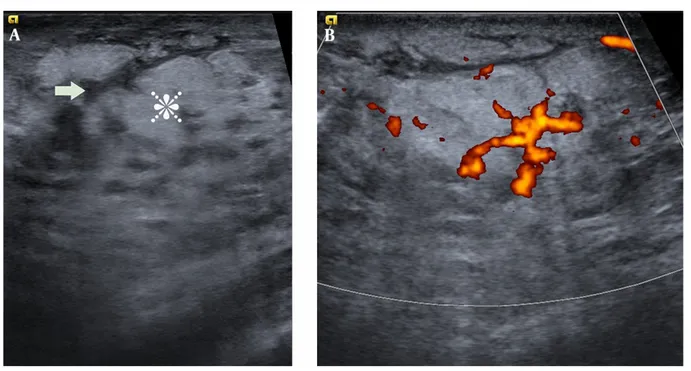
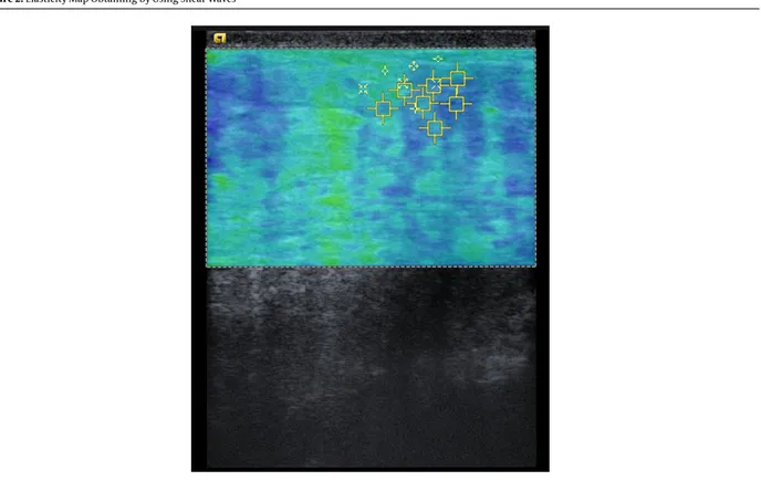
B-mode US showed an ill-defined, hyperechoic region including finger-like hypoechoic areas in the upper in-ner quadrant of the right breast without posterior acous-tic shadowing or enhancement (Figure 1A). Calcification was not seen in US. The subcutaneous fat had also in-creased echogenicity; no lymphadenopathy was shown in the right axilla; and the lesion showed increased vascu-larity by power Doppler (Figure 1B). Differential diagnosis of hyperechoic breast lesions includes both benign and malignant lesions including angiolipoma, hematoma, fat necrosis, metastasis, invasive lobular or ductal carcinoma, and angiosarcoma. On US elastography, when we set the scale to code soft lesions in blue and hard lesions in red, the lesion was coded as a mixture of blue and green areas without any red areas. The shear wave speed is coded on a color scale, ranging from 0.5 to 6.5 m/s. Shear wave US elas-tography showed low shear wave velocities, ranging from 1.89 to 2.43 m/s (Figure 2). Low shear wave velocities and color codes suggested a benign pathology.
Then, we performed breast magnetic resonance imag-ing (MRI). T1 weighted MRI showed heterogeneous sig-nal intensity, which could not be distinguished from nor-mal parenchyma. On T2 weighted MRI, the lesion was hy-perintense with irregular margins (Figure 3A). Dynamic contrast-enhanced MRI showed 3.5 cm mass enhancing
for CD31 and CD34, and negative for keratin (Figure 5). The Ki 67 index was 10%. The findings were concordant with his-tologic and immunohistochemical diagnosis of low- grade angiosarcoma. The patient went to another hospital for mastectomy, and the final diagnosis of the mastectomy specimen confirmed low-grade angiosarcoma.
Positron emission tomography (PET/CT scan) was per-formed 2 months after the surgery, and it showed a scle-rotic metastatic lesion in the T11 vertebrae when compared with the PET/CT scan taken 2 years ago. The patient was treated with radiotherapy, but she passed away 2 years af-ter surgery.
3. Discussion
Commonly, breast cancers are hypoechoic on B-mode ultrasound, and hyperechoic breast lesions are frequently thought to represent a benign finding. Differential diag-noses of hyperechoic breast masses include hematoma, abscess, fat necrosis, lactating adenoma, galactocele, my-ofibroblastoma, hamartoma, angiolipoma, hemangioma, invasive ductal and lobular carcinoma, liposarcoma, lym-phoma, metastasis, and angiosarcoma. Hyperechoic ma-lignant breast masses, which are infrequently seen, can be easily misdiagnosed as a benign pathology.
In our case, angiosarcoma had low color coded scores and velocity values by shear wave elastography, suggesting a benign lesion. Even though the patient had benign elas-tography findings, the physical examination, and patient’s history alerted us to further investigate the lesion. The strong point of our study was that the elasticity character-istics of the angiosarcoma had not been described clearly previously. In contrast to US, MRI showed early rapid en-hancement and wash-out pattern of the mass, which was highly suggestive of a malignant neoplasm. Diffusion MRI also showed restricted diffusion, supporting the diagnosis. Angiosarcomas of the breast may be primary or sec-ondary. Secondary angiosarcomas occur after breast con-servation therapy and radiation therapy. In our case, the patient had angiosarcoma of the contralateral breast
Figure 1.A, B-mode US image showing ill-defined hyperechoic lesions (star) including finger-like hypoechoic areas (arrow) classified as BI-RADS 3; B, Power Doppler image showing increased Doppler flow within the lesion.
Table 1.Summary of Imaging Findings of the Case
US and Doppler US Findings Elastography Findings MRI Findings
Non-mass like lesion without any calcification shear wave velocities ranges between 1.89-2.43 m/s hyperintense at T2-weighted MRI, hypointense at
T1-weighted MRI
Ill-defined hyperechoic lesions including finger-like hypoechoic areas
demonstrate soft lesion on color coded elasticity maps
restricted diffusion
Increased vascularity within the lesion increased echogenicity of the subcutaneous fat tissue
intense enhancement at early phase
Increased vascularity within the lesion type 3 time-intensity curve
treated with mastectomy and radiotherapy. Generally, ra-diation induced sarcomas occur between 4 to 30 years after radiation, and secondary soft tissue angiosarcomas usu-ally tend to occur in the radiation field (4). In our case, an-giosarcoma occurred in the contralateral breast 2 years af-ter radiation, and the lesion was not in the radiation field, suggesting it might not have been associated with radia-tion therapy. Instead, it might have been a second primary malignancy or a metastatic lesion. One of the weak points of our study was that it was not clear whether it was a sec-ond primary or metastases of the primary angiosarcoma. Nevertheless, the similarity between pathologic character-istics of the masses in both breasts indicated that it was probably metastases of the primary lesion. At the time of diagnosis, the patient also had vertebrae metastasis. In the
literature, metastasis to contralateral breast, bones, lung, and liver have been reported previously (5,6). Angiosarco-mas disseminates hematogenously, so axillary dissection is not commonly necessary.
As a result, on US and elastography, our case of an-giosarcoma had imaging features suggestive of benign pathology, while the MRI findings were highly suggestive of malignancy.
Footnotes
Conflict of Interests:The authors have no conflicts of in-terests to declare.
The shear wave speed is coded on a color scale ranging from 0.5 to 6.5 m/s. Soft lesions were coded as blue and hard lesions were coded as red in this scale. The lesion was coded as blue and green without any red areas. Shear wave velocities range between 1.89 - 2.43 m/s suggesting a benign pathology.
Figure 3.A, T2 weighted MRI showing hyperintense signal intensity of the lesion (black star) with irregular margins; B, Dynamic contrast-enhanced MRI shows early intense
enhancement of the lesion (black star).
from any funding agency in the public, commercial, or not-for-profit sectors.
References
1. Hodgson NC, Bowen-Wells C, Moffat F, Franceschi D, Avisar E. An-giosarcomas of the breast: a review of 70 cases. Am J Clin
On-Figure 4.A, Diffusion-weighted MRI at b value of 800 sec/mm2 showed hyperintense signal intensity of the mass (black star); B, The lesion was hypointense on ADC map and consistent with restricted diffusion (black star).
Figure 5.A, Histopathology examination of the specimen revealed a low grade angiosarcoma of the breast. There was an increased mytotic activity within the vascular
endothelial cells (Hematoxylin-eosin stain;×100 original magnification); B, The tumor cells were positive for CD31 (Immunohistochemistry×100 original magnification)
col. 2007;30(6):570–3. doi: 10.1097/COC.0b013e3181131d62. [PubMed:
18091049].
2. Glazebrook KN, Magut MJ, Reynolds C. Angiosarcoma of the breast. AJR
Am J Roentgenol.2008;190(2):533–8. doi:10.2214/AJR.07.2909. [PubMed:
18212243].
3. Yi A, Cho N, Chang JM, Koo HR, La Yun B, Moon WK. Sonoelastography for 1,786 non-palpable breast masses: diagnostic value in the decision to biopsy. Eur Radiol. 2012;22(5):1033–40. doi:10.1007/s00330-011-2341-x. [PubMed:22116557].
4. Garner HW, Kransdorf MJ, Bancroft LW, Peterson JJ, Berquist TH,
Mur-phey MD. Benign and malignant soft-tissue tumors: posttreatment MR imaging. Radiographics. 2009;29(1):119–34. doi:10.1148/rg.291085131. [PubMed:19168840].
5. Arora TK, Terracina KP, Soong J, Idowu MO, Takabe K. Primary and sec-ondary angiosarcoma of the breast. Gland Surg. 2014;3(1):28–34. doi:
10.3978/j.issn.2227-684X.2013.12.03. [PubMed:25083491].
6. Brenn T, Fletcher CD. Postradiation vascular proliferations: an in-creasing problem. Histopathology. 2006;48(1):106–14. doi: 10.1111/j.1365-2559.2005.02293.x. [PubMed:16359542].