i
FUNCTIONAL BACTERIAL AMYLOID NANOMATERIALS
A THESIS SUBMITTED TO
THE GRADUATE SCHOOL OF ENGINEERING AND SCIENCE OF BILKENT UNIVERSITY
IN PARTIAL FULFILLMENT OF THE REQUIREMENTS FOR THE DEGREE OF
MASTER OF SCIENCE IN
MATERIALS SCIENCE AND NANOTECHNOLOGY
By Tuğçe ÖNÜR September 2016
ii
FUNCTIONAL BACTERIAL AMYLOID NANOMATERIALS By Tuğçe Önür
September 2016
We certify that we have read this thesis and that in our opinion it is fully adequate, in scope and in quality, as a thesis of the degree of Master of Science.
Urartu Özgür Şafak Şeker (Advisor)
Devrim Pesen Okvur
Serkan İsmail Göktuna
Approved for the Graduate School of Engineering and Science:
Ezhan Karaşan
iii
ABSTRACT
FUNCTIONAL BACTERIAL AMYLOID NANOMATERIALS
Tuğçe ÖNÜR
M.S. in Materials Science and Nanotechnology Supervisor: Urartu Özgür Şafak Şeker
September 2016
Mis-folded or unfolded proteins tend to aggregate and aggregated structures are called as amyloids. Amyloid formation contributes to some human diseases and resulting in death in some cases. On the other hand, functional amyloids are found in nature and they are highly ordered assembled structures and they function in cellular events. Some bacteria, fungi or yeast species synthesize these kind of functional amyloids. For example, curli proteins of Escherichia coli play a role in initial attachments for biofilm formation and contribute to stiffness of the biofilm matrix. CsgA is the major subunit while CsgB is the minor subunits which nucleates CsgA polymerization. They are capable of attachment to the abiotic or biotic surfaces. Both of them share some characteristics with non-functional amyloids. For instance, their structures are dominated by sheets so they have a rigid amyloid core domain that enables to resist stress factors such as proteases and detergent treatment or pH. Their stable structures and adhesive properties make them useful in materials science. Moreover, high yield could be obtained easily by using molecular biology techniques such as cloning and protein purification so they are highly cost-effective materials.
iv
In this study, CsgA and CsgB fibers were proposed as new type of functional biomaterials to do so fiber formations of CsgA and CsgB were analyzed in detail. csgA and csgB genes were cloned into expression vectors. Their sheet rich structures were validated with CD analysis and binding capability to Thioflavin T dye were assayed which is the general property of amyloids. Self-seeding and cross-seeding strategies were applied to analyze fiber formation and quartz crystal microbalance with dissipation (QCM-D) was used. Gold coated sensors were deposited with freshly purified proteins and polymerized. Then, sensor surfaces were monitored with SEM and AFM. With self-seeding strategies long and branched fibers were obtained from CsgA proteins while sphere like structures were formed by CsgB proteins. Also, it was concluded from the cross-seeding experiments, the order of protein addition determines the final assembled structures. Furthermore, fluorescent properties of CsgA and CsgB were analyzed in detail for the first time. Finally, binding affinity of the purified proteins to different materials (gold, silica and hydroxyapatite) were determined by using QCM-D.
Keywords: biofilm proteins, biomaterials, functional amyloids, self-assembled biomaterials.
v
ÖZET
FONKSİYONEL BAKTERİYEL AMİLOİD NANOMALZEMELER
Tuğçe ÖNÜR
Malzeme Bilimi ve Nanoteknoloji Programı, Yüksek Lisans Tez Yöneticisi: Urartu Özgür Şafak Şeker
September 2016
Katlanmamış ya da doğru katlanmamış proteinler hücre içinde kümelenme eğilimindedirler ve bu kümelenmiş yapılar amiloid olarak adlandırılır. Amiloidler insanlarda bazı hastalıklara neden olurlar ve ölüme yol açabilirler. Fakat, doğada fonksiyonel amiloidler bazı bakteri, mantar ve maya türleri tarafından üretilmektedirler. Fonksiyonel amiloidler oldukça düzenli kümelenmiş yapılardır ve hücresel aktivitelerde rol alırlar. Örneğin, Escherichia coli tarafından üretilen curli amiloid proteinleri yüzeylere tutunmayı sağlayarak biyofilm oluşmasına katkı sağlarlar. Ayrıca biyofilm matriksinin dayanıklılığına da katkıda bulunurlar. CsgA asıl curli altbirimidir. CsgB ise ikincil altbirimdir ve CsgA proteinlerinin polimer oluşturmasını sağlar. İki protein de canlı veya cansız yüzeye bağlanabilir. Hastalık yapan amiloidlerle bazı ortak özellikleri vardır. Örnek vermek gerekirse, yapılarındaki fazla miktardaki yaprakları sayeside sert amiloid çekirdeği oluşur ve proteaz, deterjan ve pH gibi stres faktörlerine karşı stabil kalmalarını sağlar. Stabil kalabilme yetileri ve yapışkan özellikleri bu proteinleri malzeme bilimi için önemli kılmaktadır. Bunlara ek olarak, klonlama ve protein saflaştırma gibi moleküler biyoloji teknikleri ile büyük miktarda uygun maliyetle üretilebilirler.
vi
Bu çalışmada, CsgA ve CsgB yeni nesil fonksiyonel biyomalzeme olarak önerilmiş ve bu proteinlerin fiber oluşumları detaylıca analiz edilmiştir. csgA ve csgB genleri ekspresyon vektörüne klonlanmıştır. Amiloidlerin genel özelliği olan, yapılarındaki yoğun yaprak elementleri dairesel dikroizm spektroskopi (CD) analizi ile gösterilmiştir ve Thioflavin T adlı florasan boyaya bağlanabildikleri analiz edilmiştir. Kendi çekirdeklenme ve çapraz çekirdeklenme yöntemleri izlenerek fiber oluşumu kuvartz kristal mikroterazi (QCM-D) ile analiz edilmiştir.Altın kaplı sensör yüzeyi yeni saflaştırılan proteinlerle kaplanmış ve polimerleşmeleri için bekletilmiştir. Sensor yüzeyi TEM ve AKM ile görüntülenmiştir. Öz çekirdeklenme yöntemi ile CsgA proteinleri kullanılarak uzun ve dallanmış pollimer yapılar elde edilmiştir. CsgB proteinlerinden ise büyük küre benzeri yapılar edilmiştir. Elde edilen sonuçlara göre, proteinlerin ortama eklenme sırası oluşan yapıların şeklini belirlemektedir. Bunlara ek olarak bu çalışmada, CsgA ve CsgB proteinlerinin florasan özelliği ilk defa detaylı olarak analiz edilmiştir. Son olarak, saflaştırılan proteinlerin farklı materyallere (altın, silika ve hidroksiapatit) olan bağlanma afinitesi hesaplanmıştır.
Anahtar kelimeler: biyofilm proteinleri, biyomalzemeler, fonksiyonel amiloidler, kendiliğinden örgütlenebilen biyomalzemeler.
vii
ACKNOWLEDGEMENTS
First and foremost, I would like to express my deepest gratitude to my thesis advisor Asst. Prof. Urartu Özgür Şafak Şeker. Studying with him was a big honor for me. I learned a lot from him about research, experiments, lab equipment and life. He was always generous to share his knowledge and experiences in all fields. I believe he made a major contribution to improve myself during the last two years. He has always encouraged and motivated me. He insisted on showing the light at the end of the road when I felt down. I would like to thank for his guidance, knowledge, patience, encouragement and support.
I would like to thank my lab mates for their friendship; also, the peaceful and fruitful work environment. I want to thank Dr. Esra Yuca for her effort to organize this pleasant lab environment and I appreciate her contributions to this study. Also, special thanks to Tolga Tarkan Ölmez and Ebuzer Kalyoncu for their generous help in imaging techniques. I think they made big contribution to me while learning those imaging techniques and they were always kind to me when I asked for help.
Furthermore, I would like to thank my dear friends from my undergraduate study. I had chance to know better Çağla Eren in Ankara. We spent much time together and she has always supported and motivated me. She has found something to say me even though I was too pessimistic. Also, thanks to Behide Saltepe and Recep Erdem Ahan for their long term friendship. I believe our friendship will last and we will explore new cities and novel food. In addition, I would like to thank Onur Apaydın for his friendship.
Moreover, I am grateful to undergraduate researchers for their help and friendship. I believe they made valuable contribution to my study. I would like to
viii
thank Ilgın Buse Korkusuz, she was always ready to prepare deadly buffers. I want to thank Funda Büyükbaş and I will never forget our growth time sadnesses. Also, I want to thank Hazal Beril Çatalak for their help in various fields. In addition, thanks to Nedim Hacıosmanoğlu he also helped me during the summer internship.
Next special thanks go to the yoga team, Ebru Şahin Kehribar, Elif Ergül and Zelal Yavuz. I will never forget our yoga classes and lunch activities. I also hope our friendship and yoga practices will last in the future.
I want to thank my office mates, Aslı Çelebioğlu, Yelda Ertaş, Begüm Dikeçoğlu, Gülistan Tansık, Zeynep Aytaç and Zehra İrem Gürbüz. They made the warm and friendly study environment.
Moreover, I would like to thank my lifelong friends Büşra-Cansın Arıkoç. It was one of my biggest chance in Ankara that I met my high school friend Büşra. When I need any help, Büşra and Cansın were just a call way. I appreciate them for the big brain storms, supports and encouragements.
Finally, I present my special gratitude to my parents Aysel-Nihal Önür. More than anything, I want to thank to my parents for their unconditional love. I would like to present my special thanks to my sister Ayça Önür. She is the most special person in my life. I believe she can do anything for me no matter how far away from me. I want to thank the new member of our family Burak Gürlek. I think he is one of the main contributor of my academic career and thanks for his advices, encouragements and supports.
I would like to thank TÜBİTAK (Project number 114M163) for the financial support.
ix
CONTENTS
CHAPTER 1 ... 1
INTRODUCTION ... 1
1.1 Proteins as Building Blocks of Cells ... 1
1.2 Self-Assembly of Proteins ... 2
1.2.1 Special Class of Self-Assembled Proteins: Amyloids ... 4
1.3 Biofilm Proteins ... 6
1.4 Functional Amyloids ... 8
1.5 Functional Amyloids of Escherichia coli: curli ... 11
1.6 Major and Minor Curli Subunits ... 16
1.7 Why Amyloids are Important for Materials Science?... 18
1.7.1 Usage of Curli Proteins ... 19
CHAPTER 2 ... 21
EXPERIMENTAL ... 21
2.1 Preparation of Growth Medium and Buffers ... 21
2.2 Cloning of csgA & csgB Gene Fragments... 23
2.2.1 Plasmid Purification ... 23
2.2.2 Chromosomal DNA Isolation ... 23
2.2.3 Polymerase Chain Reaction (PCR) ... 23
2.2.4 Agarose Gel Electrophoresis ... 25
x
2.2.6 Endonuclease Digestion Reactions ... 26
2.2.7 Ligation of Double Digested DNA Fragments ... 26
2.3 Transformation ... 27
2.3.1 Escherichia coli DH5 and BL21 Competent Cell Preparation ... 27
2.3.2 Transformation of Recombinant Plasmids to E. coli Competent Cells .... 27
2.4 Protein Expression, Purification and Characterization ... 28
2.4.1 Overexpression of CsgA & CsgB Proteins ... 28
2.4.2 Purification of CsgA & CsgB Proteins ... 28
2.4.3 Determination of Protein Concentration ... 29
2.4.4 SDS-PAGE and Western Blot analysis ... 29
2.5 Thioflavin T (ThT) Assay ... 30
2.6 Secondary Structure Analysis ... 30
2.7 Quartz Crystal Microbalance with Dissipation (QCM-D) ... 31
2.7.1 Sensor Cleaning ... 31
2.7.2 Fiber Formation Assay ... 31
2.8 Immunochemistry Assay ... 32
2.9 Fluorescence Analysis of CsgA & CsgB Proteins ... 33
2.10 Kinetic Analysis of CsgA & CsgB Proteins ... 33
2.11 Imaging of CsgA & CsgB Proteins ... 34
2.11.1 Scanning Electron Microscopy Imaging ... 34
2.11.2 Atomic Force Microscopy Imaging ... 34
2.11.3 Fluorescence Microscopy Imaging ... 34
xi
RESULTS AND DISCUSSION ... 35
3.1 Cloning of csgA and csgB Gene Fragments ... 35
3.2 Purification of Recombinant Proteins ... 42
3.3 Amyloid Formation Assay ... 45
3.3 Secondary Structural Analyzes ... 46
3.4 Immunochemistry Assay ... 49
3.5 Fiber Formation Assay ... 50
3.5.1 Self-seeding and Cross-seeding of CsgA ... 52
3.5.2 Self-seeding and Cross-seeding of CsgB ... 60
3.6 Imaging of Curli Materials ... 68
3.6.1 Scanning Electron Microscopy Imaging ... 68
3.6.2 Atomic Force Microscopy Imaging ... 76
3.7 Fluorescent Analysis of CsgA & CsgB ... 78
3.8 Kinetic Analysis of CsgA & CsgB... 83
CHAPTER 4 ... 89
CONCLUSION AND FUTURE PERSPECTIVES ... 89
xii LIST OF FIGURES
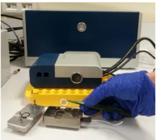
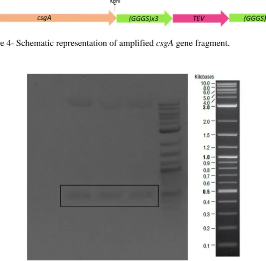
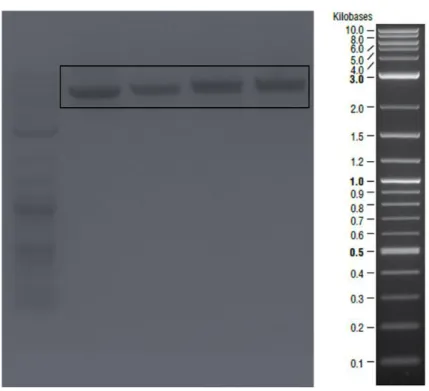
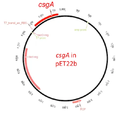
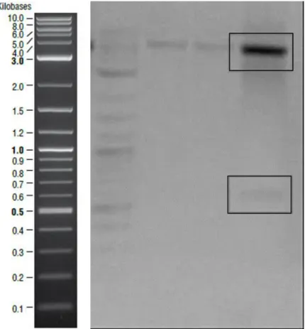
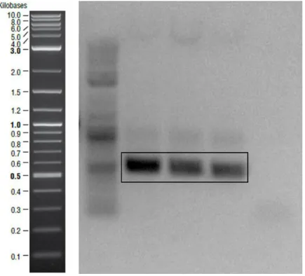
Figure 1- Transcription of curli proteins are regulated via csgBAC and csgDEFG operons. CsgA is the major curli subunit while CsgB is the minor subunit. CsgC shows chaperon activity and CsgD is the regulator protein. Other curli proteins have roles in the secretion machinery. ... 12 Figure 2- Curli secretion machinery. CsgA and CsgB proteins have sec signal sequences and are secreted to the ECM after synthesis. CsgB interacts with outer membrane and acts as template for CsgA oligomerization. CsgG forms channel on the outer membrane while CsgE and CsgF are responsible for transportation. ... 17 Figure 3- Quartz crystal microbalance with dissipation device and QCM-D sensor. 32 Figure 4- Schematic representation of amplified csgA gene fragment. ... 36 Figure 5- Bands inside the square belonged to csgA gene fragments expected as ~500 bp. Negative control was on the first lane. ... 36 Figure 6- Agarose gel image of the second PCR to obtain designed csgA gene fragments which were expected as ~600bp. Negative control was on the last lane. . 37 Figure 7- Linearized pet22b vectors expected as ~5000 bp were shown in the square. ... 37 Figure 8- Schematic representation of pET22b plasmid which contained csgA gene fragment. ... 38 Figure 9- Sequence alignment of cloned csgA gene fragment. Sequences from Genewiz were highlighted with yellow and original sequence on the bottom line. Aligned bases were marked with stars. ... 39 Figure 10- Digested plasmids that contained csgA gene. Samples digested with NcoI were on the second lane and digested with XhoI were on the third lane. On the last
xiii
lane, samples were digested with NcoI and XhoI endonucleases. Backbone ~5000 bp shown in the upper square and csgA gene ~500 bp shown in the bottom square. ... 40 Figure 11- Schematic representation of amplified csgB gene fragment. ... 40 Figure 12- Amplified csgB gene fragments ~500bp shown in the square. Negative control was on the last lane ... 41 Figure 13- Plasmid construct which is designed to express CsgB proteins. ... 41 Figure 14- Sequence analysis of csgB gene. Sequences from Genewiz were highlighted with yellow and original sequence was on the bottom line. Aligned bases were marked with stars... 42 Figure 15- CsgA monomers, dimers and trimmers shown in SDS polyacrylamide gel. ... 44 Figure 16- CsgB oligomers were indicating in red square. ... 44 Figure 17- PVDF membrane of CsgA (on the left) and CsgB (on the right). They have different assembly characteristics and this might affect antibody binding so CsgB bands are brighter than CsgA. ... 45 Figure 18- ThT assays of CsgA, CsgB and equimolar mix sample. Binding capabilities of the purified proteins to Thioflavin T dye were proved by measuring fluorescent signals. Measurements were performed during 16 hours CsgA in A), CsgB in B) and mix of CsgA and CsgB in C). The highest RFU values were recorded with CsgB fibers. ... 46 Figure 19- Secondary structural analysis of purified proteins and mix sample. CD spectra of the samples are demonstrated on the left and ratio of secondary structural elements are on the right. Data of aged CsgA is demonstrated in A), CsgB in B) and mix sample in C). Samples had low helix content in each case. Behavior of CsgA
xiv
similar to mix sample. sheet ratio of CsgB fibers were increasing with incubation time and 30 days aged CsgB sample had the highest sheet ratio. ... 48 Figure 20- Antibody binding assays of purified CsgA and CsgB proteins. Sensor surface was coated with CsgA (on the left) and CsgB (on the right), proteins were probed with fluorescent labelled secondary antibodies and frequency changes were recorded. Protein and primary antibody addition points are shown with arrows. Shifts which were caused from primary and secondary antibodies were too small compared to protein polymer shifts. Hence, their shifts were not seen clearly in this figure. Sensor surfaces were examined with laser scanning confocal microscopy. Image of probed CsgA is demonstrated on the left and CsgB is on the right. ... 50 Figure 21- Self-seeding and cross-seeding strategies are schematized. In this study, interactions of polymerized CsgA with fresh CsgA and fresh CsgB; also, polymerized CsgB with fresh CsgA and CsgB were analyzed. ... 51 Figure 22- Overnight frequency and dissipation changes of freshly purified CsgA proteins. Gold coated sensor surface was deposited with CsgA proteins overnight frequency changes were demonstrated in A) and dissipation changes in B). Initial attachments which shown in close up views causeed sharp rise and decrease in frequency and dissipation signals, respectively. ... 53 Figure 23- Freshly purified CsgA monomers were added upon polymerized CsgA fibers. Addition of fresh proteins with increasing concentrations resulted in step by step changes in frequency signals A) and dissipations signals B). Highest protein concentration caused the highest shift in this case. ... 55 Figure 24- Overnight measurements of frequency shifts in A) and dissipation changes in B) of self-seeding CsgA proteins. Changings which caused from seeding
xv
shows in close-up views. During near 25 hours measurements, dissipation signals did not change notably while frequency shift was increasing. ... 56 Figure 25- Fresh CsgB proteins flowed through the system to analyze interactions of fresh CsgB and CsgA fibers. Peaks belonged to addition point but no significant changes were recorded after the washing steps. Frequency shifts are shown in A) and dissipation changes in B). ... 58 Figure 26- Cross-seeding of CsgA fibers with fresh CsgB proteins. Frequency changes are shown in A) while dissipation changes are shown in B). CsgB addition did not cause dramatic changes. Peaks caused from the monomer addition but unbound CsgB proteins were removed with the washing steps. ... 59 Figure 27- Frequency and dissipation changes of freshly purified CsgB proteins. Purified proteins were deposited on gold coated sensor surface and incubated for overnight for polymerization. Recorded frequency changes in A) and dissipation changes in B). Sharp changes in both signals were caused from the initial attachments. Slight changes were recorded during overnight measurements in both signals. ... 61 Figure 28- Grown CsgB fibers seeded with fresh CsgB. Fresh CsgB proteins were flowed through the system with increasing concentrations but small shifts were recorded in frequency shifts in A) and dissipation shifts in B). ... 63 Figure 29- Overnight measurements of self-seeding of CsgB proteins. Fresh proteins additions shown in close-up view and slightly changes on both signals were recorded during near 20 hours measurement. Frequency shifts are shown in A) and dissipation changes are shown in B). ... 64
xvi
Figure 30- Polymerized CsgB seeded with fresh CsgA as a cross-seeding strategy. Addition of proteins with increasing concentrations cause regular changes on frequency signals in A) dissipation signals in B). ... 66 Figure 31- 20 hours measurements of cross-seeding CsgB fibers with fresh CsgA. Shifts showed the interactions between fibers and fresh proteins. Frequency shifts are shown in A) while dissipation changings are shown in B). ... 67 Figure 32- SEM image of long and branched CsgA fibers. Freshly purified CsgA proteins added upon polymerized CsgA and incubated for overnight for QCM analysis. SEM image of near 3 days aged fibers on gold coated sensor surface in A) and close up view in B). ... 69 Figure 33- CsgB monomers flowed through the system to analyze their interaction with CsgA polymers. Sensor surface was observed with SEM. Long and branch fibers are seen in A) which is similar with self-seeding of CsgA fibers and seeding at the end point is shown in B). ... 71 Figure 34- SEM images of CsgB fibers. Big sphere structures were obtained by self-seeding of CsgB proteins and shown in A); connection sites are shown in close up view in B). ... 73 Figure 35- SEM images of CsgB fibers which seeded with freshly purified CsgA. Sphere structures were observed again but in this case size of them smaller than spheres that formed by self-seeding of CsgB demonstrated in A). Small fibers elongates from the sphere surface which is shown in B). ... 75 Figure 36- After protein deposition and overnight measurements sensors were examined with AFM. Fibers formed by CsgA polymers and fresh CsgA shown in A),
xvii
CsgB polymers and fresh CsgB in B) and cross-seeding of CsgA with fresh CsgB in C). ... 77 Figure 37- Fluorescent analysis of 9 days aged samples. Bright field images and blue, green, red fibers that were imaged by using different filters of fluorescent microscope are shown. Color intensity was decreasing blue to red but brightness of the images were adjusted to show clearly. Images of CsgA fibers and emission spectra that was recorded by exciting at 338, 375 and 550 nm are demonstrated in A), CsgB fibers images and emission spectra that was recorded by exciting at 339, 413 and 550 nm are shown in B), fluorescent images and emission spectra of mix sample are demonstrated in C). Mix sample was excited at 340, 414 and 550 nm to measure emission spectra. Lines represent 100 M. ... 79 Figure 38- 18 hours aged samples were examined under fluorescent microscope. Bright field images were given as the first image and images of CsgA in A), CsgB in B) and mix sample images are placed in C). Lines represent 100 M. ... 80 Figure 39- Bright field and fluorescent images of 23 days aged samples. CsgA fibers are demonstrated in A), CsgB fibers in B) and mix samples in C). Lines represent 100 M. ... 80 Figure 40- After precipitation, CsgA fibers were dissolved in chloroform. Excited at different wavelengths (330, 338, 375 and 418 nm) in order to measure emission spectra. Emission spectra is shown in A) and dense fiber network was observed with SEM in B). ... 82 Figure 41- Frequency change vs. concentration graph of polymerized CsgA, CsgB and mix sample on the gold QCM sensors. Proteins were diluted and added to the system with increasing concentrations. Dots represent the protein addition points and
xviii
higher concentration caused the higher changes in all cases. CsgA deposition on the gold sensor resulted in lower frequency change than other samples. CsgB addition caused the highest change. Interaction of CsgA and CsgB changed the binding property and binding sites of CsgB might be covered by CsgA. ... 84 Figure 42- Frequency change vs. concentration plot of polymerized CsgA, CsgB and mix sample on silica coated QCM sensor. In this case, highest change came from the deposition of mix sample and binding capability of CsgB fibers was higher than CsgA fibers. Proteins were flowed through the system with diluted concentrations and dots represent the addition points. ... 85 Figure 43- Frequency change vs. concentration graph of CsgA, CsgB and mix of CsgA-CsgB fibers on hydroxyapatite QCM sensors. Smaller changes were obtained by binding the polymers to hydroxyapatite sensors. Binding of CsgB fibers to HAP surface caused the highest changes while lowest shifts were obtained by the mix sample. ... 86 Figure 44- Binding equilibrium constants of polymerized proteins and polymerized mix samples on gold, silica and hydroxyapatite (HAP) coated sensors. CsgA and CsgB higher tendency of binding to the gold surfaces. Mix sample had higher affinity for binding to HAP surface than others. ... 87 Figure 45- Binding free energy of aged CsgA, CsgB and mix sample on gold, silica and hydroxyapatite (HAP) surfaces were calculated. All of them had closer binding free energy. It might be cause from same assembly route of the samples. ... 88
xix LIST OF TABLES
Table 1- Components of 12% resolving gel ... 22
Table 2- Components of 4% Stacking Gel ... 22
Table 3- PCR Components ... 23
Table 4- Reaction Conditions ... 24
Table 5- Primers that were used in this study ... 25
Table 6- Digestion reaction components... 26
Table 7- Ligation reaction components... 27
xx
LIST OF ABBREVIATIONS
ECM : Extracellular Matrix
COP : Cytoplasmic Coat Proteins
CCMV : Cowpea Chlorotic Mottle Virus
ThT : Thioflavin T
PrP : Prion Protein
rPrP : Recombinant Mammalian Prion Protein CJD : Creutzfeldt–Jakob Disease
BSE : Bovine Spongiform Encephalopathy
EPS : Extracellular Polymeric Substances
DNA : Deoxyribonucleic Acid
eDNA : Extracellular Deoxyribonucleic Acid FAP : Functional Amyloid in Pseudomonas
BAP : Biofilm Associated Protein
PSMs : Phenol Soluble Modulins
MRSA : Methicillin-Resistant Staphylococcus aureus MTP : Mycobacterium tuberculosis pili
CSG : Curli Specific Gene
TAFI : Thin Aggregative Fimbrae
MFPs : Mussel Foot Proteins
LB : Luria-Bertani
TAE : Tris EDTA Acetic Acid
xxi
DMSO : Dimethyl Sulfoxide
PEG : Polyethylene Glycol
GdnHCl : Guanidine hydrochloride
SDS-PAGE : Sodium Dodecyl Sulfate Polyacrylamid Gel Electrophoresis
PBS : Phosphate Buffer Saline
TBS : Tris Buffer Saline
TBS-T : Tris Buffer Saline-Tween 20
PCR : Polymerase Chain Reaction
TEV : Tobacco Etch Virus
IPTG : Isopropyl 3-D-Thiogalacto-Pyranoside CSLM : Confocal Laser Scanning Microscopy
CD : Circular Dichroism
QCM-D : Quartz Crystal Microbalance with Dissipation
SEM : Scanning Electron Microscopy
AFM : Atomic Force Microscopy
1
CHAPTER 1
INTRODUCTION
1.1 Proteins as Building Blocks of Cells
Proteins are one of the major components of cells and they have crucial roles in cellular activities. Many chemical reactions are catalyzed and channels for transportation on the membrane are formed by proteins also they function as hormones, antibodies, fibers or luminescence.[1] 20 amino acids that have different chemical properties are coded in DNA of the organisms. These amino acids linked to each other via peptide bonds and long amino acid chains form that are called as oligopeptide or polypeptide. Proteins are polypeptide and each of them compose of unique amino acid sequences. Amino acids portions that do not contribute to peptide bonds are side chains. They could be negatively or positively charged and polar or nonpolar so they have crucial effects on the amino acid properties.[2, 3] Weak non-covalent interactions occur between these side chains and these interactions determine the stability of the protein. Hydrogen bonds, Van der Waals and electrostatic attractions are the weak interactions that affect the protein folding and also hydrophobicity is the other factor that contributes the protein conformation. Depends on energy minimization, nonpolar residues on the peptide chain tend to cluster in the interior of the molecule to reduce interactions with water. Also, hydrogen bonds are formed between side chain residues and water molecules. Hence, hydrophobicity is the major factor that determine confirmation of the proteins which has important effects on protein functionality. Proteins could be 50 and 2000 amino acids long and found in varied shapes. Large proteins are composed of domains that fold independently of each other. Hydrogen bonds between the N–H and C=O groups
2
in polypeptide backbones result in α helix and β sheet motifs. Regular and repeating conformation occur in both cases. β sheets are found parallel or anti-parallel orientation and each types of β sheets are highly rigid structure. Rigid cylinder is formed if peptide chain turns around on itself and it is called as α helix. Primary structures of the proteins are known as amino acid sequences and α helix/β sheet are constituted secondary structure of the proteins. The final three dimensional confirmation is refereed as the tertiary structure. Moreover, some proteins are composed of more than one polypeptide chain and known as quaternary structure.[2-8]
1.2 Self-Assembly of Proteins
As previously mentioned, weak bonds which require low energy are formed and dissolved in the cells during metabolic activities and weak interactions occur between protein subunits to stabilize their structure. Flat sheets or closed structures such as tube, spheres could be formed and they are much more stable because formation of those structures require more bonds. They have important roles in cellular activities. For example, cytoplasmic coat proteins (COP) are responsible for capture cargo proteins and they assemble on the membrane surface if budding occurs. They polymerize into spherical cages to capture the cargo molecules and their polymerization causes deformation of the membrane. Cargo molecules inside of the bud are transferred to their target inside the cells.[9] Different types of COP complexes are found in eukaryotic organisms which bud cargo molecules from Golgi apparatus, endoplasmic reticulum or plasma membrane.[10]
Ferritin protein is another example of cage proteins and responsible for iron storage in many species. Hollow cage structure is formed with the assembly of
3
ferritin subunits. Iron molecules are stored in 8 nm inner cavity of ferritin and autocatalytic reaction occurs naturally in the cavity. Oxidation of Fe2+ ions is an enzymatic activity of ferritin proteins. These proteins are highly stable and resistant to temperature, pH, etc. Dissociation occurs at temperatures higher than 70 °C and extreme extreme pH (2-10). [11, 12] In another study, it was proved that hydrolysis of Co (II) was achieved and products were entrapped in the horse spleen ferritin. [12]
With these properties ferritin promises a wide range usage with varies purposes in materials science, bioengineering, drug delivery.
Furthermore, capsid proteins self-assemble to protect viral genetic materials. Protein repeating units assemble around the genetic materials and they are classified according to final assembled structure. There are three types of capsid proteins: helical, icosahedral and complex. Any of them coat DNA or RNA of the viruses to protect from host immune system. In some cases, self-assembled capsid structure is surrounded by host cell membrane. Proteins, lipids which are found on the host membrane and capsid proteins are responsible for the host cell attachments, entry and release into the cells, genetic materials transportation between cells, packaging of the viral particles. Also, they provide stability against chemical or physical stresses.[13]
Identical protein subunits of capsid proteins self-assemble, in vitro. Inner and exterior surfaces have different electrochemical properties. Inner cavity forms a suitable environment for mineralization and neutral charge of the outer surface is important for localization. Cavity size changes in pH dependent manner so releasing of the entrapped molecule could be controlled by environmental stimuli.[14, 15]
In addition, self-assembled cage structures promise wide range of applications in biomedicine. For instance, encapsulation ability is important for drug
4
delivery and catalysis applications. Cowpea chlorotic mottle virus (CCMV) is a model system for switchable molecular entrapment studies.[16] CCMV virions have positively charge interior cavity while cage surfaces are not highly charged. Hence, they offer unique chemical environment. Assembly and disassembly of CCMV virions were triggered with pH changes in a study and it was stated that virions with different interior size properties were versatile strategy for molecular encapsulation.[17]
1.2.1 Special Class of Self-Assembled Proteins: Amyloids
In some cases, soluble proteins could aggregate and form insoluble and stable structures which are called as amyloid fibers. Amyloid term was firstly described by Schleiden and by Virchow in the middle of 19th century. During autopsy, iodine stained deposits were found in the liver and firstly they were thought as natural carbohydrates but their high nitrogen content was explored later. Moreover, further studies showed that those structure were capable of binding to Congo red dye. Also, their fibrillar structures were observed with electron microscopy techniques and some techniques were developed to isolate amyloids to identify their specific structure. X-ray diffraction analysis showed that they had cross-β structure.[18, 19] Amyloid cores are highly stable structure and protect the structure against stress factors. Some of them are non-functional and contribute to some human diseases such as Parkinson's disease, diabetes type 2 and Alzheimer's disease and they are the reason of the mortality for some patients. Their pathogenicity causes from deposition in the extracellular matrix and protein structure studies prove that β-sheet elements are found as predominantly in amyloids. In addition, amyloids give fluorescent shift when bind to Thioflavin T (ThT) fluorescent dye.[20] Also, cross-β-sheet structure
5
is the main characteristic of the non-functional pathogenic amyloids. According to literature, more than two dozen of plasma proteins that could form amyloids have been recognized. Although they did not share amino acid sequences identity, they have common β-sheet conformation. Hence, it was suggested that their stable and insoluble characteristics come from the β-sheet elements.[21] It was proved that amyloids cause some human diseases but it is unclear that amyloid formation is the major reason or the secondary event. Aggregation and plaque formation cause the neurofibrillary tangles and compromise synaptic connections. Hence, they are the reasons of cell loss and dementia.[22, 23]
On the other hand, prion proteins which show amyloidogenic property have been identified by Prusiner in 1982. They cause human diseases but those kind of diseases could be spread from patients to another patient unlike amyloidogenic diseases.[24] Some human and animal diseases such as Creutzfeldt–Jakob disease (CJD) in humans and bovine spongiform encephalopathy (BSE) in cattle are caused from aggregation of prion (PrP) proteins that are found on the outer surface of the plasma membrane. Properly folded proteins are converted to amyloids that have same pathological form. Because of the positive feedback loop, disease is spread easily cell to cell. Yet, it is hard to convert normal PrP to abnormal PrP form without abnormal PrP in the environment.
In addition, same polypeptide chain could formed different types of amyloid fibrils and each of them were capable of converting the normal proteins to abnormal form, according to studies in yeast cells.[25, 26]
Illumination of amyloid formation mechanism is crucial for the development of therapeutic approaches. Studies showed that partially unfolded intermediates were
6
produced during the conversion of soluble peptide to insoluble form.[27, 28] Intermediates were not thermodynamically stable so they turned immediately amyloid fibers which were energetically favorable. Biophysical techniques such as light scattering and fluorimetry were used to identify the transition kinetics. Nucleation dependent polymerization model was suggested in order to identify fibril formation.[29] In that model, a heterogeneous nucleus, which was called as seed, was formed above the critical concentration (threshold) and monomers bound to the free ends irreversibly in order to elongate amyloid fibril. Fibril growth process could be classified as lag and log phase. If seeds were added to the media, lag phase will be shorter than normal process.[30, 31]
X-ray diffraction studies were conducted to identify structure of silk proteins of the lacewing, Chrysopa, firstly. Hydrogen bonds were formed in the parallel direction to the fiber axis and β-strands were elongated perpendicular to the fiber axis. This confirmation was called as cross-β structure.[32] β-sheet elements formed the amyloid core generally and it was thought to provide stability because of the side chain interactions.[33-36] Also, β-sheets could be parallel or anti-parallel direction to the each other that depended on the precursor of the polypeptide chain.[37]
1.3 Biofilm Proteins
Firstly, microorganisms have been defined as planktonic cells that grown in rich culture media. The ability to attach abiotic surfaces of microorganisms have been identified later by van Leeuwenhoek and those surface-associated microorganisms have different growth rate and gene transcription level so distinct phenotypes are observed. This surface associated micro colonies are covered by polysaccharide layer and this structure is called as biofilm. Biofilms have
7
complicated and distinct three dimensional architecture. Micro-colonies are divided by water channels and biofilms are resistant to stress factors. Four processes occur during biofilm formation: initial attachment, biofilm development, biofilm maturation and dispersal. Microorganisms attach to surfaces by the aid of various cellular components such as pili, fimbria and adhesive proteins.[38] Then, microbial communities are covered with extracellular polymeric substances (EPS). EPS provides resistance against mechanical or chemical stress factors also stabilizes bacterial cells on the surfaces. Hence, it was thought that it has remarkable effects on most microbial infections.[39, 40] Biofilm development changes gene expression and reduces growth rate. Specific genes are up regulated while some of them are turned off. Communities in the biofilm matrix could communicate with each other via secretion of chemicals and this process is known as quorum sensing. It is important for the fate of biofilm structure by determining the attachment or detachment from surfaces and it also affects gene expression profiles. In addition, various types of biomolecules are found in biofilm matrix such as polysaccharides, proteins and extracellular DNA (eDNA), etc. Each of them contributes to the formation of hydrated and polar structural scaffold.[41] For instance, structural stability is obtained with polysaccharides and coherent cells are aligned by eDNA.[42] Different types of proteins, which are up regulated during the development process, are found in the biofilm matrix. They contribute structural stability and have role in metabolic pathways. For example, amyloid proteins are produced during biofilm development but they have specific functions for the cells unlike pathogenic amyloids. They have thought as amyloids because they share structural properties with disease causing amyloids. For instance, both types are non-branching, have cross-β structure and they
8
could resist against chemical or physical forces. In addition, they give red shift when bind to Congo red dye and fluorescent shift in the presence of ThT dye that are the main characteristics of amyloids. On the other hand, they are synthesized by a special machinery which is tightly regulated. They are beneficial for microorganisms and have specific roles in cellular behaviors. Because of these reasons they have defined as functional amyloids not cellular mishap.[43-46]
Bacterial amyloid fibers associate with each other and also bind to the small molecules such as quorum-sensing metabolites. Hence, they have crucial role in retaining the nutrients and other molecules under aquatic flow.[47] Amyloids give the high tensile strength to the biofilm structure.[48] They can resist against mechanical strengths while offer soft environment for bacterial life. Studies clarify that amyloids increase the hydrophobicity of biofilm structure and stiffness, significantly. Hence, it could be concluded that amyloids make the most crucial contribution to the robustness of biofilms.[49]
1.4 Functional Amyloids
As mentioned above amyloids are non-functional and cause many diseases. In contrast, functional amyloids were found in nature according to recent studies. They share cross-β structure but they are not misfolded or unfolded proteins. They are produced by a special machinery which is tightly regulated. Some enteric bacteria species, fungi species and also yeast synthesize them and they have role in cellular activities. For example, Pmel17 is a mammalian protein that shows fibrous structure and shares same amyloid fibril properties. It is located at the surface of melanosomes and it was suggested that Pmel17 played a role in melanin synthesis.[50] In addition, hydrophobins are amyloid like proteins that function in
9
fungal development. During sporulation, they form a coat to repel water.[51] Moreover, recombinant mammalian prion protein (rPrP) was toxic for cultured cells and cell apoptosis could be induced by fibrils of rPrP. On the other hand, monomeric form of rPrP, which was α-helices abundant structure instead of β-strands, promoted neurite development and neuron survival. According to the study, β structure dominated fibrillar form were highly toxic; also, caused cell death.[52]
Furthermore, Pseudomonas strains have ability to form biofilm and fap (Functional Amyloid in Pseudomonas) operon was discovered in Pseudomonas.[53] When it was overexpressed in Escherecia coli, biofilm formation increased. Six proteins (FapA-F) are synthesized and FapC is the main amyloid component.[54] Moreover, fap operon are found in beta- and delta- Proteobacteria not only in Pseudomonas.[55]
Staphylococcus aureus is an example of the opportunistic pathogen species and produces biofilm associated protein (Bap) that shows self-assembly properties in order to build biofilm matrix depending upon the environmental stimuli. In acidic environment and low calcium concentration, Bap proteins form amyloid like structures. Yet, cation binding to the molten globule like state that is the intermediate state prevents self-assembly and amyloid fiber formation. If calcium concentration increases in the media, Bap coordinates with cation ions so fibrillation process is hindered. It was concluded that Bap proteins function as sensors and they are scaffold proteins that promote biofilm formation according to environmental stimuli.[56] In addition, they are found at the cell surface and compose of a repetitive structure. Also, they have high molecular weight and compose of domains.[57] They are responsible for initial attachment to the different surfaces and interactions with
10
bacterial or host cells. They are capable of adhere to the mammalian epithelial cells so they prevent internal localization by immune cells. Because of the adhesive property, they have remarkable effects on the persistence of the infections.[58, 59]
Another study suggests that small peptides which are named as phenol soluble modulins (PSMs) promote biofilm integrity in S. aureus species. PSMs lacking mutants do not form robust biofilm structure. Mechanical stress and also enzymes disrupt biofilm structure of the mutant strains. While soluble forms of PSMs are disperse in the biofilm matrix, aggregated fibrils, which have amyloid characteristics, function in metabolic activities in the biofilm.[60] Their antimicrobial acticity against other bacteria was also proved and they are responsible for recruiting and lysing neutrophils hence they prevent host immune response. In addition, community-associated Methicillin-resistant Staphylococcus aureus (MRSA) strains synthesize virulence factors that contain PSMs.[61, 62]
Also, Mycobacterium tuberculosis produces functional amyloid proteins. It produces special types of pili that are known as M. tuberculosis pili (MTP) and they are formed by protein subunits that have low molecular weights. Adhesive property of MTP was suggested in a study and binding to the laminin that is an extracellular matrix protein was showed. Hence, it is important for host colonization. Morphological, functional and biochemical characteristics of MTP are similar with curli amyloids.[63, 64]
Furthermore, Bacillus subtilis is capable of endospore production. Endospores are coated by complex proteins so they could resist against stress factors such as protease treatment, pH changes, etc. Exopolysaccharides and TasA proteins are the main components of the matrix and TasA is associated with spores and
11
functions as antimicrobial. Later studies proved that TasA proteins were the major protein components and formed amyloid fibers that were crucial for the biofilm integrity.[65, 66] Spores of tasA lacking mutant strains have asymmetric material distribution and altered outer coat unlike wild-type strains. Another study suggested that TasA and GerE acted synergistically for endospore formation. Also, maturation of TasA depends on another protein which is called as SipW. It has bifunctional activity: signal peptidase and regulatory roles. Signal peptidase activity is not required for the biofilm development but it is still required for that process. Hence, its regulatory role was suggested in a recent study.[67] If mother cell engulf the pre-spore cell, precursor TasA is translocated to the pre-spore membranes in order to nucleate the organization of the under endospore coat.[68] TasA proteins self-assemble and form stable fiber that are similar with functional amyloids. TapA is the assistant protein of TasA. Bacillus subtilis biofilm formation relies on the assembly of a fibrous scaffold which is formed by TasA protein. TasA polymerizes into highly stable fibers. 8 amino acids residues at the N term of TapA are essential for TasA fibrillation. In the absence of those residues, biofilm formation delays, in vivo. [69] 1.5 Functional Amyloids of Escherichia coli: curli
The most studied functional amyloid is curli that is produced by Escherichia coli and discovered in the 1980s. Salmonella species also produce curli fimbria. Both type of curli have similar roles such as cell to cell contact, host cell adhesion and invasion.
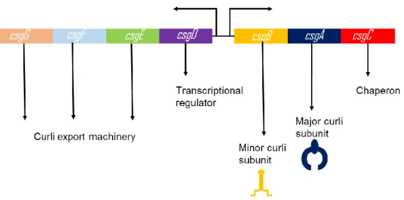
In E. coli, transcription of curli subunits, other regulatory and transporter proteins are regulated by curli specific gene operons: csgBAC and csgDEFG which are represented in Figure 1.[70]
12
Figure 1- Transcription of curli proteins are regulated via csgBAC and csgDEFG operons. CsgA is the major curli subunit while CsgB is the minor subunit. CsgC shows chaperon activity and CsgD is the regulator protein. Other curli proteins have roles in the secretion machinery.
On the other hand, agfBA and agfDEFG opreons regulate curli production in Salmonella spp. Mutations on those genes have similar effects with csg opreons. Tafi (thin aggregative fimbriae) is the special name of curli fimbriae that is synthesized by Salmonella typhimurium.[71]. CsgB is the minor subunit while CsgA is the major curli subunits. Chaperone activity of CsgC protein was recently identified. During translocation to the extracellular environment, CsgC prevents aggregation of curli subunits in the periplasmic space.[72] Major and minor subunits have 30% sequence identity. Also, both of them compose of five imperfect repeating units.[73] CsgA polymerization requires CsgB minor subunit. CsgB and CsgA could be synthesized by different cells. If csgA lacking mutants and csgB lacking mutants are found in the same media, polymerization occurs because of the interbacterial complementation. Yet, this process do not occur in Salmonella enterica.[74] Accessory proteins are also required for assembly and they are encoded by csgDEFG operon. While CsgD
13
regulates the other operon positively, synthesis of CsgD proteins is not regulated by itself.[70, 75] Because of helix-turn-helix motif at C terminal, DNA binding capability of CsgD was suggested but there was no experimental evidence to prove its potential. Also, it was proposed that its activation is triggered by phosphorylation event but mechanisms that control expression and activation are not clear.[75] CsgG forms a channel on the outer membrane so it is required for secretion of curli subunits.[76, 77] CsgG protein is analog of other outer membrane channel-forming proteins.[78, 79] According to studies, fiber formation did not occur in csgG lacking strains. Also, those strains were not capable of functional major or minor curli subunit production.[80] CsgE is found in the periplasmic space and little amount of fiber formation was observed in mutant strains. They were less stable than wild-type subunits and they did not compensate polymerization by using wild-type subunits from wild-type strains. Although those mutants synthesized a few CsgA fibers, they were not similar with wild-type fibers, morphologically.[76] Another important point is the interaction of CsgE and CsgG at the outer membrane. It was thought that CsgE or CsgG act cooperatively because CsgE or CsgG deficient strains had similar phenotypes. Yet, exact role of CsgE on curli assembly is still unknown. However, CsgF is a periplasmic protein which interacts with CsgG, phenotype of csgF lacking strains is different from csgE lacking mutants. On the other hand, csgF lacking mutants had similar phenotype with csgB deficient mutants since soluble and unpolymerized CsgA produced by mutant strains.[76-79]
As mentioned before, curli subunits are functional amyloids and they are synthesizes by a special machinery that is controlled tightly. In addition to CsgD control on csgBAC operon, environmental factors affect curli expression. Below
14
30°C curli expression is promoted; also, many E. coli strains could synthesize curli at 37°C.[81, 82] Contents of growth media also affect curli expression. For example, in the absence of salt, curli proteins are transcribed maximally. Limitations of phosphate, iron and nitrogen trigger curli biosynthesize.[83] Oxygen content of the environment also affects, maximal CsgD expression occurs at low oxygen environment.[84]
Furthermore, curli expression level changes during bacterial growth. Studies showed that maximum transcription of curli genes occurred during stationary phase and that process depended on RpoS that was the stationary-phase sigma factor.[85, 86] Moreover, Crl protein was proposed as thermal sensor and it increased curli protein synthesize at low temperature. Crl is more stable at low temperature so it was thought that its stability could have positive effects on curli transcription.[81, 87] In addition, RpoS and Crl act cooperatively to regulate csgBAC operon. Also, RpoS activates MlrA expression that regulates CsgD biosynthesis, positively.[88, 89]
Additionally, there are dual regulatory systems which regulate transcription of curli protein and they affect dramatically curli gene expression more than other factors. One of them is OmpR/EnvZ system and it regulates curli expression according to osmolarity of the environment. EnvZ is sensor kinase protein that senses changings to activate OmpR regulator protein. Furthermore, expression of CsgD that is another curli operon regulator protein is regulated positively by OmpR.[83, 90] The other two component regulator system is the CpxA/R system which is activated by overexpression of csgA when csgG gene is not overexpressed. CpxA is a sensor kinase like EnvZ and CpxR is the regulator like OmpR but it regulates negatively. In addition, membrane and misfolded proteins in the periplasm activate that system and
15
cause the upregulation of periplasmic chaperones and proteases. It was suggested that curli subunits were found transiently in the periplasmic space due to the CpxA/R system.[91-93] Moreover, outer membrane stress results in the activation of Rcs pathway which regulates csgD transcription negatively. Rcs pathway has important roles in biofilm formation so it could be concluded that regulation of biofilm formation is linked to the regulation of curli operons. It was suggested that curli had roles in initial stages of biofilm development. They might be responsible for the initial adhesion. It was proposed that curli expression was turned off by Cspx and Rcs pathways.[94-96] Curli gene expression was triggered in non-adherent strains of Escherichia coli to form biofilm, according to a study. Biofilm formation ability was identified as CsgA dependent process in that strain.[97] Morphology of the biofilm which was developed by curli lacking mutants was analyzed in another study. They formed flat biofilm which was not similar with wild-type biofilm.[98]
Although they are beneficial for microorganisms, they could contribute human diseases. For instance, curli proteins contribute to the host cell attachment and invasion, induce host immune response by interacting with host proteins.[82, 86, 99] Also, interactions with host proteins enable to spread in host organisms.[82] CsgA proteins that are major subunits of curli were detected in sera of sepsis patients.[82] In addition, it was reported that polymerization of the disease-associated amyloid protein A was induced by injection of curli proteins to the mice. Curli might act as a seed for disease-associated amyloid protein A polymerization.[100] Furthermore, curli proteins are capable of binding to the plant cells but this ability changes strains to strains because other cellular components also have roles in attachment according
16
to mutant studies. Moreover, adherence of alfalfa sprouts decreased in csgB lacking strains of S. enterica but not in csgA deficient mutants.[101, 102]
1.6 Major and Minor Curli Subunits
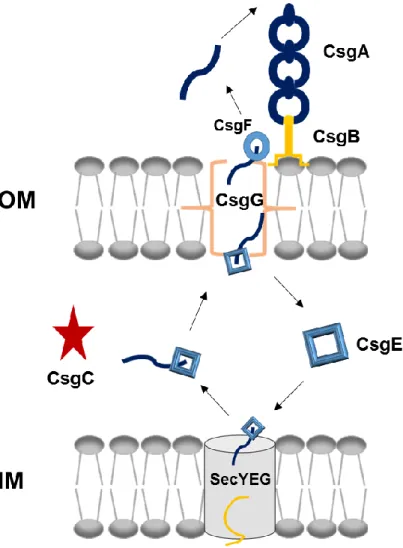
Major and minor curli subunits (CsgA and CsgB) have similar characteristics. For instance, they have sec signal sequence at N terminals and following 22 amino acids in CsgA and 23 amino acids in CsgB important for secretion. Five imperfect repeating units that are responsible for β sheet formation follow these sites. Also, they have %49 sequence similarity and 30% sequence identity. Amyloid core which is resistant to many stress factors are formed by these repeating units.[73, 103] According to studies, CsgA and CsgB proteins are synthesized as monomers which are soluble forms and then they are translocated to the extracellular media because sec signal sequences at N terminals direct them to the periplasmic space and they are cleaved here. CsgB anchors the outer membrane via fifth repeating unit and acts as a template for CsgA polymerization. Other CsgA proteins bind to each other for elongation and secretion machinery is represented in Figure 2. csgB lacking mutants are capable of soluble CsgA secretion. Without CsgB subunits CsgA do not form oligomers, polymerization could be achieved by interbacterial complementation, in vivo.[73, 76] On the other hand, self-seeding occurs for CsgA and CsgB, in vitro. Also, CsgA polymerization is induced by addition of preformed CsgA fibers to the environment. The first and fifth repeating units of CsgA serve as interaction sites.[104] Moreover, Ser-X5-Gln-X4-Asn-X5-Gln residues are conserved in the five
repeating units of CsgA so they may contribute the amyloid formation. Studies proved that specific side chain interactions are important for self-seeding and CsgB nucleation. Polar side chains participate CsgB nucleation while self-seeding of CsgA
17
does not depend on polar side chains. Also, aromatic residues that found in CsgA do not affect positively amyloid formation.[105] Predicted structure of CsgA was identified as β strand-loop-β strand and it could be right or left handed that were energetically equal. Also, β strands elongated as parallel. Interactions between β sheet layers resulted in rectangular core domains which were highly hydrophobic. This predicted structure was in agreement with previous studies.[106]
Figure 2- Curli secretion machinery. CsgA and CsgB proteins have sec signal sequences and are secreted to the ECM after synthesis. CsgB interacts with outer membrane and acts as template for CsgA oligomerization. CsgG forms channel on the outer membrane while CsgE and CsgF are responsible for transportation.
18
1.7 Why Amyloids are Important for Materials Science?
Amyloids are dominated by β-sheet structures which are highly stable and
energetically favorable form. According to microscopic studies, they are few micrometers with 7–10 nm diameters. Another important property is their stiffness which is comparable to silk. Based on the physical measurements, it was proved that they were as strong as steel. Much as described above, they could resist stress factors such as detergent or protease treatment, temperature, etc. Molecular biomaterials such as wires, scaffolds, liquid crystals could be fabricated by using amyloids with bottom-up strategy. They assemble into highly ordered structures and they are produced easily with low cost. Also, they could be tuned easily with protein engineering techniques. All of these properties make them useful source for materials science. For example, nanoscale electrical circuits could be designed according to a study. Protein fibers with 10 nm wide, which were highly stable, produced from prion proteins of Saccharomyces cerevisiae that were known as Sup35p. Length of the fiber were controlled easily and could be reached to hundreds micrometers. Silver and gold wires by deposition of ions on colloidal surface were achieved and near 100 nm wide conductive wires were obtained.[50] Also, water filled nanotubes may be produced from amyloid assembly which may be useful for nanowire production.[107] Amyloid proteins could be formed liquid-crystal phases like many polymers. It was suggested that liquid-crystal phases were observed with hen lysozyme at low pH. New type of soft-solid like materials that is biodegradable and also biocompatible could be obtained with hen lysozyme.[108, 109] Natural amyloids have adherent properties and they function in attachment to the biotic or abiotic surfaces. In a study, hydrophobins were used to change chemical properties
19
of surfaces to improve biocompatibility. It was possible with their amphipathic nature. Hence, they may serve scaffolds for tissue engineering.[110] Furthermore, amphiphilic peptides were produced artificially which could undergo ordered assembled structure in liquid medium. Hence, those novel artificial peptides may support cell growth and they have big potential to use in tissue engineering.[111]
Also, amyloid peptides were designed to increase carbon dioxide capture capacity and gene transfer efficiency.[112] In addition, protein film was created to form hierarchically assembled amyloids fibers.[113]
To sum up, protein based materials could be easily manipulated and recent studies proved that they could be controlled and stable structures could be obtained. Molecular biology techniques and protein engineering have been developing day by day to improve recombinant protein production strategies, decrease cost and increase the yield. As discussed above, amyloids have big potential to use in various fields such as tissue regeneration, sophisticated three-dimensional scaffolds formation, cage for controlled drug release, coating materials, conductive wires, biosensors or bioswitches.[114-116]
1.7.1 Usage of Curli Proteins
Curli subunits are highly adhesive proteins which function in surface attachment. They were suggested as underwater adhesives and CsgA proteins fused with mussel foot proteins (Mfps) of Mytilus galloprovincialis in a study. The highest underwater adhesion energy was determined from the fused proteins.[117] Furthermore, they are good candidate for enzyme immobilization. CsgA were used to immobilize α-amylase enzyme via covalent bonds. Stability was tested at different pH and with organic solvents.[118] In another study, genetic circuits were designed
20
to produce CsgA which depended on environmental signals. CsgA fibrils were interfaced with quantum dots and gold nanoparticles. With this strategy gold nanowires or nanorods could be produced.[119] Peptides were fused with CsgA and new recombinant proteins synthesized and secreted by E. coli cells. CsgA proteins were assembled onto the outer membrane without losing peptide functionality. This system was called as Biofilm-Integrated Nanofiber Display (BIND).[120]
21
CHAPTER 2
EXPERIMENTAL
2.1 Preparation of Growth Medium and Buffers
To prepare 1 L of Luria-Bertani (LB) broth media, 10 g tryptone, 5 g sodium chloride and 5 g yeast extract were dissolved in deionized water and final volume was adjusted to 1 L. Also, for 1 L agar media, 15 g agar was added to LB media.
50X Tris-Acetic acid-EDTA (TAE) media was prepared and diluted to use in agarose gel electrophoresis. 242 g Tris was dissolved in deionized water and 100 mL of EDTA from 0,5 M EDTA solution was added. Finally, 57,1 mL of 100% acetic acid was added. Deionized water was added to reach 1 L.
To prepare Transformation & Storage Solution (TSS), 5 g polyethylene glycol (PEG) mixed with 1,5 mL of 1 M MgCl2 and 2,5 mL of dimethyl sulfoxide
(DMSO). LB was added to reach 50 mL. Then, solution was filtered with 0,22 m filter.
10X SDS (Sodium Dodecyl Sulfate) running buffer was prepared with 30 g Tris, 10 g SDS and 144 g glycine. All components were dissolved in deionized water and final volume was adjusted to 1 L. It was diluted to 1X before use in SDS-PAGE (Sodium Dodecyl Sulfate-Polyacrylamide Gel Electrophoresis).
2X Laemmli sample buffer was prepared by mixing 65,8 mM Tris-HCl, 2,1% SDS, 26,3% (w/v) glycerol and 0,01% bromophenol blue.
TGX Stain-Free FastCast acrylamide solutions were used to prepare 10% SDS gel according to manufacturer instructions.
Furthermore, SDS-PAGE was performed with 12% resolving gel and 4% stacking gel. Components of them given the following tables.
22 Table 1- Components of 12% resolving gel
Deionized water 3.2mL Acrylamide/Bis-acrylamide (30%/0.8% w/v) 4 mL 1,5M Tris(pH=8.8) 2,6 mL 10% (w/v)SDS 0,1 mL 10% (w/v) ammonium persulfate (AP) 100 L TEMED 10 L
Table 2- Components of 4% Stacking Gel
Deionized water 2,975 mL Acrylamide/Bis-acrylamide (30%/0.8% w/v) 1,25 mL 0,5M Tris(pH=8.8) 0,05 mL 10% (w/v)SDS 0,67 mL 10% (w/v) ammonium persulfate (AP) 50 L
TEMED 5 L
10X phosphate buffer saline (PBS) was prepared with 80 g sodium chloride, 1 g potassium chloride, 14,4 g sodium dihydrogen phosphate and 2,4 g potassium dihydrogen phosphate. All of them dissolved in deionized water and final volume was adjusted to 1 L. Before use, stock solution was diluted.
Recombinant proteins were purified in denaturing conditions. To prepare wash buffer, 6 M GdnHCl was dissolved in phosphate buffer saline (PBS) and 10 mM imidazole was added while 150 mM imidazole added for elution buffer. Wash buffer used as lysis buffer to dissolve cells. Also, 8 M GdnHCl in PBS was used as extraction buffer.
Tris buffered saline (TBS) was used for western blot analysis. 20 mM Tris and 150 mM sodium chloride was dissolved in deionized water. 5% (w/v) non-fat milk powder was added and it was used to block membrane. Antibodies were also diluted in that solutions. Tris buffered saline with Tween 20 solution was used to wash western blot membrane by adding 0,1% Tween 20 to TBS.
23 2.2 Cloning of csgA & csgB Gene Fragments 2.2.1 Plasmid Purification
Plasmid isolation was performed by using Qiagen miniprep kit according to manufacturer’s instructions. Plasmid DNA was eluted with preheated water (65°C). Plasmid concentrations were determined with Nanodrop. They were stored at -20°C. 2.2.2 Chromosomal DNA Isolation
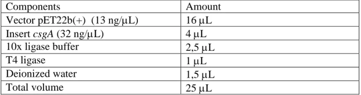
Qiagen genomic DNA isolation kit was used to purify E. coli genome according to manufacturer’s instructions.
2.2.3 Polymerase Chain Reaction (PCR)
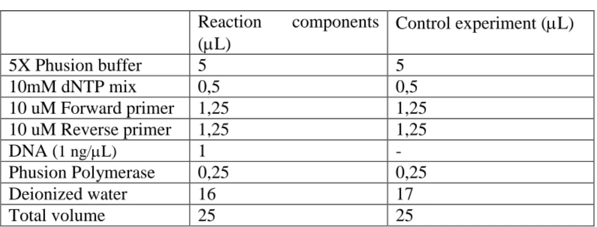
csgA gene fragments were amplified from E. coli genome and GS soft linker sequences that compose of three repeats of GGGS amino acids (G for glycine and S for serine), Tobacco Etch Virus (TEV) protease restriction site and same soft linker sequence were added to the 3’ prime of csgA genes. Two different PCR reactions were performed. First PCR components and conditions are given in Table 3 and 4, respectively. For two-setp PCR, first annealing temperature was calculated as 70°C and second was 89 °C.
Table 3- PCR Components Reaction components (L) Control experiment (L) 5X Phusion buffer 5 5 10mM dNTP mix 0,5 0,5 10 uM Forward primer 1,25 1,25 10 uM Reverse primer 1,25 1,25 DNA (1 ng/L) 1 - Phusion Polymerase 0,25 0,25 Deionized water 16 17 Total volume 25 25
24 Table 4- Reaction Conditions
Temperature (°C) Time Cycle Initial denaturation 98 30 sec
Denaturation 98 10 sec 5 cycle Annealing 70 20 sec Extension 72 20 sec Denaturation 98 10 sec 30 cycle Annealing 89 20 sec Extension 72 20 sec Final Extension 72 7 min
Second reaction was set up according to Table 3. PCR products of the first reaction were used as template in the second reaction. Same PCR conditions by just changing annealing temperatures were set. First annealing temperature was calculated as 60°C and the second was 82°C.
Furthermore, csgB gene was amplified from E. coli genome and reactions were set up according to Table 3 and PCR conditions are given in Table 4. Annealing temperature were calculated as 59°C and 72°C. Primers are listed in Table 5.
25 Table 5- Primers that were used in this study
Primers Oligonucletotide sequences
The first reaction of csgA Forward GCTCCTCGCTGCCCAGCCGGCGATGGCCATGGAT GAAACTTTTAAAAGTAGCAGCAATTGCAGCAAT CGT Reverse AAAATACAGGTTTTCGCCGCTACCGCCTCCGCTA CCGCCACCGCTACCGCCGGTACCGTACTGATGAG CGGTCGCGTTGTT The second reaction of csgA Forward GCTGCCGACCGCTGCTGCTGGTCTGCTGCTCCTC GCTGCCCAG Reverse GTGCTCGAGGCCGCTACCGCCTCCGCTACCGCCA CCGCTACCGCCGCCCTGAAAATACAGGTTTTCGC CGCT The reaction of csgB Forward GCTCCTCGCTGCCCAGCCGGCGATGGCCATGGAT GAAAAACAAATTGTTATTTATGATGTT Reverse CTCCGCTACCGCCACCGCTACCGCCGGTACCACG TTGTGTCACGCGA
2.2.4 Agarose Gel Electrophoresis
1% (w/v) agarose gel was used to observe DNA fragments. 1 g of agarose was dissolved in 1X TAE and boiled. After cooling, SYBR® Safe was added and it was poured into the gel cast. PCR products were loaded to the agarose gel and 1X TAE buffer was used as a running buffer. Horizontal electrophoresis apparatus was used to carry out experiments. Purple loading dye (NEB) was added to the samples before loading to gel and 100 Volts were applied for 40 minutes to carry out gel electrophoresis. 2-log DNA ladder marker was used to determine molecular weights of the PCR products.