(Director: Prof.Dr. Mahir Pamukçu) and
Department of Internal Medicine (Director: Prof. Dr. Selahattin N. Yalkı)
INTRACRANIAL NERVE ROOTS TUMOR IN A DOG.
Hüseyin K. Urman* and Cahit Özcan**
Tumors of intracranial nerve root in dogıesticated animals have been infrequently reported in dogs (LA), cattle (3), and horse (z). These int-racranial tumors observed were neurofibromas in dogs, neurilemmomas in. cattle, and in one horse.
This paper describes a neoplasm which arose from the nervi glosso-pharngicus, vagus, accessorius and hypoglossus.
Clinically: A six year-old, male gorden setter dog was admİtted to the clinic with a tentatiye diagnosis .of encephalitis and was under observation for one and a half months. The owner stated that the dog was vomiting, coughing and had sjgns of intermİttent incoordination on both hind legs. The animal leaned against objects, apparently in an attempt to support itself. No circling movemets was observed, but there was a tendency to the left while he was walking. Unilateral mydriasis, and absence of pupillary reflex to light of the left eye were observed. There was slight lowering of the left side of the lower lips.
In spite of the treatment with antibiotics and calments the dog's health deteriorated graduaııy. Finally he feıı down repeatedly when he was for-ced to move, and appeared depressed euthanasia was performed at the ow. ner's reques~.
Necropsy findings: Post mortem examination of the brain revealed a peanut-shape mass involving the nervi glossopharyngicus, vagus, ac-cessorius and hypoglossus lying in the angle formed by the cerebellum and meduııa. This grayish-white rather firm mass protruted through the hyopg-lossus and jugular canals to from a second enlargement between the bulla tympanica and occipitale basis. The intracranial portion of the tumor had
* Doç. Dr. Med. Vet. Dept. of. Pathology
**
Doç. Dr. Med. Vet. Dept. of. Internal MedicineHO
Intracranial tumor
slightly displaced the cerebellum and the medulla laterally. In comparison \vith the normal right side of the aboral part of the cranium the hypoglossal, and jugular channel of the left side was widen due to com pres sion of the tumor to the bony wall (Fig. ı). The tumor produced a depression on the inner side of the left bulla tympanica. The whole bony \ValI of the bulla was atrophied, and contained small osteophyts on its surface.
Fig. i - The jugular and hypoglossal channcls are widen in comparison with the ri-ght side. The waıı of the buııa tympanica is atrophied and pcrforctcd.
The tumor had arather hard consistenel'. On cut section the tumor was pale, grayish-white and homogencous.
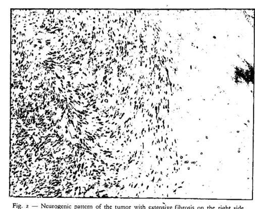
Microscopically: The tumor is not encapsulated, and in so me areas nerve fibers were diffusely incorporated within its substance. Some large cells, resembling nerve celles, were seen in one part of the tumor. Multinucleated cells were also seen in a different part of the tumor tissue. Some focal areas suggested atypical tactile corpuscles which were in fact whorled arrange-ments and interlacing bundles of fibers. Verocay bodies ,and unquestioned palisading of nuclei were not recogı:ıized. Most cells and their nuclei were elongated (Fig. 2), and well-formed fibers of collegan arranged in interlacing fasciculi were numerous. Mitotic figures were relatively common. There are extensive areas of necrosis and fibrosis. The walls of some of the vessels were thickened and contained dense hyalin material; some of the m showed a necrotic endarteritis.
" o
Fig. 2 - Ncurogcnic pattcm of the tumor with cxtcn;ivc fibrosis on the right side. j-Iematoxyline and eosin x 15°
Diseussion: The tumor described appears to represent a nerve - sheath tumor, it is not morphologieaIly identical with the typical neurilemmoma, as is so eharaeteristicaIly observed involving the aeoustie nerve of man,
and animaIs. This tumor eould be designeted as NEUROFIBROSARCOMA on the basis of eell and anaplasia, and diffuse involvement of the multiple
nerves, laek of eneapsulation, paIisading of nucIei and Veroeay bodies. Altough the loeatlon of the tumor was in the eerebeHo-medullar angle no circIing movements \Vas observed; may be the innerpart of the tumor have not reaehed sufficient size to eause this type of movement whieh is eharae-teristic in eerebellopontine angle lesions. However, there was atendeney to the left when he was foreed to walk.
We are not able to explain the absenee of pupiIIary reflex of the left eye whieh is aetuaHy innerveted by the n. oeulomotorius. No lesions were ob-served at this part of the brain nor at the n. oeulomotorius.
St. C!air and Safanie (I) have reported a somewhat asimilar ease in a 3-year-old dog, exeept that only the root of the left trigeminal nerve was in-volved and maIignaney was perhaps not as cIear eut as in this ease. Verner and van den Akker (4) have reported two ne.urofibromas in dogs arising
112
Intracranial tumor
from the VII th and VIII th nerve lying beside the medulla oblongata. The reported "akusticusneurinom" at the left side of a horse by Potel (2)is pos-sibly related to this group rather than to the acoustic neurinomas. Though' palisading of the nuclei has been mentioned the figures do not confirm this statement.
Summary
A tumor arising of the in~racranial portion of the IX th, Xth, Xlth, and XIIth nerve and extending through the ca_nalsto form a second enlar-ge ment outside the cranium is reported. The tumor is classified as a Neu-rofibrosarcoma.
ClinicaIIy, vomiting, coughing, and signs of intermittent incoordina-tion on both hind legs were observed.
Appreciation is expressed for the consultation of Dr. Charlie N. Barron
References
i - Clair, L. E. St., and Safanie, A. H. Intracranial nerve root tumorus-Report of a neoplasm (neurofibroma) of the trigeminal nerve in a dog.
J.
A. V. M. A. ,1;1, 188-191, 1957. ,2 _ Potel, K.: Ober ein Akustikusneurinom beim pferd. BerI. Münch. Tierarzt. Wschr., 72, 166-168, 1959.
; Sullivan, D.J., and Anderson,W. A.: Tumors of the bovine acous-tic nerve-A-report of two cases.Am.
J.
Vet. Res., XIX, 848-852, ,1958. 4 - Vemer, M. A.J.,
and Akker S. van den: Intracranial tU!Jıorsindogs. Proc. 16th. Int. Vet. Congr. II, 289-291, 1959.
Öze t
Köpeke intrakranial Sinirlerden Orijinini Alan N~urofibrosarcoma Olayı
Tümör Glossopharyngicus, Vagus, Accessorius ve Hypoglosus, sinirlerinden gelişmiştir
Klinik belirtiler: Altı yaşında, erkek gorden setter köpek kusma, ök-sürük ve arka ayaklarında zaman zaman görülen inkoordinasyondan dolayı iç hastalıklar kliniğinde birbuçuk ay müşahede ve tedavi denemelerine tabi tutulmuştur. Klinikman "encephalitis" teşhisi konulan köpeğin durumu -gün geçtikçe ağırlaştığı sebeple, sahibinin rızası alınarak itlM edilmiştir.
Nekropsi bulgusu; Yukarıda adı geçen sinirler üzerinde gelişen tümör biri intrakranial ve diğeri extrakranial iki kitle halinde görülmüştür. For.
cerum ve for. ni hypoglossi tümörün basıncından ötürü genişlemişlerdi (Fig. ı). Tümör oldukça katı kıvamda ve kesit yüzü homojen boz beyaz renkte idi.
Mikroskopik: Tümör kapsüllenmemiş, bazı bölgelerde sınır iplikleri bağ dokusu içinde diffuz olarak yayılmıştı. Değişik bölgelerde gangılon hücrelerine benzeyen hücreler ve multinuklear hücreler müşahede edilmiş-tir. Verocay cisimcikleri ve nukleusların bir seviyede sıralanmaları (palisa-ding) görülmemiştir. Mitotik figürlere rastlanmıştır. Yukarıdaki kiritederin temeli üzerinde tümör, bir neurofibrosarcoma olarak teşhis edilmiştir. Tet-kik edebildiğimiz veteriner literatüründe bu tüdü tümüdere sedece 3 olay-da rastlanmıştır.