Gastrointestinal endoscopy in pregnancy
Nurten Savas
Nurten Savas, Department of Gastroenterology, Baskent
Uni-versitesi Hastanesi, 06990 Istanbul, Turkey
Author contributions: Savas N designed and performed the
study, and wrote the paper.
Correspondence to: Dr. Nurten Savas, Department of Gastro-enterology, Baskent Universitesi Hastanesi, Oymacı Sokak No. 7, Altunizade, 06990 Istanbul, Turkey. [email protected] Telephone: +90-216-5541500 Fax: +90-216-4749596 Received: January 10, 2014 Revised: May 25, 2014 Accepted: July 29, 2014
Published online: November 7, 2014
Abstract
Gastrointestinal endoscopy has a major diagnostic and therapeutic role in most gastrointestinal disorders; however, limited information is available about clini-cal efficacy and safety in pregnant patients. The major risks of endoscopy during pregnancy include potential harm to the fetus because of hypoxia, premature labor, trauma and teratogenesis. In some cases, endoscopic procedures may be postponed until after delivery. When emergency or urgent indications are present, endoscopic procedures may be considered with some precautions. United States Food and Drug Administra-tion category B drugs may be used in low doses. Endo-scopic procedures during pregnancy may include upper gastrointestinal endoscopy, percutaneous endoscopic gastrostomy, sigmoidoscopy, colonoscopy, enteroscopy of the small bowel or video capsule endoscopy, scopic retrograde cholangiopancreatography and endo-scopic ultrasonography. All gastrointestinal endoendo-scopic procedures in pregnant patients should be performed in hospitals by expert endoscopists and an obstetrician should be informed about all endoscopic procedures. The endoscopy and flexible sigmoidoscopy may be safe for the fetus and pregnant patient, and may be performed during pregnancy when strong indications are present. Colonoscopy for pregnant patients may be considered for strong indications during the second trimester. Although therapeutic endoscopic retrograde cholangiopancreatography may be considered during
pregnancy, this procedure should be performed only for strong indications and attempts should be made to minimize radiation exposure.
© 2014 Baishideng Publishing Group Inc. All rights reserved.
Key words: Pregnancy; Endoscopy; Colonoscopy;
En-doscopic retrograde cholangiopancreatography; Safety
Core tip: Gastrointestinal endoscopy has a major
diag-nostic and therapeutic role in most gastrointestinal dis-orders; however, limited information is available about clinical efficacy and safety in pregnant patients. Endo-scopic procedures during pregnancy may include upper gastrointestinal endoscopy, percutaneous endoscopic gastrostomy, sigmoidoscopy, colonoscopy, enteroscopy of the small bowel or video capsule endoscopy, scopic retrograde cholangiopancreatography and endo-scopic ultrasonography. All gastrointestinal endoendo-scopic procedures in pregnant patients should be performed in hospitals by expert endoscopists and an obstetrician should be informed about all endoscopic procedures.
Savas N. Gastrointestinal endoscopy in pregnancy. World J
Gas-troenterol 2014; 20(41): 15241-15252 Available from: URL:
http://www.wjgnet.com/1007-9327/full/v20/i41/15241.htm DOI: http://dx.doi.org/10.3748/wjg.v20.i41.15241
INTRODUCTION
Although gastrointestinal (GI) endoscopy is usually safe, the safety of this procedure during pregnancy must be evaluated. The best option may be to postpone the pro-cedure until the third trimester or postpartum. When therapeutic intervention is necessary in specific clinical situations, GI endoscopy may be a safe alternative to ra-diography or surgical intervention.
However, many potential risks are associated with endoscopy during pregnancy[1]. Over sedation may cause maternal hypotension, maternal hypoxia and potentially,
REVIEW
fetal hypoxia. The fetus may be exposed to potentially teratogenic drugs, radiation and premature birth risk[1-6]. In addition, the pregnant woman’s uterus may apply pressure to the inferior vena cava, causing decreased uterine blood flow and fetal hypoxia. Therefore, the American Society for Gastrointestinal Endoscopy has issued guidelines for endoscopy in pregnant women (Table 1)[2].
The purpose of this article was to review GI endos-copy in pregnancy including indications and treatment options.
FETAL SAFETY OF DRUGS USED IN
ENDOSCOPIC PROCEDURES
To prevent hypoxia and hypotension during GI endosco-py, pregnant patients may be positioned in the left lateral position and given prompt intravenous hydration with normal saline or other high osmolar solutions. The use of analgesics and sedatives should be minimized and the endoscopic procedure may be terminated prematurely when necessary[5-6]. A major challenge for anesthesiolo-gists is sedation in pregnant women. Inhalational or local anesthetic drugs have no proven teratogenic effects in humans; however benzodiazepines are associated with congenital anomalies. Any drugs that are given during pregnancy must be used with caution. Antidepressant drugs may affect the fetus because they could cross the placental barrier[5-7].
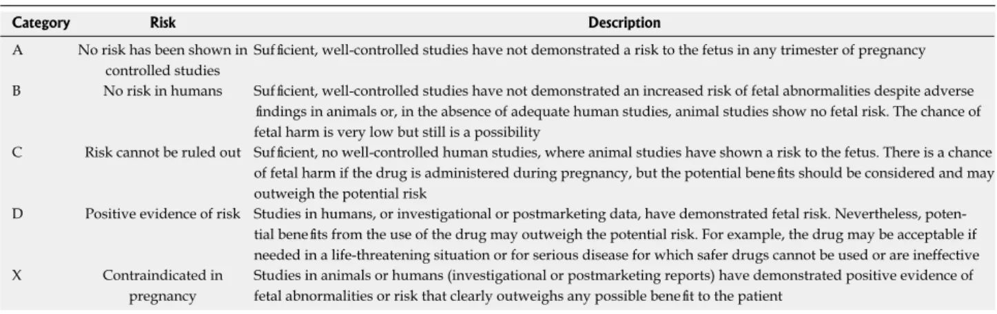
Endoscopic procedures are associated with terato-genic risk in the first trimester and premature labor in the third trimester. Therefore, endoscopic procedures should be considered with caution in pregnant patients with anesthesiology assistance. The United States Food and Drug Administration (FDA) have defined five cat-egories of drugs in terms of safety to pregnant women (Table 2)[8]. Category A drugs are considered safe during pregnancy and category B drugs also may be used during pregnancy (Table 2). Category C drugs may be used when required during pregnancy, but there may be risks to the fetus. Category D drugs usually are contraindicated dur-ing pregnancy and are used only with extreme caution.
Category X drugs are absolutely contraindicated during pregnancy (Table 2)[8].
There are no category A drugs that are used for endoscopy. During endoscopic procedures, category B and when necessary, category C drugs may be recom-mended (Table 3). Category D drugs may be used when the benefits outweigh the risks. These categories are of limited use in determining the safety of one-time use; therefore, consultation with an obstetrician about drugs should be considered. For most procedures, anxiolytic drugs or moderate sedation may be adequate. Heavy sedation, when necessary, should be administered by an anesthesiologist[2].
The opiate analgesic meperidine was commonly used for GI endoscopy for the general population; however, it has been replaced by short-acting analgesics because of adverse events (respiratory depression and seizures) (Table 3). After intravenous administration, meperidine is transferred rapidly across the human placenta and is metabolized to normeperidine, which has a longer half-life than meperidine. Repeated administration of meperi-dine at high doses may cause progressive accumulation of normeperidine, maternal respiratory depression and maternal seizures. Meperidine is a drug in category B for regular use, but is category D for prolonged use at high doses. Meperidine use should be limited to 50-75 mg for endoscopic procedures in pregnant women[4,5,9]. Fentanyl (category C) is a potent narcotic that has a rapid onset of action and a shorter recovery time than meperidine; fentanyl is usually safe in low doses (< 125 mg) during pregnancy (Table 3)[2-5].
Benzodiazepines (diazepam and midazolam) are com-monly used before GI endoscopy to reduce anxiety, in-duce brief amnesia and proin-duce muscle relaxation. Pro-longed use of diazepam during early pregnancy may be associated with cleft palate malformations; however, this association is unproven[10-13]. The use of diazepam in the first trimester of pregnancy, however, is not safe because of a strong relation between diazepam use and mental retardation or neurological defects, cardiac defects and Mobius syndrome (a neurological disorder with normal intelligence but sixth and seventh nerve palsies)[11,14,15]. There are limited data about the use of midazolam, but there is no known association of midazolam with oral cleft palate. However, midazolam may be associated with transient depression of neonatal neurobehavioral respon-siveness during labor[16,17]. When meperidine cannot be used, the preferred benzodiazepine is midazolam (cat-egory D) because associated fetal abnormalities have not been reported.
Propofol (category B) is commonly used for anesthe-sia during endoscopy. It is a short-acting anesthetic agent with a short recovery period. It is usually administered by anesthesiologists because of its narrow therapeutic index and potential for respiratory depression. Endoscopy soci-eties have recommended the use of propofol for patients who are difficult to sedate or have complicated clinical situations. Propofol is considered safe during pregnancy, but there are insufficient data available about the use of Table 1 General principles for endoscopy in pregnant
women1
1 Always have a strong indication, particularly in high-risk pregnancies 2 Endoscopy should be postponed to second trimester whenever possible 3 Lowest effective dose of sedative medications should be used 4 Especially category A or B drugs should be used
5 Procedure time should be very short
6 To avoid vena caval or aortic compression, pregnant women should be positioned in the left pelvic tilt or left lateral position
7 Fetal heartbeat should be detected before sedation and also after the endoscopic procedure
8 Obstetric support should be available whenever pregnancy-related complications occur
9 Placental abruption, imminent delivery, ruptured membranes, or eclampsia are defined as obstetric complications of endoscopy
propofol in the first trimester[4,5,18].
Ketamine (category B) can be used for endoscopy when there is insufficient sedation with propofol. Ket-amine has a rapid onset of action and a short duration of effect, but data are limited about use of ketamine during the first trimester of pregnancy, prolonged use or overdose[19].
Naloxone (category B) is a fast-acting narcotic antag-onist that may be administrated to reverse narcotic over-dose during endoscopy. Naloxone crosses the placenta within 2 min[2,20]. It is used to treat respiratory depression, systemic hypotension or unresponsiveness in closely monitored settings during or after endoscopy. Naloxone is given in small, graded doses and titrated to the required effect during pregnancy, because there has been one neonatal fatality that was attributed to naloxone use[21]. Naloxone is contraindicated in narcotic-dependent preg-nant patients because of the risk of opiate withdrawal syndrome[22].Flumazenil (category C) is a benzodiazepine antagonist that is used to reverse over sedation from benzodiazepines that are administered during endoscopy. Its fetal risks during pregnancy are unknown and is used
only to reverse benzodiazepine overdose. Flumazenil overdose may cause maternal seizures, especially when given to patients who are chronically habituated to ben-zodiazepines. The risk of benzodiazepine overdose may be minimized by careful and slow titration of minimal doses of benzodiazepines required for endoscopy[19-23].
UPPER GASTROINTESTINAL
ENDOSCOPY
It would be ideal to postpone endoscopic procedures until after delivery; however, pregnant patients may de-velop conditions that require urgent upper endoscopy. The most common indications for esophagogastroduo-denoscopy (EGD) in pregnant patients include major or continued GI hemorrhage, dysphagia, and refractory nausea and vomiting (Table 4). The EGD procedure is reasonably safe for the fetus and may be performed when strongly indicated during pregnancy.
In a multicenter retrospective study of 83 pregnant women concerning the safety and clinical efficacy of EGD in pregnant patients, indications for endoscopy in-Table 2 United States food and drug administration categorization of drug safety during pregnancy1
Category Risk Description
A No risk has been shown in controlled studies
Sufficient, well-controlled studies have not demonstrated a risk to the fetus in any trimester of pregnancy B No risk in humans Sufficient, well-controlled studies have not demonstrated an increased risk of fetal abnormalities despite adverse
findings in animals or, in the absence of adequate human studies, animal studies show no fetal risk. The chance of fetal harm is very low but still is a possibility
C Risk cannot be ruled out Sufficient, no well-controlled human studies, where animal studies have shown a risk to the fetus. There is a chance of fetal harm if the drug is administered during pregnancy, but the potential benefits should be considered and may outweigh the potential risk
D Positive evidence of risk Studies in humans, or investigational or postmarketing data, have demonstrated fetal risk. Nevertheless, poten-tial benefits from the use of the drug may outweigh the potenpoten-tial risk. For example, the drug may be acceptable if needed in a life-threatening situation or for serious disease for which safer drugs cannot be used or are ineffective X Contraindicated in
pregnancy
Studies in animals or humans (investigational or postmarketing reports) have demonstrated positive evidence of fetal abnormalities or risk that clearly outweighs any possible benefit to the patient
1Adapted from Food and Drug Administration (1980)[8].
Table 3 Safety of anesthetics commonly used in gastrointestinal endoscopy
Drug FDA category in
pregnancy Key points about drug safety
Narcotics
Meperidine B, but D at term Repeated use of high dose and prolonged administration can cause respiratory depression and seizures Fentanyl C It is safe in low doses
Propofol B Generally suggested for use in patients who are sedated with difficulty and in complicated clinical situations General anesthetics
Ketamine B Data are limited with humans; animal data suggest prolonged use is not safe Sedatives
Diazepam D Some congenital malformations and mental retardation may be associated with diazepam, the use of diazepam during pregnancy is restricted
Midazolam D As a benzodiazepine member, its use is restricted during pregnancy, especially in the first trimester Reversing agents
Naloxone B It probably is safe but should be used only in respiratory depression, systemic hypotension, or unresponsiveness in a closely monitored pregnant woman after endoscopy
Flumazenil C Fetal risks are unknown, but it should be given carefully in small doses FDA: United States Food and Drug Administration.
women had lower frequencies of blood transfusion, hypovolemic shock or EGD than nonpregnant women. The proportion of EGD procedures that led to thera-peutic intervention was similar for pregnant (8.9%) and nonpregnant women (7.2%). The frequency of maternal mortality and fetal loss were < 1% and it was concluded that it was appropriate to defer endoscopy in most pa-tients who were hemodynamically stable and who had self-limited NVUGB[29].
Patients who have cirrhosis are not likely to become pregnant because they may have hypothalamic-pituitary dysfunction and associated disturbance of estrogen and endocrine metabolism. The exact incidence of pregnancy in cirrhosis is not known, but only 45 cases of cirrhosis occur in every 100000 women of reproductive age. On the other hand, women with noncirrhotic portal hyperten-sion have normal frequency of fertility and these patients may have 45% incidence of variceal bleeding during pregnancy and 18%-50% associated mortality. The vari-ceal bleeding typically occurs during the second or third trimester. The high severity of variceal bleeding in preg-nancy may be attributed to increased fluid retention and cardiac output in pregnancy. Women who have esophageal varices or severe liver disease should be advised about the high risk of variceal bleeding and hepatic decompensation during pregnancy. Nonselective β-blockers may be given to patients who have esophageal varices or severe liver dis-ease, but the safety of β-blockers is controversial because of reports of premature labor, fetal growth restriction, neonatal apnea, bradycardia and hypoglycemia. Further-more, myometrial relaxation of the gravid uterus is a β2 -receptor-mediated process and nonselective β-blockers, such as propranolol, may counteract the effect of β2 -receptor stimulation. The pregnant patient should be in-formed about the possible benefits and adverse effects of β-blockers during pregnancy.
THERAPEUTIC ENDOSCOPY
Endoscopic hemostatic techniques for nonvariceal bleed-ing include injection therapy (epinephrine, sclerosbleed-ing agents, thrombin or cyanoacrylate), ablative therapy (elec-trocoagulation, thermocoagulation, photocoagulation or argon plasma coagulation), and compression (hemoclips, detachable snares, graspers or sutures)[30]. Although there are numerous techniques, there are few case reports about the fetal safety of endoscopic hemostasis for NVUGB, including epinephrine injection, thermocoagulation or electrocoagulation. In the available reports, hemostatic techniques were successful in all patients except for one patient who required surgery. The fetal outcomes were all healthy infants without any fetal malformations[24,27,30,31].
Epinephrine (category C) may cause a decrease in uterine blood flow. Although there are limited data from case reports, no adverse events from epinephrine injec-tion have been reported and the benefits (cessainjec-tion of hemorrhage and prevention of rebleeding) may outweigh the risks[1,2,26,32]. Electrocautery is safe when used for he-mostasis, but amniotic fluid may conduct electrical cur-cluded GI bleeding, abdominal pain and vomiting[24]. The
most common causes of the GI bleeding were Mallory-Weiss tear and peptic ulcer, which were significantly lower than the reported frequencies in non-pregnant patients The diagnostic yield for upper GI bleeding was 95% and there were no patients who had premature labor or con-genital fetal malformation.
During pregnancy, increased progesterone and estro-gen levels mediate lower esophageal sphincter relaxation, with 50% decreased lower esophageal sphincter pressure during, compared with before, pregnancy and decreased gastric emptying that may cause symptoms of gastro-esophageal reflux disease (GERD). As pregnancy pro-gresses, the frequency and intensity of GERD symptoms may increase because of changes in GI motility during pregnancy and the physical effects of the gravid uter-us[25,26]. The EGD procedure is rarely helpful or indicated for nausea or vomiting during pregnancy or hyperemesis gravidarum. In patients who have major upper GI bleed-ing, severe nausea and vomiting accompanied by abdomi-nal pain that is refractory to medical treatment or signs of gastroduodenal obstruction, EGD may be appropriate for diagnosis of major peptic ulcers, diagnosis of gastric outlet obstruction or treatment of a bleeding site. Debby
et al[27] reported a study of patients who had EGD in the first trimester of pregnancy, in that study, 49 patients had intractable nausea with or without epigastric pain and 11 patients had nausea and upper GI bleeding; the diagnos-tic yield of EGD was similar for patients who had GI bleeding or intractable vomiting. The endoscopic findings changed the treatment of patients who had nausea and vomiting minimally, and they concluded that EGD may be useful for treatment of upper GI bleeding, but not nausea, vomiting or hyperemesis gravidarum[27].
Acute nonvariceal upper GI bleeding (NVUGB) is a common clinical emergency that causes 50-160 hospi-talizations per 100000 adults annually. Mortality may be decreasing but remains at 10%-14%[28]. Endoscopy for NVUGB may provide an assessment of the risk of bleeding and enable therapeutic hemostasis that could re-duce bleeding, frequency of surgery and risk of death. In a population-based study of NVUGB, there were 1210 pregnant women and 6050 nonpregnant women who had NVUGB. The most common causes of NVUGB were Mallory-Weiss tear in pregnant women and peptic ulcer disease and gastritis in nonpregnant women[29]. Pregnant
Table 4 Indications for endoscopy in pregnancy
No. Indication
1 Major or continued bleeding
2 Severe or refractory nausea and vomiting or abdominal pain 3 Dysphagia or odynophagia
4 High suspicion of colonic mass 5 Severe diarrhea with negative evaluation 6 Biliary pancreatitis, CBD stones, or cholangitis 7 Biliary or pancreatic ductal injury
rent to the fetus; therefore, during electrocoagulation, a grounding pad should be placed such that the uterus is not between the electrical cord and the grounding pad, and bipolar electrocautery should be used to minimize the risk of stray current going through the fetus.
There are limited data about hemostasis for nonvari-ceal bleeding in pregnant patients, and the therapeutic technique is chosen from expert opinion that is based on results of clinical studies in nonpregnant patients. Pro-phylactic or urgent endoscopic injection sclerotherapy (EIS) and endoscopic band ligation (EBL) are safe pro-cedures during pregnancy. When bleeding is not stopped endoscopically in cirrhotic patients, an emergency tran-sjugular intrahepatic portosystemic shunt (TIPSS) is indicated, but data about pregnant cirrhotic women are limited[33-39]. There are only a few case reports about the treatment options for esophageal varices, and further studies are needed about the treatment of hemorrhage during pregnancy. In the early 1980s, EIS was a first-line treatment procedure for bleeding esophageal varices. However, only a few cases of EIS with sclerosing agents (polidocanol, absolute alcohol or sodium tetradecyl sul-fate) have been reported during pregnancy[36-39]. There are no studies available about the effects of these sclerosing agents on the fetus; however, the procedure is considered safe and effective in controlling active variceal bleeding. Vasoactive drugs that are used to achieve hemostasis are contraindicated during pregnancy because these drugs (vasopressin and terlipressin) may induce labor or fetal malformations.
EBL may be an effective treatment option for active variceal hemorrhage and prophylaxis for this severe com-plication during pregnancy. There are several case reports that describe successful hemostasis without fetal compli-cations[40-41]. When EBL is used, there is no risk of migra-tion of a toxic substance to the placenta. Studies of EBL
vs EIS in nonpregnant patients have shown improved
reduction in rebleeding and mortality with EBL[40,42]. However, there are no studies that directly compare EBL to EIS in pregnant patients.
In a previous study, 17 patients had acute variceal bleeding during pregnancy because of noncirrhotic portal hypertension that was caused by extrahepatic portal vein obstruction or portal fibrosis. These patients underwent EIS with either absolute alcohol or sodium tetradecyl sulfate[43]. There were two patients who required EBL after failure of EIS to obliterate esophageal varices[43]. In an another report, 10 patients underwent EIS with absolute alcohol for treatment of active variceal bleeding (five patients) or prophylaxis against variceal bleeding (five patients). Hemostasis was achieved in the five patients who had active variceal bleeding and all 10 patients de-livered healthy infants[44]. In pregnant patients, EBL may be a reasonable option for the treatment of acute variceal bleeding and prophylaxis against variceal bleeding. EIS may be a secondary choice for acute variceal bleeding be-cause of probable detrimental effects on fetal safety.
When endoscopic and pharmacologic therapy fail, TIPSS may be a salvage procedure for pregnant women
who have variceal bleeding that is recurrent, difficult to treat or unresponsive to endoscopic or pharmacologic treatment. However, adequate controlled trials are lack-ing, and this procedure should be limited to a selected group of patients. TIPSS placement is associated with radiation exposure to the patient and fetus because the procedure usually requires prolonged fluoroscopy. There are several reported cases of TIPSS placement in preg-nancy in which the fetal dose of radiation was 5.2 mSv to 2.1 mGy[35,45,46].
The average person in the United States receives 0.0036 Sv (0.36 rem) ionizing radiation annually, includ-ing 0.0006 Sv (0.06 rem) from manmade sources, such as diagnostic radiography. Fetal radiation exposure may cause developmental abnormalities, especially when the exposure occurs during the first trimester. Fetal radiation exposure should not exceed 0.001 Sv (0.1 rem) during the first trimester and later exposures > 0.001 Sv (0.1 rem) during neuron development and migration may be asso-ciated with microcephaly, seizures, decline in mental abil-ity and childhood cancer. The maximum permitted dose of ionizing radiation to the fetus during the entire preg-nancy is 0.005 Sv (0.5 rem)[47-49]. Therefore, in patients who have upper GI bleeding all therapeutic procedures that are used in nonpregnant patients can also be used in pregnant patients. In cirrhotic patients, pregnancy is not an absolute contraindication for TIPSS placement for the treatment of relapsing bleeding varices, but minimizing the duration of radiation exposure is important to pre-vent toxic radiation exposure to the fetus.
PERCUTANEOUS ENDOSCOPIC
GASTROSTOMY
During pregnancy, optimal nutrition is important to minimize maternal and neonatal morbidity[50,51]. Nausea and vomiting are observed in 80% pregnancies but are usually mild and self-limited. Patients who have severe hyperemesis gravidarum with dehydration and ketonuria should be hospitalized and treated with intravenous hy-dration and antiemetic drugs. When the hospitalization is prolonged and there is no oral intake, supportive nutri-tion with enteral feeding or total parenteral nutrinutri-tion may be considered. Long-term nasogastric feeding is limited by patient intolerance and nasal septal necrosis. Adverse events may limit the use of long-term total parental nutri-tion during pregnancy[52].
Percutaneous endoscopic gastrostomy (PEG) is an important option for long-term enteral feeding. Place-ment of PEG tubes in pregnant women may be limited because of risks of uterine damage, fetal injury, prema-ture labor and infection, but there were no major com-plications associated with PEG tube placement in several reported cases[53-60]. In previous studies, PEG enteral nutritional support was provided for an average 14 wk. During pregnancy, PEG tube placement is feasible for optimal enteral nutrition in the critical care setting and in the third trimester of pregnancy. A major risk of PEG
during pregnancy is puncture of the uterus or fetus dur-ing transabdominal needle insertion, but this risk may be minimized by demarcating the upper border of the uterus before PEG and inserting the PEG needle ≥ 5 cm cephalad.
Placement of a PEG tube is reserved for severe re-fractory cases of impaired nutrition of the mother and fetus. The pregnant woman should be informed about the risks of the procedure and potential placental injury. If possible, less invasive alternative techniques, such as a nasoenteric feeding tube or peripherally inserted catheter for parenteral nutrition, should be considered, and PEG tube placement may be offered when other methods are unsuccessful or declined by the patient. When refractory nausea and vomiting persist despite PEG tube placement, and the risk of aspiration pneumonia is increased, the PEG may be converted to a percutaneous endoscopic gastrojejunostomy[50,61].
SIGMOIDOSCOPY
Most pregnant patients are young, healthy women and the gestational period is 40 wk. Therefore, most patients do not need to have flexible sigmoidoscopy or colonos-copy during pregnancy. Lower GI endoscolonos-copy is avoided for weak indications during pregnancy and deferred until after the first trimester or postpartum[62]. However, sigmoidoscopy or colonoscopy is indicated for the evalu-ation of major lower GI bleeding, suspicion of colonic mass or severe diarrhea.
Sigmoidoscopy is usually safe during pregnancy and indications include rectal bleeding, chronic diarrhea, abdominal pain and rectal pain. Guidelines for colonos-copy during pregnancy are limited because of insuffi-cient data, but colonoscopy is typically safe and effective when obstetrical consultation and close monitoring are performed[63,64].
The safety and efficacy of flexible sigmoidoscopy during pregnancy was studied in a case controlled study of 45 patients undergoing sigmoidoscopy[65]. In that study, the most common clinical indication was hematochezia in 29 patients, diarrhea in 10 patients and abdominal pain in 4 patients. The most common sigmoidoscopic diagnoses were reactivated or newly di-agnosed inflammatory bowel disease, bleeding internal hemorrhoids and other types of colitis. In 29 patients who had hematochezia, 8 patients had de novo or
recur-rent episodes of ulcerative colitis, 7 patients had de novo
or recurrent episodes of Crohns disease, 3 patients had proctosigmoiditis, 2 patients had bleeding internal hem-orrhoids, 1 patient had pseudomembraneous colitis and 1 patient had a sigmoid colon adenoma. Hematochezia gave the highest diagnostic yield compared with other clinical indications. Therapeutic changes because of the sigmoidoscopic findings occurred in 24 patients, includ-ing changinclud-ing or startinclud-ing drugs for inflammatory bowel disease in 15 patients, steroid enemas for nonspecific proctitis in 2 patients, avoiding surgery in 2 patients and
treatment of hemorrhoids in 2 patients.
Other studies of sigmoidoscopy performed dur-ing pregnancy have included case reports and a mailed survey[26,66-70]. Multiple case reports describing flexible sigmoidoscopy in pregnant patients have confirmed the safety of this procedure. These studies suggested that sigmoidoscopy during pregnancy may not induce labor or cause congenital malformations. Thus, sigmoidoscopy is not contraindicated and may be considered in medically stable patients who have important indications. Sigmoid-oscopy should be performed with maternal monitoring (electrocardiography and pulse oximetry) after obstetric consultation and after medical stabilization. Medical stabilization may include blood transfusion and supple-mental oxygen[62,64]. For evaluation of a change in bowel habits, abdominal pain, family history of colon cancer or routine screening or surveillance, sigmoidoscopy is not recommended during pregnancy but is deferred until > 6 wk postpartum[63,64].
COLONOSCOPY
There are insufficient data about the safety of perform-ing a colonoscopy durperform-ing pregnancy. The largest case control study about colonoscopy in pregnancy included 20 patients who were evaluated for symptoms including hematochezia, diarrhea, bloody diarrhea and abdominal pain[71]. In that study, colonoscopy was performed in 16 patients in the second trimester and in 4 patients in the first or third trimester; colonoscopic diagnoses included ulcerative colitis, Crohn disease, ischemic colitis and lymphocytic colitis. Colonoscopy resulted in a change in therapy in seven (35%) patients. Most patients had favor-able fetal outcomes (18 healthy infants) and there was one involuntary abortion and one infant who was born with a cardiac defect (septum secundum)[71].
In another study of eight pregnant women who had colonoscopy (10 different medical centers) there were 6 healthy infants born, 1 elective abortion and one fetal death that was unrelated to colonoscopy[65]. Outcomes were independent of the trimester during which colo-noscopy was performed. In addition, several case reports about colonoscopy during pregnancy have shown 8 healthy births, 2 stillbirths unrelated to colonoscopy and 1 unknown fetal outcome[72-80].
With limited data about safety and adverse events, colonoscopy should be limited to patients who have strong indications or life-threatening emergencies dur-ing the second trimester. However, colonoscopy may be considered in lieu of surgery during the first and third trimester for evaluation of suspected colon cancer, co-lonic mass, uncontrolled severe coco-lonic hemorrhage, colonic stricture of unknown cause or colonic pseudo-obstruction. When required before urgent colonic sur-gery, colonoscopy should be considered, even in the first and third trimester. Otherwise, colonoscopy for elective indications, such as surveillance for prior history of colon cancer or colonic polyps usually is deferred in any
trimes-ter until aftrimes-ter delivery.
When colonoscopy is performed, especially in late pregnancy, patients should not be placed in the decubitus or prone position. External abdominal pressure should be avoided and when required, applied pressure should be minimal and directed away from the uterus. Limited information is available about the safety of bowel cleans-ing agents durcleans-ing pregnancy. The systemic absorption of polyethylene glycol is minimal and abdominal bloating and gas symptoms are less common with polyethylene glycol than with other laxatives[81]. However, polyethylene glycol solutions (category C) have not been studied dur-ing pregnancy. Sodium phosphate solutions (category C) may cause fluid and electrolyte disturbance, and should be avoided during pregnancy. In addition, newborns may have bone demineralization and bone growth failure be-cause of maternal phosphate overload[82], but one-time use in pregnancy may not be detrimental. Furthermore, sodium phosphate preparations may be associated with the risk of phosphate nephropathy[83]. Bowel preparation with phosphate enemas before flexible sigmoidoscopy may be safe, but has not been studied in pregnancy. Un-derprepared sigmoidoscopy is generally not recommend-ed because of the risk of overlooking lesions; instead, sigmoidoscopy with tap water enemas may be sufficient.
Therefore, flexible sigmoidoscopy with tap water en-emas is preferred instead of colonoscopy. However, in patients who have strong indications or life-threatening emergencies or when the alternative treatment is surgical decompression, colonoscopy may be considered, even during the first and third trimesters.
THERAPEUTIC COLONOSCOPY
Therapeutic colonoscopy is applied for the manage-ment of lower GI bleeding, colonoscopic polypectomy and colonic stenting. All the hemostatic techniques that are mentioned above for upper GI bleeding can be ap-plied during lower GI bleeding. These are mainly injec-tion therapies, ablative therapies, hemoclips, detachable snares, graspers, or sutures[30]. Epinephrine is commonly used to treat GI bleeding and may cause hemostasis by vasoconstriction. Numerous studies have confirmed the fetal safety of epinephrine administration during labor, and epinephrine is commonly added to spinal epidural anesthesia. A previous study showed no congenital defect in 35 infants who had first trimester in utero exposure to epinephrine[10]. However, the dosage of epinephrine (category C) during pregnancy is kept low because of α-adrenergic effects and decreased uterine blood flow.
Electrocautery may provide hemostasis during lower GI bleeding and is used during polypectomy or biopsy. Electrocautery of lesions may be required, in which case bipolar electrocautery should be used. Removal of non-bleeding polyps may be postponed until after delivery[4,5,62]. Colonic tattooing is performed with India ink or methylene blue in nonpregnant patients, and India ink may persist for the entire life of the patient. A literature
search showed no reports of long-term complications of India ink tattooing. Although methylene blue tattoo-ing durtattoo-ing pregnancy has not been studied, there are reports of methylene blue examination during amnio-centesis and in the detection of ruptured membranes. In these reports, fetal death and jejunal atresia were report-ed and methylene blue has been labelreport-ed as teratogenic. Although the safety of colonic injection with methylene blue has not been studied, its use should be avoided dur-ing pregnancy[62,84,85].
ENTEROSCOPY AND VIDEO CAPSULE
ENDOSCOPY
Enteroscopy of the small bowel is a procedure with long duration and anesthesia time. There are no case reports of enteroscopy during pregnancy and the safety of enter-oscopy to the fetus is unknown.
Video capsule endoscopy (VCE) is a major advance in the investigation of small bowel diseases. The main indications include obscure GI hemorrhage, Crohn’s disease, celiac disease, small bowel tumors and polyposis syndromes. The main contraindications include known or suspected GI obstruction, strictures, fistulas, cardiac pacemakers and swallowing disorders[86]. During pregnan-cy, the growing gravid uterus pushes and compresses the GI tract, and GI motility decreases because of inhibition of the intestinal smooth muscle by gestational progestin. These effects raise concerns about capsule impaction during VCE in pregnant women[87]. According to the United States FDA, pregnancy is a relative contraindica-tion for VCE.
There is a report of VCE use in a young, acutely bleeding pregnant patient in whom endoscopy and colo-noscopy showed no lesion except fresh blood exiting the terminal ileum. On VCE, an actively bleeding jejunal lesion was shown and pathological examination showed that this lesion was a jejunal carcinoid tumor. After the procedure the pregnant patient and fetus did well[88]. Therefore, VCE may be considered during pregnancy for strong indications, and it is not absolutely contraindicated during pregnancy.
ENDOSCOPIC RETROGRADE
CHOLANGIOPANCREATOGRAPHY
Pregnancy is associated with an increased risk of gall-stone formation. Complications of cholelithiasis, such as cholecystitis, common bile duct (CBD) stones and pan-creatitis are uncommon, and are frequently treated non-operatively. However, patients may develop complications of gallstones that require intervention during pregnancy, and these complications are among the most frequent in-dications for nonobstetric surgery during pregnancy[89-93].
There is controversy about the safety of endoscopic retrograde cholangiopancreatography (ERCP) during pregnancy, and data are limited. Major concerns are
asso-ciated with radiation exposure to the fetus and the risk of the procedure on the outcome of pregnancy. In women who have an acute biliary tract disorder during pregnancy, it is advisable to provide nonoperative treatment whenev-er possible and delay surgwhenev-ery until aftwhenev-er pregnancy or the second trimester, when the surgical risks of pregnancy are lowest. There are numerous reports about ERCP dur-ing pregnancy, especially durdur-ing the past 10 years. The largest series included 65 pregnant patients, and the most common indications for ERCP during pregnancy were recurrent biliary colic, abnormal liver function tests and a dilated bile duct on ultrasonography[94]. There were 68 ERCP procedures performed in 65 pregnant patients (trimester: first, 17 patients; second, 20 patients; third, 31 patients). The median fluoroscopy time was 1.45 min and most patients had a therapeutic procedure. Pancre-atitis after ERCP developed in 11 patients (16%), but no patient had a severe course. Most patients achieved term pregnancy (89%); only 5 babies (8%) were born prema-turely or with low birth weight, and there were no con-genital malformations[94].
In another series of 23 patients who had ERCP (ther-apeutic, 20 patients; diagnostic, 3 patients), complications included pancreatitis after ERCP (1 patient), spontaneous abortion (1 patient) and neonatal death at 26 h after de-livery (1 patient)[95]. The neonatal death and post-ERCP pancreatitis occurred in the same patient who had three ERCP procedures (2 during the first trimester; 1 dur-ing the third trimester) with pancreatic duct stentdur-ing for treatment of pancreatic orifice stenosis after a previous surgical sphincteroplasty.
In a study of 18 pregnant women who had biliary sphincterotomy for CBD stones during pregnancy (tri-mester: first, 4 patients; second, 6 patients; third, 8 pa-tients), short-term complications occurred in 2 patients (postsphincterotomy bleeding, 1 patient; mild post-ERCP pancreatitis and preterm labor, 1 patient); however, no long-term maternal complications were observed after a median of 6 years (range, 1-11 year)[96]. In 11 families that were contacted retrospectively, all 11 children were healthy at a mean of 6 years postpartum[96].
In a prospective study of therapeutic ERCP during pregnancy, a single 10-French stent was placed without sphincterotomy and all patients had uncomplicated preg-nancy and delivery of healthy infants[97]. All women had ERCP with sphincterotomy and stent extraction postpar-tum: eight patients had stones extracted. In two patients, the 10-French stent remained in place for 7 to 8 mo and no patient developed cholangitis[97].
During ERCP, radiation exposure to the fetus may in-crease the risk of intrauterine fetal death, malformations, disturbance of growth and development, mutations and cancer. Therefore, these risks should be discussed with the pregnant patient and her family before ERCP. Lead shielding should be used to minimize radiation exposure to the uterus. When the radiation source is underneath the patient, the lead apron shield must be placed un-derneath the patient and not draped over the abdomen.
External shielding may not completely eliminate fetal exposure because of internally scattered radiation, and efforts should be made to avoid performing ERCP dur-ing the first trimester. Although the harmful effects of radiation exposure are unlikely to develop below a thresh-old radiation dose, the threshthresh-old associated with the risk of childhood cancers, such as leukemia, is unknown and no long-term studies (10-20 years after exposure during pregnancy) are available.
The use of ERCP without fluoroscopy has been reported, including a 2-step procedure with (1) biliary sphincterotomy and stenting without fluoroscopy and (2) definitive ERCP with stone extraction after delivery[98,99]. In this study, initial CBD cannulation was performed with a double lumen sphincterotome; deep cannulation was achieved and bile was aspirated to confirm CBD position[98]. After deep CBD cannulation, the guide wire was passed and complete biliary sphincterotomy was performed over the guide wire. When deep CBD cannu-lation was not possible, the sphincterotome was removed and needle knife sphincterotome was used. After the biliary orifice was identified, a complete biliary sphincter-otomy was performed using a double lumen sphinctero-tome. A 7-French double pigtail stent was placed in the CBD. After delivery the stent was removed and definitive ERCP was performed[98].
In another study of ERCP without fluoroscopy, the procedure included cannulation of the bile duct and sphincterotomy[99]. The endoscopist controlled the wire-guided cannulation, and the cannula was not advanced into the duct unless the endoscopist was confident that the CBD had been cannulated (as assessed by the pres-ence of bile flowing around the wire from the papillary orifice). After biliary cannulation was confirmed, a wire-guided biliary sphincterotomy was performed using a papillotome. When bile was not observed flowing around the guide wire, the catheter was not advanced to aspirate fluid, but a 5-French stent was inserted over the wire and drainage from the stent was observed. The color of the draining fluid was used to assess whether the stent was in the bile or pancreatic duct. When the stent showed bile flow, a stent-guided biliary sphincterotomy using a needle knife was performed. The stent was removed after biliary sphincterotomy[99].
Although these techniques may be less risky for the pregnant woman and fetus, ERCP should be avoided for weak indications, such as preoperative cholangiography in patients who have low probability of having CBD stones. All women of childbearing age should be asked about the possibility of pregnancy and a pregnancy test should be ordered based on clinical history. Other methods of diagnosis without radiation exposure should be consid-ered. Magnetic resonance cholangiopancreatography may provide diagnostic information for various hepatobiliary conditions, and endoscopic ultrasonography is highly sensitive and specific for CBD stones. However, ERCP with or without fluoroscopy is indicated in patients who have CBD stones, biliary pancreatitis, cholangitis and bile
duct dilation on abdominal ultrasonography with known gallstones and abnormal liver function tests.
ENDOSCOPIC ULTRASONOGRAPHY
Endoscopic ultrasonography is commonly performed for the diagnosis of GI and pancreatobiliary diseases. Endoscopic ultrasonography may reduce unnecessary interventions in patients who have a low or moderate probability of developing CBD stones and it is a safe alternative to fluoroscopy for the evaluation of biliary disorders during pregnancy. Case reports about the use of endoscopic ultrasonography for pregnant patients are available. The largest study included endoscopic ultra-sonography performed in six pregnant patients for sus-pected CBD stones[99] Endoscopic ultrasonography find-ings in this study included CBD stones (two patients), biliary sludge (two patients) and nonspecific findings (two patients). All six patients had ERCP after endoscopic ultrasonography and there were no maternal complica-tions; fetal outcome was favorable for five infants and unknown for one infant[99].In another report, endoscopic ultrasonography was performed for acute pancreatitis of unknown cause in three pregnant patients. Biliary pancreatitis without CBD stones was observed in two patients and pancre-atitis caused by an unspecified pancreatic anomaly was observed in one pregnant patient. There were no re-ported maternal complications and two healthy infants were delivered; however, there was one fetal death be-cause of recurrent cholangitis at 10 wk after endoscopic ultrasonography[100].
Endoscopic ultrasonography may prolong the evalu-ation. However, when endoscopic ultrasonography is normal, ERCP intervention may be avoided. In addi-tion, endoscopic ultrasonography may provide other useful information, and the added time for endoscopic ultrasonography may be only several minutes. Further studies are required to evaluate the potential benefits of endoscopic ultrasonography in the treatment of pregnant patients. It may be acceptable to perform endoscopic ultrasonography when CBD stones are suspected, the diagnosis is unproven and magnetic resonance cholangio-pancreatography is an undesirable alternative.
CONCLUSION
All GI endoscopic procedures in pregnant patients should be performed in hospitals by expert endoscopists, and an obstetrician should be informed about all endo-scopic procedures. GI endoscopy may be performed safely in pregnant patients when there are strong indi-cations. To minimize fetal risks from drugs during en-doscopy, category D drugs should be avoided, drug use should be minimized and an anesthesiologist should at-tend at endoscopy. The EGD and flexible sigmoidoscopy may be safe for the fetus and pregnant patient and may be performed during pregnancy when strong indications
are present. Colonoscopy for pregnant patients may be considered for strong indications during the second tri-mester. Although therapeutic ERCP may be considered during pregnancy, this procedure should be performed only for strong indications and attempts should be made to minimize radiation exposure.
REFERENCES
1 O’mahony S. Endoscopy in pregnancy. Best Pract Res
Clin Gastroenterol 2007; 21: 893-899 [PMID: 17889814 DOI: 10.1016/j.bpg.2007.05.007]
2 Qureshi WA, Rajan E, Adler DG, Davila RE, Hirota WK,
Jacobson BC, Leighton JA, Zuckerman MJ, Hambrick RD, Fanelli RD, Baron T, Faigel DO. ASGE Guideline: Guidelines for endoscopy in pregnant and lactating women. Gastrointest Endosc 2005; 61: 357-362 [PMID: 15758903 DOI: 10.1016/ S0016-5107(04)02780-4]
3 Kammerer WS. Nonobstetric surgery during pregnancy.
Med Clin North Am 1979; 63: 1157-1164 [PMID: 529882] 4 Cappell MS. Risks versus benefits of gastrointestinal
endos-copy during pregnancy. Nat Rev Gastroenterol Hepatol 2011; 8: 610-634 [PMID: 21970872 DOI: 10.1038/nrgastro.2011.162] 5 Gilinsky NH, Muthunayagam N. Gastrointestinal
en-doscopy in pregnant and lactating women: emerging standard of care to guide decision-making. Obstet Gynecol Surv 2006; 61: 791-799 [PMID: 17107628 DOI: 10.1097/01. ogx.0000248745.10232.bb]
6 Nurten SA. Endoscopy in pregnant patients. Endoscopy. In:
Amornyotin S, editor. Croatia: InTech, 2013: 321-348 7 Morgan GE, Mikhail SM, Murray JM. Clinical
anesthesiol-ogy. New York: McGrow-hill, 2000: 819-846
8 Food and Drug Administration. Federal Register 1980; 44: 37434-37467
9 Jiraki K. Lethal effects of normeperidine. Am J Forensic Med
Pathol 1992; 13: 42-43 [PMID: 1585886 DOI: 10.1097/00000433 -199203000-00009]
10 Briggs GC, Freeman RK, Yaffe SJ. Drugs in pregnancy and lactation: a reference guide to fetal and maternal risks. 8th ed. Philadelphia: Lippincott, Williams & Wilkins, 2008 11 Rothman KJ, Fyler DC, Goldblatt A, Kreidberg MB.
Exog-enous hormones and other drug exposures of children with congenital heart disease. Am J Epidemiol 1979; 109: 433-439 [PMID: 443241]
12 Ornoy A, Arnon J, Shechtman S, Moerman L, Lukashova I. Is benzodiazepine use during pregnancy really terato-genic? Reprod Toxicol 1998; 12: 511-515 [PMID: 9763242 DOI: 10.1016/S0890-6238(98)00035-5]
13 Czeizel A. Lack of evidence of teratogenicity of benzodiaze-pine drugs in Hungary. Reprod Toxicol 1987; 1: 183-188 [PMID: 2980381 DOI: 10.1016/S0890-6238(87)80031-X]
14 Laegreid L, Olegård R, Walström J, Conradi N. Terato-genic effects of benzodiazepine use during pregnancy. J Pediatr 1989; 114: 126-131 [PMID: 2562851 DOI: 10.1016/ S0022-3476(89)80619-5]
15 Arduini D, Rizzo G, Dell’Acqua S, Mancuso S, Romanini C. Effect of naloxone on fetal behavior near term. Am J Obstet Gynecol 1987; 156: 474-478 [PMID: 3826187 DOI: 10.1016/000 2-9378(87)90313-9]
16 Bland BA, Lawes EG, Duncan PW, Warnell I, Downing JW. Comparison of midazolam and thiopental for rapid sequence anesthetic induction for elective cesarean section. Anesth Analg 1987; 66: 1165-1168 [PMID: 3662061 DOI: 10.121 3/00000539-198711000-00016]
17 Ravlo O, Carl P, Crawford ME, Bach V, Mikkelsen BO, Nielsen HK. A randomized comparison between midazolam and thiopental for elective cesarean section anesthesia: II. Neonates. Anesth Analg 1989; 68: 234-237 [PMID: 2919759]
18 Lazzaroni M, Bianchi Porro G. Preparation, premedica-tion, and surveillance. Endoscopy 2005; 37: 101-109 [PMID: 15692924 DOI: 10.1055/s-2004-826149]
19 Cappell MS. Sedation and analgesia for gastrointesti-nal endoscopy during pregnancy. Gastrointest Endosc Clin N Am 2006; 16: 1-31 [PMID: 16546020 DOI: 10.1016/ j.giec.2006.01.007]
20 Fassoulaki A, Theodoraki K, Melemeni A. Pharmacology of sedation agents and reversal agents. Digestion 2010; 82: 80-83 [PMID: 20407249 DOI: 10.1159/000285351]
21 Gross D, Grassino A, Ross WR, Macklem PT. Electromyo-gram pattern of diaphragmatic fatigue. J Appl Physiol Respir Environ Exerc Physiol 1979; 46: 1-7 [PMID: 457515]
22 Gibbs J, Newson T, Williams J, Davidson DC. Naloxone hazard in infant of opioid abuser. Lancet 1989; 2: 159-160 [PMID: 2567922 DOI: 10.1016/S0140-6736(89)90214-6] 23 Brogden RN, Goa KL. Flumazenil. A reappraisal of its
pharmacological properties and therapeutic efficacy as a benzodiazepine antagonist. Drugs 1991; 42: 1061-1089 [PMID: 1724638 DOI: 10.2165/00003495-199142060-00010]
24 Cappell MS, Colon VJ, Sidhom OA. A study of eight medi-cal centers of the safety and clinimedi-cal efficacy of esophagogas-troduodenoscopy in 83 pregnant females with follow-up of fetal outcome with comparison control groups. Am J Gastro-enterol 1996; 91: 348-354 [PMID: 8607505]
25 Baron TH, Ramirez B, Richter JE. Gastrointestinal motil-ity disorders during pregnancy. Ann Intern Med 1993; 118: 366-375 [PMID: 8257464 DOI: 10.7326/0003-4819-118-5-19930 3010-00008]
26 Bruno JM, Kroser J. Efficacy and safety of upper endos-copy procedures during pregnancy. Gastrointest Endosc Clin N Am 2006; 16: 33-40 [PMID: 16546021 DOI: 10.1016/ j.giec.2006.01.008]
27 Debby A, Golan A, Sadan O, Glezerman M, Shirin H. Clini-cal utility of esophagogastroduodenoscopy in the manage-ment of recurrent and intractable vomiting in pregnancy. J Reprod Med 2008; 53: 347-351 [PMID: 18567280]
28 Barkun AN, Bardou M, Kuipers EJ, Sung J, Hunt RH, Martel M, Sinclair P. International consensus recommendations on the management of patients with nonvariceal upper gastro-intestinal bleeding. Ann Intern Med 2010; 152: 101-113 [PMID: 20083829 DOI: 10.7326/0003-4819-152-2-201001190-00009] 29 Nguyen GC, Dinani AM, Pivovarov K. Endoscopic
man-agement and outcomes of pregnant women hospitalized for nonvariceal upper GI bleeding: a nationwide analysis. Gastrointest Endosc 2010; 72: 954-959 [PMID: 20875639 DOI: 10.1016/j.gie.2010.07.018]
30 Cappell MS. Therapeutic endoscopy for acute upper gas-trointestinal bleeding. Nat Rev Gastroenterol Hepatol 2010; 7: 214-229 [PMID: 20212504 DOI: 10.1038/nrgastro.2010.24] 31 Brunner G, Meyer H, Athmann C. Omeprazole for peptic
ulcer disease in pregnancy. Digestion 1998; 59: 651-654 [PMID: 9813388 DOI: 10.1159/000007570]
32 Marcus MA, Vertommen JD, Van Aken H, Wouters PF. Hemodynamic effects of intravenous isoproterenol versus epinephrine in the chronic maternal-fetal sheep preparation. Anesth Analg 1996; 82: 1023-1026 [PMID: 8610860]
33 Russell MA, Craigo SD. Cirrhosis and portal hyperten-sion in pregnancy. Semin Perinatol 1998; 22: 156-165 [PMID: 9638910 DOI: 10.1016/S0146-0005(98)80048-7]
34 Homburg R, Bayer I, Lurie B. Bleeding esophageal varices in pregnancy. A report of two cases. J Reprod Med 1988; 33: 784-786 [PMID: 3262745]
35 Lodato F, Cappelli A, Montagnani M, Colecchia A, Festi D, Azzaroli F, Compagnone G, Cecinato P, Golfieri R, Mazzella G. Transjugular intrahepatic portosystemic shunt: a case re-port of rescue management of unrestrainable variceal bleed-ing in a pregnant woman. Dig Liver Dis 2008; 40: 387-390 [PMID: 17420158 DOI: 10.1016/j.dld.2007.02.013]
36 Starkel P, Horsmans Y, Geubel A. Endoscopic band
liga-tion: a safe technique to control bleeding esophageal varices in pregnancy. Gastrointest Endosc 1998; 48: 212-214 [PMID: 9717793 DOI: 10.1016/S0016-5107(98)70169-5]
37 Dhiman RK, Biswas R, Aggarwal N, Sawhney H, Chawla Y. Management of variceal bleeding in pregnancy with endo-scopic variceal ligation and N-butyl-2-cyanoacrylate: report of three cases. Gastrointest Endosc 2000; 51: 91-93 [PMID: 10625810 DOI: 10.1016/S0016-5107(00)70398-1]
38 Iwase H, Morise K, Kawase T, Horiuchi Y. Endoscopic injec-tion sclerotherapy for esophageal varices during pregnancy. J Clin Gastroenterol 1994; 18: 80-83 [PMID: 8113592 DOI: 10.1097/00004836-199401000-00018[]
39 Ghidirim G, Mishin I, Dolghii A, Lupashcu A. Prophylac-tic endoscopic band ligation of esophageal varices during pregnancy. J Gastrointestin Liver Dis 2008; 17: 236-237 [PMID: 18568152]
40 Stiegmann GV, Goff JS, Michaletz-Onody PA, Korula J, Lieberman D, Saeed ZA, Reveille RM, Sun JH, Lowenstein SR. Endoscopic sclerotherapy as compared with endo-scopic ligation for bleeding esophageal varices. N Engl J Med 1992; 326: 1527-1532 [PMID: 1579136 DOI: 10.1056/ NEJM199206043262304]
41 Gimson AE, Ramage JK, Panos MZ, Hayllar K, Harrison PM, Williams R, Westaby D. Randomised trial of variceal banding ligation versus injection sclerotherapy for bleed-ing oesophageal varices. Lancet 1993; 342: 391-394 [PMID: 8101900 DOI: 10.1016/0140-6736(93)92812-8]
42 de la Peña J, Rivero M, Sanchez E, Fábrega E, Crespo J, Pons-Romero F. Variceal ligation compared with endoscopic sclerotherapy for variceal hemorrhage: prospective ran-domized trial. Gastrointest Endosc 1999; 49: 417-423 [PMID: 10202052 DOI: 10.1016/S0016-5107(99)70036-2]
43 Aggarwal N, Sawhney H, Vasishta K, Dhiman RK, Chawla Y. Non-cirrhotic portal hypertension in pregnancy. Int J Gynaecol Obstet 2001; 72: 1-7 [PMID: 11146070 DOI: 10.1016/ S0020-7292(00)00263-0]
44 Kochhar R, Kumar S, Goel RC, Sriram PV, Goenka MK, Singh K. Pregnancy and its outcome in patients with noncir-rhotic portal hypertension. Dig Dis Sci 1999; 44: 1356-1361 [PMID: 10489918 DOI: 10.1023/A:1026687315590]
45 Sanyal AJ, Freedman AM, Luketic VA, Purdum PP, Shiff-man ML, Tisnado J, Cole PE. Transjugular intrahepatic por-tosystemic shunts for patients with active variceal hemor-rhage unresponsive to sclerotherapy. Gastroenterology 1996;
111: 138-146 [PMID: 8698192 DOI: 10.1053/gast.1996.v111.
pm8698192]
46 Tesdal IK, Filser T, Weiss C, Holm E, Dueber C, Jaschke W. Transjugular intrahepatic portosystemic shunts: adjunctive embolotherapy of gastroesophageal collateral vessels in the prevention of variceal rebleeding. Radiology 2005; 236: 360-367 [PMID: 15955858 DOI: 10.1148/radiol.2361040530] 47 Kahaleh M, Hartwell GD, Arseneau KO, Pajewski TN,
Mul-lick T, Isin G, Agarwal S, Yeaton P. Safety and efficacy of ERCP in pregnancy. Gastrointest Endosc 2004; 60: 287-292 [PMID: 15278066 DOI: 10.1016/S0016-5107(04)01679-7] 48 Wagner L, Lester R, Saldana L. Exposure of the pregnant
pa-tient to diagnostic radiations: a guide to medical management. 2nd ed. Madison (WI): medical Physics Publishing, 1997 49 Campbell N, Sparrow K, Fortier M, Ponich T. Practical
radi-ation safety and protection for the endoscopist during ERCP. Gastrointest Endosc 2002; 55: 552-557 [PMID: 11923771 DOI: 10.1067/mge.2002.122578]
50 Pereira JL, Velloso A, Parejo J, Serrano P, Fraile J, Garrido M, Pizarro A, Romero H, García-Luna PP. [Percutaneous endo-scopic gastrostomy and gastrojejunostomy. Experience and its role in domiciliary enteral nutrition]. Nutr Hosp 1998; 13: 50-56 [PMID: 9578687]
51 Merialdi M, Carroli G, Villar J, Abalos E, Gülmezoglu AM, Kulier R, de Onis M. Nutritional interventions during pregnancy for the prevention or treatment of impaired fetal
growth: an overview of randomized controlled trials. J Nutr 2003; 133: 1626S-1631S [PMID: 12730476]
52 Wong M, Apodaca CC, Markenson MG, Yancey M. Nutri-tion management in a pregnant comatose patient. Nutr Clin Pract 1997; 12: 63-67 [PMID: 9155403 DOI: 10.1177/01154265 9701200263]
53 Koh ML, Lipkin EW. Nutrition support of a pregnant coma-tose patient via percutaneous endoscopic gastrostomy. JPEN J Parenter Enteral Nutr 1993; 17: 384-387 [PMID: 8271365 DOI: 10.1177/0148607193017004384]
54 Shaheen NJ, Crosby MA, Grimm IS, Isaacs K. The use of percutaneous endoscopic gastrostomy in pregnancy. Gastro-intest Endosc 1997; 46: 564-565 [PMID: 9434231 DOI: 10.1016/ S0016-5107(97)70019-1]
55 Godil A, Chen YK. Percutaneous endoscopic gastrostomy for nutrition support in pregnancy associated with hyper-emesis gravidarum and anorexia nervosa. JPEN J Parenter Enteral Nutr 1998; 22: 238-241 [PMID: 9661126 DOI: 10.1177/ 0148607198022004238]
56 Serrano P, Velloso A, García-Luna PP, Pereira JL, Fernádez Z, Ductor MJ, Castro D, Tejero J, Fraile J, Romero H. Enteral nutrition by percutaneous endoscopic gastrojejunostomy in severe hyperemesis gravidarum: a report of two cases. Clin Nutr 1998; 17: 135-139 [PMID: 10205331 DOI: 10.1016/ S0261-5614(98)80008-3]
57 O’Connell MP, Wilson OF, Masson EA, Lindow SW. Preg-nancy outcome in a patient with chronic malnutrition: case report. Hum Reprod 2000; 15: 2443-2445 [PMID: 11056150 DOI: 10.1093/humrep/15.11.2443]
58 Wejda BU, Soennichsen B, Huchzermeyer H, Mayr B, Cirkel U, Dormann AJ. Successful jejunal nutrition therapy in a pregnant patient with apallic syndrome. Clin Nutr 2003; 22: 209-211 [PMID: 12706140 DOI: 10.1054/clnu.2002.0633] 59 Irving PM, Howell RJ, Shidrawi RG. Percutaneous
endo-scopic gastrostomy with a jejunal port for severe hypereme-sis gravidarum. Eur J Gastroenterol Hepatol 2004; 16: 937-939 [PMID: 15316422 DOI: 10.1097/00042737-200409000-00021] 60 Ceccaldi PF, Bazin A, Gomis P, Ducarme G, Chaufer
AL, Gabriel R. Persistent vegetative state with encepha-litis in a pregnant woman with successful fetal outcome. BJOG 2005; 112: 843-844 [PMID: 15924551 DOI: 10.1111/ j.1471-0528.2004.00543.x]
61 Senadhi V, Chaudhary J, Dutta S. Percutaneous endoscopic gastrostomy placement during pregnancy in the critical care setting. Endoscopy 2010; 42 Suppl 2: E358-E359 [PMID: 21181630 DOI: 10.1055/s-0030-1256052]
62 Siddiqui U, Denise Proctor D. Flexible sigmoidoscopy and colonoscopy during pregnancy. Gastrointest Endosc Clin N Am 2006; 16: 59-69 [PMID: 16546023 DOI: 10.1016/ j.giec.2006.01.009]
63 Cappell MS, Sidhom O. Multicenter, multiyear study of safety and efficacy of flexible sigmoidoscopy during preg-nancy in 24 females with follow-up of fetal outcome. Dig Dis Sci 1995; 40: 472-479 [PMID: 7851214 DOI: 10.1007/ BF02065437]
64 Cappell MS. The fetal safety and clinical efficacy of gastro-intestinal endoscopy during pregnancy. Gastroenterol Clin North Am 2003; 32: 123-179 [PMID: 12635415 DOI: 10.1016/ S0889-8553(02)00137-1]
65 Cappell MS, Colon VJ, Sidhom OA. A study at 10 medical centers of the safety and efficacy of 48 flexible sigmoidosco-pies and 8 colonoscosigmoidosco-pies during pregnancy with follow-up of fetal outcome and with comparison to control groups. Dig Dis Sci 1996; 41: 2353-2361 [PMID: 9011442 DOI: 10.1007/ BF02100127]
66 Huang WS, Lin PY, Wang JY, Chin CC, Hsieh CC. Urgent colectomy and caesarean section of a pregnant familial ade-nomatous polyposis: a case report. Int J Colorectal Dis 2007; 22: 847-848 [PMID: 16479367 DOI: 10.1007/s00384-006-0087-8] 67 Ishijima N, Ojima E, Tonouchi H, Suzuki H, Fukunishi S.
Delivery of a normal newborn after intensive medical treat-ment for an acute exacerbation of ulcerative colitis during pregnancy: a case report. Surg Today 1999; 29: 1257-1259 [PMID: 10639707 DOI: 10.1007/BF02482218]
68 Minter A, Malik R, Ledbetter L, Winokur TS, Hawn MT, Saif MW. Colon cancer in pregnancy. Cancer Control 2005; 12: 196-202 [PMID: 16062167]
69 Mirza MS, Mulla M, Hall RI. Large bowel obstruction in pregnancy: a rare entity, an unusual cause. Arch Gynecol Ob-stet 2009; 279: 177-178 [PMID: 18437404 DOI: 10.1007/s00404-008-0656-x]
70 Seubert DE, Puder KS, Goldmeier P, Gonik B. Colonoscopic release of the incarcerated gravid uterus. Obstet Gynecol 1999;
94: 792-794 [PMID: 10546731]
71 Cappell MS, Fox SR, Gorrepati N. Safety and efficacy of colonoscopy during pregnancy: an analysis of pregnancy outcome in 20 patients. J Reprod Med 2010; 55: 115-123 [PMID: 20506671]
72 Bashir RM, Montgomery EA, Gupta PK, Nauta RM, Crock-ett SA, Collea JV, al-Kawas FH. Massive gastrointestinal hemorrhage during pregnancy caused by ectopic decidua of the terminal ileum and colon. Am J Gastroenterol 1995; 90: 1325-1327 [PMID: 7639239]
73 Gonsoulin W, Mason B, Carpenter RJ. Colon cancer in preg-nancy with elevated maternal serum alpha-fetoprotein level at presentation. Am J Obstet Gynecol 1990; 163: 1172-1173 [PMID: 1699415 DOI: 10.1016/0002-9378(90)90682-W] 74 Rojansky N, Shushan A, Livni N, Jurim O, Sulam M, Galun
E. Pregnancy associated with colon carcinoma overexpress-ing p53. Gynecol Oncol 1997; 64: 516-520 [PMID: 9062163 DOI: 10.1006/gyno.1996.4549]
75 Van Voorhis B, Cruikshank DP. Colon carcinoma complicat-ing pregnancy. A report of two cases. J Reprod Med 1989; 34: 923-927 [PMID: 2685290]
76 Woods JB, Martin JN, Ingram FH, Odom CD, Scott-Conner CE, Rhodes RS. Pregnancy complicated by carcinoma of the colon above the rectum. Am J Perinatol 1992; 9: 102-110 [PMID: 1590863 DOI: 10.1055/s-2007-994680]
77 Chan YM, Ngai SW, Lao TT. Colon cancer in pregnancy. A case report. J Reprod Med 1999; 44: 733-736 [PMID: 10483546] 78 Montes H, Wolf J. Cecal volvulus in pregnancy. Am J
Gas-troenterol 1999; 94: 2554-2556 [PMID: 10484025 DOI: 10.1111/ j.1572-0241.1999.01394.x]
79 Rausch ME, Troiano NH, Rosen T. Use of neostigmine to relieve a suspected colonic pseudoobstruction in pregnancy. J Perinatol 2007; 27: 244-246 [PMID: 17377607 DOI: 10.1038/ sj.jp.7211669]
80 Rozen P, Schreiber L, Brazowski E. Endometriosis, preg-nancy, and colonoscopy. Endoscopy 2003; 35: 975 [PMID: 14606025 DOI: 10.1055/s-2003-43477]
81 Prather CM. Pregnancy-related constipation. Curr Gastro-enterol Rep 2004; 6: 402-404 [PMID: 15341717 DOI: 10.1007/ s11894-004-0057-7]
82 Rimensberger P, Schubiger G, Willi U. Connatal rickets following repeated administration of phosphate enemas in pregnancy: a case report. Eur J Pediatr 1992; 151: 54-56 [PMID: 1728548 DOI: 10.1007/BF02073893]
83 Desmeules S, Bergeron MJ, Isenring P. Acute phosphate ne-phropathy and renal failure. N Engl J Med 2003; 349: 1006-1007 [PMID: 12954755 DOI: 10.1056/NEJM200309043491020] 84 Kidd SA, Lancaster PA, Anderson JC, Boogert A, Fisher CC,
Robertson R, Wass DM. A cohort study of pregnancy out-come after amniocentesis in twin pregnancy. Paediatr Perinat Epidemiol 1997; 11: 200-213 [PMID: 9131711]
85 Cragan JD. Teratogen update: methylene blue. Teratology 1999; 60: 42-48
86 Waterman M, Eliakim R. Capsule enteroscopy of the small intestine. Abdom Imaging 2009; 34: 452-458 [PMID: 18575929 DOI: 10.1007/s00261-008-9431-5]
in human pregnancy: prolongation in the second and third trimesters followed by postpartum normalization. Gastroen-terology 1985; 89: 996-999 [PMID: 4043680]
88 Hogan RB, Ahmad N, Hogan RB, Hensley SD, Phillips P, Doolittle P, Reimund E. Video capsule endoscopy detec-tion of jejunal carcinoid in life-threatening hemorrhage, first trimester pregnancy. Gastrointest Endosc 2007; 66: 205-207 [PMID: 17521645 DOI: 10.1016/j.gie.2006.11.021]
89 Glenn F, McSherry CK. Gallstones and pregnancy among 300 young women treated by cholecystectomy. Surg Gynecol Obstet 1968; 127: 1067-1072 [PMID: 5681352]
90 Printen KJ, Ott RA. Cholecystectomy during pregnancy. Am Surg 1978; 44: 432-434 [PMID: 686528]
91 Amos JD, Schorr SJ, Norman PF, Poole GV, Thomae KR, Mancino AT, Hall TJ, Scott-Conner CE. Laparoscopic sur-gery during pregnancy. Am J Surg 1996; 171: 435-437 [PMID: 8604838 DOI: 10.1016/S0002-9610(97)89626-2]
92 Curet MJ, Allen D, Josloff RK, Pitcher DE, Curet LB, Miscall BG, Zucker KA. Laparoscopy during pregnancy. Arch Surg 1996; 131: 546-550; discussion 550-551 [PMID: 8624203 DOI: 10.1001/archsurg.1996.01430170092017]
93 Graham G, Baxi L, Tharakan T. Laparoscopic cholecystec-tomy during pregnancy: a case series and review of the lit-erature. Obstet Gynecol Surv 1998; 53: 566-574 [PMID: 9751939 DOI: 10.1097/00006254-199809000-00024]
94 Tang SJ, Mayo MJ, Rodriguez-Frias E, Armstrong L, Tang L, Sreenarasimhaiah J, Lara LF, Rockey DC. Safety and util-ity of ERCP during pregnancy. Gastrointest Endosc 2009; 69:
453-461 [PMID: 19136111]
95 Jamidar PA, Beck GJ, Hoffman BJ, Lehman GA, Hawes RH, Agrawal RM, Ashok PS, Ravi TJ, Cunningham JT, Troiano F. Endoscopic retrograde cholangiopancreatography in pregnancy. Am J Gastroenterol 1995; 90: 1263-1267 [PMID: 7639227]
96 Gupta R, Tandan M, Lakhtakia S, Santosh D, Rao GV, Reddy DN. Safety of therapeutic ERCP in pregnancy - an Indian experience. Indian J Gastroenterol 2005; 24: 161-163 [PMID: 16204904]
97 Farca A, Aguilar ME, Rodriguez G, de la Mora G, Arango L. Biliary stents as temporary treatment for choledocholithia-sis in pregnant patients. Gastrointest Endosc 1997; 46: 99-101 [PMID: 9260726]
98 Sharma SS, Maharshi S. Two stage endoscopic approach for management of choledocholithiasis during pregnancy. J Gas-trointestin Liver Dis 2008; 17: 183-185 [PMID: 18568140] 99 Shelton J, Linder JD, Rivera-Alsina ME, Tarnasky PR.
Commitment, confirmation, and clearance: new techniques for nonradiation ERCP during pregnancy (with videos). Gastrointest Endosc 2008; 67: 364-368 [PMID: 18226705 DOI: 10.1016/j.gie.2007.09.036]
100 Roumieu F, Ponchon T, Audra P, Gaucherand P. Acute pancreatitis in pregnancy: place of the different explorations (magnetic resonance cholangiopancreatography, endoscopic ultrasonography) and their therapeutic consequences. Eur J Obstet Gynecol Reprod Biol 2008; 140: 141-142 [PMID: 18096296 DOI: 10.1016/j.ejogrb.2007.10.012]
P- Reviewer: Akyuz U, Farmer AD, Tang D S- Editor: Qi Y L- Editor: Stewart G E- Editor: Wang CH
8226 Regency Drive, Pleasanton, CA 94588, USA
Telephone: +1-925-223-8242
Fax: +1-925-223-8243
E-mail: [email protected]
Help Desk: http://www.wjgnet.com/esps/helpdesk.aspx
http://www.wjgnet.com
I S S N 1 0 0 7 - 9 3 2 7
9 7 7 1 0 07 9 3 2 0 45 4 1