Address for Correspondence / Yazışma Adresi: Hilal Erinanç, MD Baskent University, Faculty of Medicine, Pathology Department , Konya Research and Training Center Selcuklu Konya, Turkey E-mail: [email protected]
©Telif Hakkı 2019 Gazi Üniversitesi Tıp Fakültesi - Makale metnine http://medicaljournal.gazi.edu.tr/ web adresinden ulaşılabilir. ©Copyright 2019 by Gazi University Medical Faculty - Available on-line at web site http://medicaljournal.gazi.edu.tr/
doi:http://dx.doi.org/10.12996/gmj.2019.87
Gastric Polyps: A Retrospective Analysis of Endoscopic Biopsies: A Single Center Study in
Central Anatolia
Gastrik Polipler: İç Anadolu'da Tek Merkeze ait Endoskopik Biyopsilerin Retrospektif Analizi
Hilal Erinanç
1, Gulhan Kanat Ünler
2, Hüseyin Savaş Göktürk
2, Gulsum Teke Özgür
31Baskent University, Faculty of Medicine, Department of Pathology, Konya Research and Training Center, Konya , Turkey 2Baskent University, Faculty of Medicine, Department of Gastroenterology, Konya Research and Training Center, Konya, Turkey 3Baskent University, Faculty of Medicine , Department of Family Medicine, Konya Research and Training Center, Konya, Turkey
ABSTRACT
Objective: Gastric polyps accounts for %6 of gastrointestinal endoscopy. Geographical differences occur in the prevalence of them. We aimed to determine the spectrum of gastric polyps .
Matherial and method: Data including demographics, medical history and histological characteristics of polyps and Helicobacter pylori were obtained retrospectively.
Results: Of the 24568 endoscopies performed, 184 patients were identified as having gastric polyps. The most frequently encountered were hyperplastic polyps (62%), followed by fundic gland polyps (19,3%) and adenomatous polyps (3, 8%). The corpus (36,4 % ) was the most common site and the antrum (25%) was the second. Female patients were more affected than male patients (64,7%). Majority of the polyps (88%) were smaller than 1 cm in diameter. The mean age was 62,10+13,4 (range:30-90) years. Fundic gland polyps were observed at younger age (mean age: 56,58) compared to other gastric polyps. Intestinal metaplasia was more frequently encountered in hyperplastic polyps than in fundic gland polyps.
Conclusion: In this study, hyperplastic polyps are the most common in our population however there was no association between the hyperplastic polyps and H. pylori infection. H. pylori infection ratio was similar in both hyperplastic polyps and fundic gland polyps however all of the patient have chronic gastritis. This findings suggest chronic gastritis also play role in hyperplastic polyps development. The study also showed that H. Pylori infection is still common in our country and further studies need to find efficacy of H.pylori treatment.
Key Words: Gastric polyps, H. Pylori, epidemiology, retrospective Received: 01.28.2018 Accepted:08.29.2018
ÖZET
Amaç: Gastrik polipler gastrointestinal endoskopilerin% 6 'sında izlenmektedir. Gastrik polip prevalansı coğrafi farklılıklar gösterir. Çalışmamızda gastrik polip spektrumunun belirlenmesi amaçlanmıştır. Yöntem: Gastrik poliplerin histopatolojik özellikleri, olgulara ait demografik özellikler ve klinik öykü ile Helicobacter pylori varlığı retrospektif olarak tespit edilmiştir.
Bulgular: 24568 endoskopinin 184'ünde gastrik polip tespit edildi. En sık rastlanan polip; hiperplastik polip (% 62) olup bunu fundik gland polibi (% 19,3) ve adenomatoz polipler (%3, 8) izlemekteydi. Gastrik polipler en sık korpus (% 36,4) ve ikinci sıklıkla antrumda (% 25) lokalizeydi. Kadın hastaların(% 64,7) erkek hastalardan daha fazla etkilendiği görüldü. Gastrik poliplerin çoğunluğu (% 88) 1 cm'den küçüktü. Yaş ortalaması 62,10 + 13,4 (yaş dağılım: 30-90yaş) idi. Fundik gland polibinin diğer gastrik poliplere göre daha genç yaşta (ortalama yaş 56,58) ortaya çıktığı gözlendi. Hiperplastik poliplerde fundik gland
poliplerinden daha sık intestinal metaplazi görüldü.
Sonuç: Bu çalışmada, hiperplastik poliplerin popülasyonumuzda en yaygın olduğu bulunmakla birlikte hiperplastik polipler ve H. pylori enfeksiyonu arasında bir ilişki bulunamamıştır. H. pylori enfeksiyon hem hiperplastik poliplerde hem de fundik bez poliplerde benzer orandadır ve tüm poliplerde kronik gastrit varlığı izlenmiştir Bu bulgu, hiperplastik polip gelişiminde kronik gastritin de rolünü desteklemektedir. Çalışma ayrıca H. Pylori enfeksiyonunun ülkemizde hala yaygın olduğunu ve H. pylori eradikasyon tedavisinin ülkemizdeki etkinliğinin belirlenmesi için daha ileri çalışmalara ihtiyaç olduğunu göstermiştir.
Anahtar Sözcükler: Gastric polip, H. Pylori, epidemioloji
Geliş Tarihi:28.01.2018 Kabul Tarihi:29.08.2018
Original Investigation / Özgün Araştırma
INTRODUCTION
Gastric polyps are usually discovered incidentally in approximately 2 % of upper gastrointestinal endoscopies (1). Widespread use of endoscopy has resulted in increased detection of gastric polyps. Polyps are defined as mucosal proliferations that grow from the normal mucosa and project into the lumen. Gastric polyps may develop because of epithelial or stromal cell hyperplasia, inflammation, ectopia or neoplasia. They can be classified according to predominant cell types: epithelial polyps which are the "classical" gastric polyps (hyperplastic polyps, fundic gland polyps and adenomatous polyps), mesenchymal proliferations (including gastrointestinal stromal tumors (GIST), leiomyoma and lipoma ), clusters of endocrine cells (carcinoids), and infiltrates (xanthomas, and lymphoid proliferations). Although the vast majority of gastric polyps consist of non-neoplastic lesions some gastric polyps tend to have malignant transformation to cancer. Gastric cancer is one of the most frequently occurring cancers in the world and the second most common cause of cancer-related death (2). Thus, the diagnosis and treatment of gastric polyps should become important. In addition frequency and the types of gastric polyps vary depending upon the population being studied.
In the present study we aim to evaluate the spectrum of gastric polyps in the our patient population by reviewing a 10-year database of histopathologic reports. We also hypothesized that their relation with specific features such as age, sex, subsite distribution and prevalence. We also investigated the correlations between the "classical" gastric epithelial polyps and underlying gastric conditions such as H. pylori infection, gastric atrophy and intestinal metaplasia.
MATERIALS and METHODS
Between 2002 and 2014, 24568 upper gastrointestinal endoscopies were performed at Baskent University, Konya Hospital, Gastroenterology Department. Among these endoscopic biopsy specimens, a total of 184 biopsies were identified as gastric polyps. The specimens were received in 10% buffered formalin and processed in auto processors. Paraffin embedded sections were stained using the routine hematoxylin and eosin method. We analyzed the pathology reports of all patients who had undergone endoscopic polypectomy or biopsy sampling and who were diagnosed with gastric polyps. The specific data including subsite localization, size, sex and age of the patients were obtained from the patient reports and recorded according to histologic types of gastric polyps. Beside gastric polyp biopsy , multiple biopsies were taken from normal gastric mucosain each patient and the histological features were scored in accordance with the Sydney system for the classification of chronic gastritis.
H.pylori colonization were determined with Giemsa staining. The presence of H pylori infection and environmental metaplastic atrophic gastritis were also recorded. A detailed data having been recorded when patients come to the gastroenterology policlinic were reviewed. Data includes also medication of PPIs, initiation of PPIs, mean duration of PPI use and H Pylori eradication.
Gastric polyps were categorized as "classical" gastric polyps, namely gastric epithelial polyps ( hyperplastic polyps, fundic gland polyps and adenomatous polyps) or other types of polyps (leiomyoma, xanthoma, ectopic pancreas, carcinoid, and GIST) according to histopathological diagnosis.
Statistical analyses were performed using the SPSS software ( version 20.0) . Descriptive statistics were presented using frequencies and ratios for categorical variables and for "age" variable. The results are presented in tables. Between group comparisons were not performed, since the number of patients are not distributed equally.
RESULTS
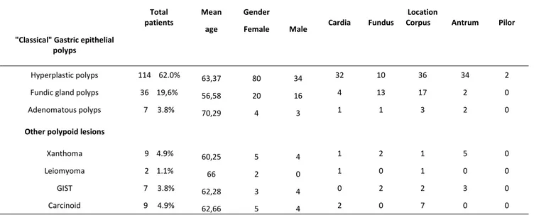
Types of polyps: In this study, we identified gastric polyps in 184 patients, which represented 0,81 % of all patients who underwent upper gastrointestinal endoscopy (24568 patients ). In the "classical" gastric epithelial polyps group, hyperplastic polyps were the most common (62%), followed by fundic gland polyps (19,6%) and adenomatous polyps (3,8%). Among other polypoid lesions group, gastric carcinoid (4,9%) and xanthoma (4,9%) were the most common lesions.
Sex: In our study we found that female patients were more frequently affected than male patients. Of the 184 patients, 119 (64,7%) were women and 65 (34,3%) were men. There was also a female predominance in "classical" gastric epithelial polyps (105 women vs 52 men). The female/male ratio for hyperplastic polyps and fundic gland polyps was 2.5:1 and 1.4:1, respectively.
Age: The statistical analysis indicates that the mean age of the patients was 62,10+13,4 (range:30-90) years. Our results also showed that fundic gland polyps were observed at younger age (mean age: 56,58) compared to the age at which hyperplastic polyps (mean age:63,37) and adenomatous polyps (mean age: 70,29) occurred .
Site of polyps: In our study, the most frequent site of gastric polyps was corpus 36,4 % (n:67) and followed by antrum %25 (n:46), cardia 22,3 % (n: 41), fundus 15, 2% (n:28) and pylorus 1,1%(n:2). We also found that all types of gastric epithelial polyps mostly located in the corpus (31,5% of hyperplastic polyps, 47,2% of fundic gland polyps, 42,8 % of adenomatous polyps ) The specific characteristics of gastric polyps such as age, sex and location are shown in Table 1.
Table I: Specific charecteristic of gastric polypoid lesions according to histopathologic diagnosis. Total
patients Mean age
Gender
Female Male Cardia Fundus
Location
Corpus Antrum Pilor "Classical" Gastric epithelial
polyps
Hyperplastic polyps 114 62.0% 63,37 80 34 32 10 36 34 2
Fundic gland polyps 36 19,6% 56,58 20 16 4 13 17 2 0
Adenomatous polyps 7 3.8% 70,29 4 3 1 1 3 2 0
Other polypoid lesions
Xanthoma 9 4.9% 60,25 5 4 1 2 1 5 0
Leiomyoma 2 1.1% 66 2 0 1 0 1 0 0
GIST 7 3.8% 62,28 3 4 0 2 2 3 0
Carcinoid 9 4.9% 62,66 5 4 2 0 7 0 0
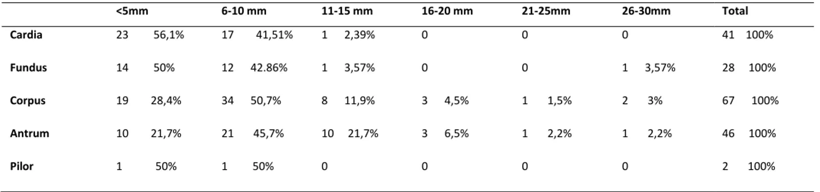
Size: A majority of polyps (88%, n:152) were smaller than 1 cm in diameter, while only six polyps were larger than 2cm in diameter. We noticed that fundic gland polyps tend to be smaller than hyperplastic polyps (94% of fundic gland polyps <1 cm, 84% of hyperplastic polyps <1 cm ). Adenomatous polyps were bigger in size than other types of polyps.
The polyps which are smaller than 5mm in diameter, were mostly seen in the cardia and most of them were hyperplastic polyps. We also determined that larger hyperplastic polyp incidence increases with patients age however there were no statistically significant correlations between polyp size and age. We have no found any association between the types of polyps and age, sex or size. Table 2 shows the distribution of gastric polyps size according to histopathogic diagnosis and Table 3 shows the distribution of gastric polyps size according to anatomic location of polypoid lesions.
Gastric polyps
Erinc et al.
337
GMJ
2019; 30: 336-340
Table 2: Relationship between gastric polypoid lesions and size; the values indicate number of cases
Table 3: Relationship between polyp size and anatomic location ; the values indicate number of cases and percentage.
H. Pylori: Among the114 patients with hyperplastic gastric polyps and fundic gland polyps, H. pylori colonization was detected in 26 patients (22,8%) with hyperplastic polyps and 9 patients (25%) with fundic gland polyps.
Intestinal metaplasia: Intestinal metaplasia was detected in the adjacent gastric mucosa in 24,6% of hyperplastic polyps, 13,9 % of the fundic gland polyps and 42,9 % of adenomatous polyps.
Chronic gastritis and Atrophy: In the present study, we determined that all gastric polyp patients exhibited chronic gastritis histopathologically. We found that the adjacent gastric mucosa have exhibited atrophy in 29,82% of hyperplastic polyps and in 57,14 % of adenomatous polyps. Table 4 shows the rates of H.pylori infection and intestinal metaplasia as well as gastric atrophy status.
Table 4: Histopathologic features of adjacent gastric mucosa according to histologic type of gastric polyps
There was no statistically significant relation between the polyp types and
H:pylori infection, gastric atrophy, intestinal metaplasia. hyperplastic polyps and fundic gland polyps however high-grade dysplasia Dysplasia: In our study, no dysplastic change or malignancy was found in was only seen in one of adenomatous polyps.
DISCUSSION
In the current study we presented the findings of a retrospective analysis of gastric polyps, which were diagnosed at the Baskent University Hospital, over a ten year period. Gastric polyps are found in approximately 6 percent of upper gastrointestinal endoscopic procedures in the United States(3). However, lower rates have been reported in other countries. Frequency and the types of gastric polyps vary depending upon the population being studied. In our study, the prevalence of gastric polyps (0,83%, 184 patients with gastric polyps in 24568 endoscopies) was similar to the findings of studies from Greek and Brasil, 1,2% ( 157 patients with gastric polyps in 12974 endoscopies) and 0.58% (153 patients with gastric polyps in 26,000 endoscopies), respectively (4,5). Similar prevalance rate has also been reported in Turkey, befeore (6,7). Recent investigation in China showed that the detection rate of gastric polyps was is 3,1% ( 2125 patients with gastric polyps in 69575 endoscopy) (8).
"Classical" gastric epithelial polyps were the most frequently assessed polyps in our study. We found that the majority of polyps were hyperplastic polyps(62%) followed by fundic gland polyps (19,6%) and adenomatous polyps (3,8% ). Other studies fromTurkey also reported that hyperplastic polyps were the most commonly observed polpys however there are differences in prevalence rates (9-11).
Hyperplastic polyps are thought to result from excessive regeneration of foveolar epithelium after mucosal damage. They account for up to 75 % of gastric polyps in the geographic areas where H. pylori infection is common (12). Hyperplastic Polyps (n) Fundic gland polyps (n) Adenomatous
Polyps (n) Xantoma (n) Leiomyoma (n) GIST (n) Carcinoid
0-5 mm 37 19 0 9 0 0 2 6-10 mm 59 15 3 0 2 1 5 11-15 mm 14 1 2 0 0 2 1 16-20 mm 3 1 1 0 0 0 1 21-25 mm 0 0 1 0 0 1 0 26-30 mm 1 0 0 0 0 3 0 114 36 7 9 2 7 9 <5mm 6-10 mm 11-15 mm 16-20 mm 21-25mm 26-30mm Total Cardia 23 56,1% 17 41,51% 1 2,39% 0 0 0 41 100% Fundus 14 50% 12 42.86% 1 3,57% 0 0 1 3,57% 28 100% Corpus 19 28,4% 34 50,7% 8 11,9% 3 4,5% 1 1,5% 2 3% 67 100% Antrum 10 21,7% 21 45,7% 10 21,7% 3 6,5% 1 2,2% 1 2,2% 46 100% Pilor 1 50% 1 50% 0 0 0 0 2 100%
H.Pylori % Atrophy % Intestinal
metaplasia
%
Hyperplastic polyps 26/114 22,8 34/114 29,8 28/114 24,6
Fundic gland polyps 9/36 25,0 5/36 13,9 5/36 13,9
Adenomatous polyps 2/7 28,6 4/7 57,1 3/7 42,9
Erinc et al.
Beside arising in a background of H. pylori infection, hyperplastic polyps also occur in previously damaged gastric mucosa, reactive or chemical gastritis when adjacent to ulcer erosions and around gastroenterostomy stomas or autoimmune gastritis (7). Conversly, fundic gland polyps can be sporadic or associated with familial adenomatous polyposis (FAP) and affect patients receiving long-term treatment with proton-pump inhibitors (PPi).
Recent studies showed that the gastric polyp spectrum has changed, with an increasing prevalence of fundic gland polyps. It is believed that H.pylori eradication and use of PPi is associated with higher incidence of fundic gland polyps in western populations. Regression of hyperplastic polyps has as many as 71% of patients with H. Pylori infection after eradication of bacteria (13-15) . In United States, latest published retrospective study confirmed that fundic gland polyps were the highest prevalence of gastric polyps, and hyperplastic polyps were the second, 7.72% and 1.79% of patients, respectively (16) . Similar changes in the spectrum of gastric polyp has also been reported in the Northern Chinese population (17). Recently, a study reported that there is also a decrease in H. Pylori seroprevalence in Turkey (18). On the other hand, in the study from Korea, authors pointed out that the incidence of fundic gland polyp are lower than Western reports, but that of gastric adenoma and gastric cancer is relatively high, similar to previous Asian reports(19). In East Asian where H. pylori infection remains high, larger proportions of gastric polyps are still hyperplastic(20).
Our results showed that H. pylori colonization was detected in 26 patients (22,8%) with hyperplastic polyps and in 25% of fundic gland polyps. H. pylori infection rates in patients with hyperplastic polyps were not significantly different than in patients with fundic gland polyps and adenomatous polyps. In our study more than %70 of gastric polyp patients had previously undergone eradication therapy to eliminate H. pylori. In addition, most patients had a prolonged history of gastric symptoms with history of treatment with PPi, antacids, or H2 receptor antagonists. While we believe that PPi administration has become common in our patients however we cannot obtain precise data about the duration of PPI treatment.
Chronic gastritis was a constant feature in the adjacent mucosa in all of the our cases. In literature gastric hyperplastic polyps have reported to be the mark of the beginning of a progression from chronic gastritis to intestinal metaplasia and gastric atrophy, however gastric adenoma reflect later stages of this process. Although hyperplastic polyps were believed to be benign lesions not associated with the risk of malignant transformation authors have reported dysplastic changes and/or foci of gastric adenocarcinoma harboured within hyperplastic polyps, recently(21,22). It is important to know that both polyps and gastric mucosa adjacent the polyps have an increased risk of cancer development. The risk of focal gastric cancer is five-fold higher in gastric adenomatous polyps than in the hyperplastic ones (10% vs 2.1%), and 2-fold higher in gastric mucosa surrounding the adenomatous than hyperplastic polyps (13.3% vs 7.1%). Therefore performing biopsy of the adjacent gastric mucosa should be performed for further assessment. Our results indicated that atrophy and intestinal metaplasia in the adjacent gastric mucosa is more common in adenomatous polyps and hyperplastic polyps compare to fundic gland polyps however we have no found dysplasia in hyperplastic polyps and fundic gland polyps. The risk of dysplasia in sporadic fundic gland polyps is rare, while it occurs in 30%-50% of FAP-associated polyps (23,24,25).
In our study, most of the hyperplastic polyps and fundic gland polyps were approximately 1cm in diameter while gastric adenomas were approximately 5 to 8 cm in diameter. We determined that fundic gland polyps tend to be smaller than hyperplastic polyps. Our results also showed that the size of polyps increases with increasing age, in hyperplastic polyps. A size greater than 1 cm and pedunculated morphology have been identified as risk factors for dysplasia in hyperplastic polyps. Guidelines recommend polypectomy of all gastric hyperplastic polyps greater than 0,5 cm to 1 cm (26). Similarly adenomatous polyps that are greater than 2 cm and retain a villous histology have a higher risk for developing neoplasia (28 %-40%) (27). The statistical analysis indicates that the mean age of the patients was 62,10+13,4 (range:30-90) years. Our results also showed that fundic gland polyps were observed at younger age (mean age: 56,58) compared to the age at which hyperplastic polyps (mean age:63,37) and adenomatous polyps (mean age: 70,29) occurred. Authors found that age spectrum has changed in gastric polyps, they reported that patients aged 45-59 have currently twice more gastric polyps than 10 years ago, but the inverse relationship is observed for patients aged 60 years and over(28). Authors also reported that location of gastric polyps has changed in the past 10 years; the incidence of polyps has increased in the stomach body (19% vs 32%) and decreased in the antrum (46% vs 24%) (17). In our patient population, most gastric polyps were detected in the corpus.
In the present study, we observed that gastric epithelial polyps were more frequently detected in women; however it is known that men and women are equally affected. The female to male ratio was more than two-thirds in hyperplastic polyps, and was equal in fundic gland polyps.
These findings are similar to other studies that have reported a slight predisposition in women, where in women comprised 58-70,5% of patients (12) however there are also studies that have indicated a male predominance (14).
We have also investigated lesions with a polypoid appearance which includes gastrointestinal stromal tumors (GISTs), leiomyomas, lipomas, and other subepithelial lesions. In this group, our results showed that carcinoids were the most common lesions (49%) followed by gastric xantomas(22%). These lesions are commonly associated with chronic gastritis. In our study all patients experienced chronic gastritis however carcinoids may also be associated with chronic autoimmune atrophic gastritis and pernicious anemia or Zollinger-Ellison syndrome and multiple endocrine neoplasia (29,30).
There are some limitations in of our study due to its retrospective nature, limiting the access to patient’s endoscopic and clinical data. Although the diagnosis of polyps was confirmed histologıcally in all cases, no exact / certain data were available regarding use of PPI status and H. Pylori eradication therapy.
In conclusion, we evaluated the spectrum of gastric polyp developments in our study population by reviewing a 10-year database . Although this study presents the results of single center, it involves a relatively large number of patients. Knowing the spesific and characteristic features of gastric polyps may help the clinicians to evaluate and management of gastric polyps. In the current study showed that overwhelming majority of gastric polyps were hyperplastic polyps. H. pylori infection rates were similar in patients with hyperplastic polyps and fundic gland polyps. On the other all gastric epithelial polyps arise in a background of chronic gastritis. Chronic gastritis may also related to intestinal metaplasia and atrophy therefore clinicians should consider that the excision of the polypoid lesion always should be accompanied by additional sampling of the unaffected mucosa to obtain reliable information about severity of the background gastritis.
Further prospective or retrospective multicenter studies may allow us to explore mechanisms of the origin and the spectrum of gastric polyps in the population of our country. We belive that retrospective studies performed in long term period should provide more information about the gastric polyp spectrum and effect of geographical differences on the prevalence of gastric polyps.
Conflict of interest
No conflict of interest was declared by the authors.
REFERENCES
1-Park do Y, Lauwers GY, Gastric polyps: Classification and management. Arch Pathol Lab Med. 2008;132:633-40.
2- Yakirevich E, Resnick MB. Pathology of gastric cancer and its precursor lesions. Gastroenterol Clin North Am 2013; 42: 261-84.
3- Carmack SW, Genta RM, Schuler CM, Saboorian MH. The current spectrum of gastric polyps: a 1-year national study of over 120,000 patients. Am J Gastroenterol 2009; 104:1524-32.
4-Archimandritis A, Spiliadis C, Tzivras M, et al. Gastric epithelial polyps: a retrospective endoscopic study of 12974 symptomatic patients. Ital J Gastroenterol 1996; 28:387-90.
5- Morais DJ, Yamanaka A, Zeitune JM, Andreollo NA. Gastric polyps: a retrospective analysis of 26,000 digestive endoscopies. Arq Gastroenterol 2007; 44:14-7.
6-Vatansever S,Akpınar Z,Alper E, İpek S, Yazıcıoğlu N, Ekinci N et al. Gastricpolyps and polypoid lesions: Retrospective analysis of 36650 endoscopic procedures in 29940 patients. Turk J Gastroenterol. 2015;26:117-22.
7-Abraham SC, Singh VK, Yardley JH, Wu TT. Hyperplastic polyps of the stomach: association with histological patterns of gastritis and gastric atrophy. Am J Surg Pathol 2001;25:500
8-Zheng E, Ni S, Yu Y, Wang Y, Weng X, Zheng L. Impact of gender and age on the occurrence of gastric polyps: data analysis of 69575 southeastern Chinese patients. Turk J Gastroenterol. 2015;26:474-9.
9-Buyukasik K, Sevinc MM, Gunduz UR, Ari A, Gurbulak B, Toros AB et al. Upper gastrointestinal tract polyps: what do we know about them? Asian Pac J Cancer Prev. 2015;16:2999-3001
10-Karaman A, Deniz K, Karaman H., Gürsoy S,Baskol M, Güven K et al. Prevalence and histopathological condition of gastric polyps in Central Anatolia, Endoscopy 2011;19:56-8.
11-Atalay R, Solakoğlu T, Ozer Sarı S, Köseoğlu H, Akın FE, Demirezer Bolat A et al. Evaluation of gastric polyps detected by endoscopy: a single-center study of a four-year experience in Turkey. Turk J Gastroenterol. 2014 ; 25:370- 12-Stolte , Sticht T, Eidt S, Ebert D, Finkenzeller G. Frequency, location, and age and sex distribution of various types of gastric polyp. Endoscopy 1994; 26: 659-65.
Gastric polyps
Erinc et al.
339
GMJ
13- Ohkusa T, Miwa H, Kumagai J, Tanizawa T, Asaoka D, Terai T et al. Endoscopic, histological and serologic findings of gastric hyperplastic polyps after eradication of Helicobacter pylori: Comparison between responder and non-responder cases. Digestion 2003;68:57-62
14-Ljubicic N, Banic M, Kujundzic M, Antic Z, Vrkljan M, Kovacevic I et al. The effect of eradicating Helicobacter pylori infection on the course of adenomatous and hyperplastic gastric polyps. Eur J Gastroenterol Hepatol. 1999, 11: 727-30.
15- Ohkusa T, Takashimizu I, Fujiki K, Suzuki S, Shimoi K, Horiuchi T et al. Disappearance of hyperplastic polyps in the stomach after eradication of Helicobacter pylori. A randomised, clinical trial. Ann Intern Med 1998;129:712-5.
16- Sonnenberg A, Genta RM. Prevalence of benign gastric polyps in a large pathology database. Dig Liver Dis. 2015;47:164-9.
17- Cao H, Wang B, Zhang Z, Zhang H, Qu R. Distribution trends of gastric polyps: an endoscopy database analysis of 24 121 northern Chinese patients. J Gastroenterol Hepatol 2012; 27: 1175-80.
18- Ozden A, Bozdayı G, Ozkan M, Köse KS, Changes in the seroepidemiological pattern of Helicobacter pylori infection over the last 10 years Turk j Gastroenterol 2004;15: 156-8.
19- Park SY, Ryu JK, Park JH, Yoon H, Kim JY, Yoon YB et al Prevalence of gastric and duodenal polyps and risk factors for duodenal neoplasm in korean patients with familial adenomatous polyposis. Gut Liver. 2011;5:46-51. 20 - Shaib YH, Rugge M, Graham DY, Genta RM. Management of gastric polyps: an endoscopy-based approach. Clin Gastroenterol Hepatol. 2013;11:1374-84.
21-Orlowska J, Kupryjanczyk J. Malignant transformation of gastric hyperplastic polyps. Am J Clin Pathol. 2002;117:165-6.
22-Taniuchi K, Okada M, Sakaeda H. Focal Intramucosal Adenocarcinoma Occurring in Gastric Hyperplastic Polyps: Two Case Reports. Case Rep Gastrointest Med. 2015;2015:201042
23-Bertoni G, Sassatelli R, Nigrisoli E, Pennazio M, Tansini P,Arrigoni A et al. Dysplastic changes in gastric fundic gland polyps of patients with familial adenomatous polyposis. Ital J Gastroenterol Hepatol 1999; 31:192-7. 24-Declich P, Ambrosiani L, Bellone S,Tavani E, Ferrara A, Galati F et al Fundic gland polyps: a not so innocuous entity worth a careful evaluation. Am J Gastroenterol 1998; 93: 2641.
25-Wu TT, Kornacki S, Rashid A, Yardlet JH, Hamilton SR. Dysplasia and dysregulation of proliferation in foveolar and surface epithelia of fundic gland polyps from patients with familial adenomatous polyposis. Am J Surg Pathol 1998; 22: 293-8.
26- Evans JA, Chandrasekhara V, Chathadi KV, Decker GA, Early DS, Fisher DA et al. ASGE Standards of Practice Committee, The role of endoscopy in the management of premalignant and malignant conditions of the stomach. Gastrointest Endosc. 2015;82:1-8.
27- Oberhuber G, Stolte M. Gastric polyps: an update of their pathology and biological significance. Virchows Arch. 2000; 437:581-90.
28- Fan NN, Yang J, Sun G, Lu ZS, Ling Hu EQ, Wang XD et al. Changes in the spectrum of gastric polyps in the Chinese population. World J Gastroenterol 2015; 21: 9758-64
29- Kokkola A, Sjoblom S, Haapiainen R, Sipponen P, Puolakkainen P, Jarvinen H. The risk of gastric carcinoma and carcinoid tumours in patients with pernicious anaemia: A prospective follow-up study. Scand J Gastroenterol 1998;33:88-92.
30- Laine L, Ahnen D, McClain C, Solcia E, Walsh JH, Potential gastrointestinal effects of long-term acid suppression with proton pump inhibitors. Aliment PharmacolTher 2000;14:651-68.