Genome composition analysis of
multipartite BnYVV reveals the
occurrence of genetic re-assortment
in the isolates of Asia Minor and
thrace
canan Yüksel Özmen
1, Saber Delpasand Khabbazi
1, Afsaneh Delpasand Khabbazi
2,
Songül Gürel
3, Rıza Kaya
4, Muhammet Çağrı oğuz
1, ferzat turan
1, fereshteh Rezaei
1,6,
Umut Kibar
5, ekrem Gürel
3& Ali ergül
1*Beet necrotic yellow vein virus (BnYVV) is the cause of rhizomania, an important disease of sugar beet
around the world. The multipartite genome of the BNYVV contains four or five single-stranded RNA that has been used to characterize the virus. Understanding genome composition of the virus not only determines the degree of pathogenicity but also is required to development of resistant varieties of sugar beet. Resistance to rhizomania has been conferred to sugar beet varieties by conventional breeding methods or modern genome engineering tools. However, over time, viruses undergo genetic alterations and develop new variants to break crop resistance. Here, we report the occurrence of genetic reassortment and emergence of new variants of BnYVV among the isolates of thrace and Asia Minor (modern-day Turkey). Our findings indicate that the isolates harbor European A-type RNA-2 and RNA-3, nevertheless, RNA-5 is closely related to East Asian J-type. Furthermore, RNA-1 and RNA-4 are either derived from A, B, and P-types or a mixture of them. The RNA-5 factor which enhance the pathogenicity, is rarely found in the isolates studied (20%). The creation of new variants of the virus emphasizes the necessity to develop new generation of resistant crops. We anticipate that these findings will be useful for future genetic characterization and evolutionary studies of BNYVV, as well as for developing sustainable strategies for the control of this destructive disease.
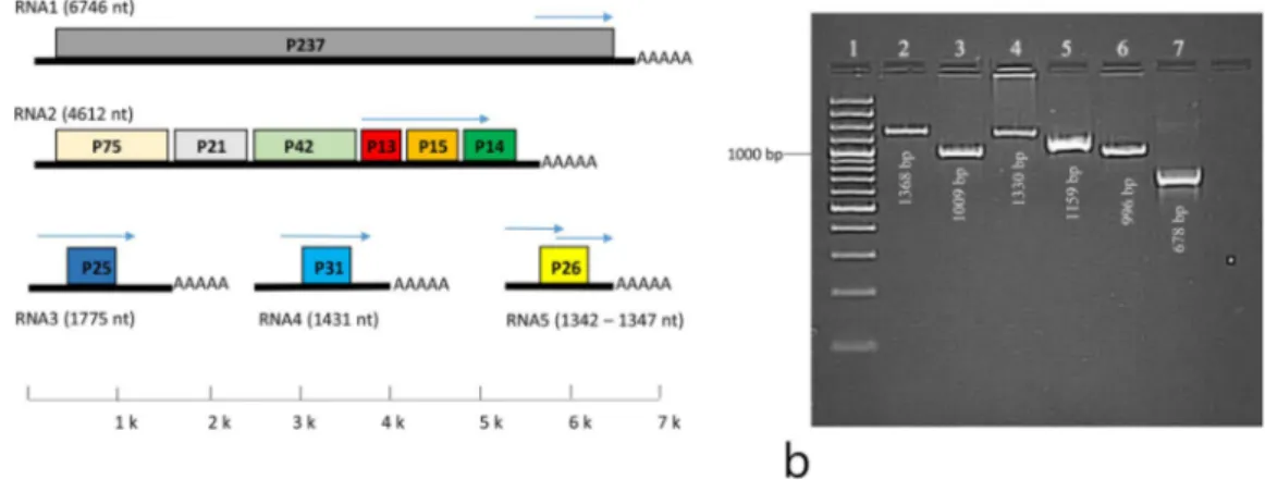
Rhizomania is one of the most destructive soil-borne diseases of sugar beet (Beta vulgaris L.) worldwide. Since the first report of rhizomania1 numerous studies have reported the worldwide infection of sugar beet fields with this disease. Tamada and Baba2 first identified Beet necrotic yellow vein virus (BNYVV) as the cause of rhizomania when they isolated the virus from infected plants of sugar beet fields in Japan. This disease reduces sugar content by 8%, root yield up to 90%, and sugar yield up to 80%3,4. The BNYVV genome is multipartite and composed of four single-stranded RNA species designated as RNA-1, RNA-2, RNA-3, and RNA-4, coating with a 21-kDa pro-tein5. In addition, a fifth RNA species (RNA-5) has been identified in some of the European and Asian BNYVV isolates6–12. RNA-1 and RNA-2, which contain 6746 and 4612 nt-long RNA species, respectively, encode viral “housekeeping” genes involved in virus replication, assembly, cell-to-cell movement and suppression of post tran-scriptional gene silencing13,14. Therefore, when the virus vector Polymyxa betae Keskin15 is not present, RNA-1 and RNA-2 are required for the maintenance of BNYVV in the environment8,14,16. RNA-3 consisting of a 1775 nt-long RNA species, is involved in viral pathogenicity7,10,11,17,18. RNA-4 (1431 nt) plays a key role in transmission of the virus by P. betae7,11,13,19. RNA-5 (1342–1347 nt in length) is associated with rhizomania severity, but is not
1Ankara University, Biotechnology Institute, 06135, Ankara, Turkey. 2University of Tabriz, Department of Plant
Protection, 51666, Tabriz, Iran. 3Bolu Abant İzzet Baysal University, Department of Biology, 14030, Bolu, Turkey. 4Sugar Institute, Department of Phytopathology, Etimesgut, 06930, Ankara, Turkey. 5Republic of Turkey Ministry of
Agriculture and Forestry, Agriculture and Rural Development Support Institution, 06550, Ankara, Turkey. 6Present
address: Başkent University, Institute of Transplantation and Gene Sciences, 06980, Kahramankazan, Ankara, Turkey. *email: [email protected]
required for virus survival20,21. Comparative studies revealed that the RNA-1, RNA-4, and RNA-5 contribute to the development of different rhizomania symptoms7. Moreover, the interaction between RNA-3, RNA-4 and RNA-5 increases rhizomania symptoms11,20. BNYVV is mainly divided into three pathogenic types designated as A, B and P types4,8,12,17. A and B are the most widespread types such that A-type is more prevalent than B-type virus and the P is a rare type throughout the world3. Type A is found in European countries as well as in the USA, China, and Japan, whereas, Type B occurs mainly in countries such as France, Germany, Sweden, Poland, China and Iran6,10,22,23. Type P is associated with RNA-5 and was first isolated from the city of Pithiviers, France24. However, RNA-5 species was previously described in East Asian isolates20. Studies also revealed that the J-type East Asian BNYVV isolates was different from French isolates for the length and the sequence of RNA-57,25. With more than 340 thousand hectares of sugar beet harvesting area, Turkey is the world’s fifth largest sugar beet producing country (FAO stat, 2017) and rhizomania is known to cause serious economic losses in sugar beet production26.
In the current study, the BNYVV strains isolated from different provinces of Turkey with long history of sugar beet cultivation have been subjected to comprehensive genomic analyses of all the RNA components and further phylogenetic studies were carried out. According to our findings genetic reassortment between the European and East Asian A, B and P types has led to the emergence of new variants of BNYVV in the region of Thrace and Asia Minor (modern-day Turkey).
Results
DAS-eLiSA and Rt-pcR-based detections of BnYVV isolates.
The double-antibody sandwich enzyme-linked immunosorbent assay (DAS-ELISA) results confirmed BNYVV infection of only 38 bait plants grown in collected soil samples (57.6% of all bait plants). The highest OD value was recorded 2.160 for samples of Kütahya; however, the other values of positive isolates ranged between 0.068–1.609 (Table 1). The DAS-ELISA assessments for all the plants grown in soil samples collected from Eskişehir, Kahramanmaraş, Kırşehir, Konya, Kütahya, Niğde, Sakarya, Tokat, Edirne and Elazığ provinces were positive. Among three provinces of Iğdır, Çorum and Bursa where the most number of sampling performed (7 samples from each), only 4, 3 and 2 soil samples were infected by BNYVV, respectively (Table 1).Reverse transcription-polymerase chain reaction (RT-PCR) assay was successfully optimized to amplify the desired fragments using positive control isolates (Fig. 1b). Based on RT-PCR, BNYVV isolates were detected in only 34 of the bait plants (51% of all bait plants). The majority of the BNYVV isolates (27 isolates) contained RNA-1-4, while only 7 isolates (20%) contained all five RNA species, indicating the rarity of RNA-5 in Turkish BNYVV isolates (Table 1).
Genome analysis and typology of the BnYVV isolates.
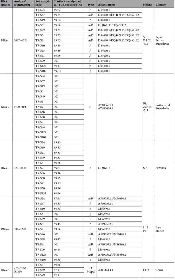
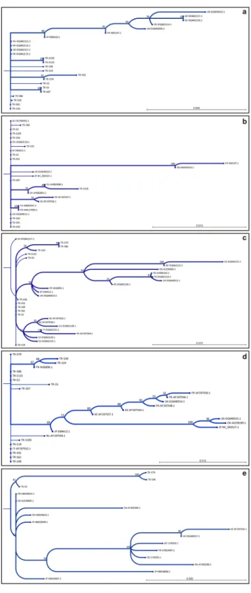
RNA-1. Isolates representing different regions (TR-S24, TR-S67, TR-S19, TR-S61, TR-S49, TR-S5, TR-S2, TR-S86, TR-S58, TR-S91, TR-S79, TR-S125, TR-S105) were selected to study the nucleotide sequences of RNA-1 (Table 1). Analysis of RNA-1 (P237) in the coding region of 5427–6520 and comparing the results with available sequences in the NCBI database revealed the incident of novel nucleotide polymorphism among the isolates in 8 positions (Fig. 2a). Similarity assessments indicated that most of the isolates are highly similar to the A-type isolates of S, F-Pi76 and Yu2 earlier reported from Japan (D84410.1), France (DQ462115) and Yugoslavia (DQ462112), respectively. Isolates such as TR-S24, TR-S86, TR-S58, TR-S91, TR-S125, TR-S105 and TR-S79 are highly similar to the A-type isolates S, F-Pi76 and Yu2 (99.44–100%). However, in other isolates (TR-S67, TR-S61, TR-S49, TR-S5, TR-S2) a complex structure was revealed as these resemble both the P and A-type isolates of S, F-Pi76 and Yu2 (99.35–99.66%) (Table 2). Phylogenetic studies indicated that the new isolates are in close relationship with Japanese Isolate S (bootstrap value 68%), and isolates S8, Yu2, F-Pi72 and F-Pi76 from Sweden, Yugoslavia and France with the support of bootstrap lower than 50% (Fig. 3a; Sup. 1).RNA-2. Isolates of TR-S49, TR-S58, TR-S61, TR-S91, TR-S105, TR-S5, TR-S24, TR-S2, TR-S19, TR-S86, TR-S125, TR-S67 were studied for P13, P14 and P15 open reading frames (ORF) corresponding to transport and suppressor associated proteins in the positions of between 3700–4140 nt. Although no novel nucleotide variation was identified in P13, ORF sequences of P14 and P15 contained several nucleotide variations that were not previously reported (Fig. 2b). RNA-2 sequence analyses designated the isolates as A-type isolates (Table 2). According to the matrix analyses of RNA-2, isolates were identical to the Rio Zurich and A14 isolates reported from Switzerland (AY682691.1) and Yugoslavia (AY682698.1). Phylogenetic analyses displayed that TR-S67 and TR-S125 are related to the isolates A142 (AY682698.1) and Rio Zurich (AY682691.1) with the support of boot-strap value over 70%. In addition, the remaining isolates are closely related to the isolates of Japan (S), Switzerland (Ch23) and UK (MH) with the bootstrap value lower than 50% (Fig. 3b; Sup. 1).
RNA-3. Studying the isolates of TR-S5, TR-S19, TR-S2, TR-S24, TR-S125, TR-S61, TR-S86, TR-S49, TR-S79, TR-S91, TR-S58 for RNA-3 (P25) revealed the occurrence of unique nucleotide polymorphisms between the nucleotide positions of 420–1000 (Fig. 2c). Based on matrix analyses similarity ratios calculated and accord-ingly isolates highly (99.16–99.83%) resemble Sl7 isolate reported from Slovakia (DQ462127.1) (Table 2). Results designated RNA-3 as A-type and furthermore, a close phylogenetic relationship was identified between current isolates and isolates of Slovakia (bootstrap < 50%), UK and Spain (bootstrap > 50%) (Fig. 3c).
RNA-4. RNA-4 (P31) analyses of the selected isolates (TR-S49, TR-S58, TR-S61, TR-S91, TR-S105, TR-S5, TR-S24, TR-S2, TR-S19, TR-S86, TR-S125 and TR-S67) unveiled the incidence of unique polymorphism in the nucleotide position of 381–1280 (Fig. 2d). According to matrix analyses of RNA-4 sequences, the isolates were either highly resembling or identical to the Italian isolate I-12 (AF197552.1) and French isolate F2 (M36896.1).
No.
Code Soil sampling area
DAS-ELISA (Average
Absorbance Value) RT-PCR
Province (district) (+/−) RNA-1 RNA-2 RNA-3 RNA-4 RNA-5
1 TR-S1 Afyon (Şuhut) − − − − − − 2 TR-S2 Afyon (Çobanlar) + (0.217) + + + + − 3 TR-S3 Afyon (Çay) + (0.186) + + + + − 4 TR-S12 Aksaray (Center) − − − − − − 5 TR-S13 Aksaray (Yeşilova) − − − − − − 6 TR-S14 Aksaray (Yeşilova) − − − − − − 7 TR-S15 Aksaray (Yeşilova) + (0.214) + + + + − 8 TR-S16 Amasya (Aydınca) − − − − − − 9 TR-S17 Amasya (Suluova) − − − − − − 10 TR-S19 Amasya (Suluova) + (0.689) + + + + − 11 TR-S20 Amasya (Center) + (1.518) − − − − − 12 TR-S21 Ankara(Ayaş) − − − − − − 13 TR-S22 Ankara (Ayaş) + (0.577) + + + + − 14 TR-S23 Ankara (Ayaş) + (0.598) + + + + − 15 TR-S24 Ankara (Polatlı) + (0.755) + + + + − 16 TR-S28 Ankara (Temelli) + (0.410) + + + + − 17 TR-S31 Burdur (Gölhisar) − − − − − − 18 TR-S32 Burdur (Gölhisar) − − − − − − 19 TR-S33 Burdur (Gölhisar) + (0.279) + + + + − 20 TR-S37 Bursa (Yenişehir) + (1.384) − − − − − 21 TR-S39 Bursa (Yenişehir) + (0.594) − − − − − 22 TR-S40 Bursa (Karacabey) − − − − − − 23 TR-S42 Bursa (Karacabey) − − − − − − 24 TR-S43 Bursa(Mustafa Kemal) − − − − − −
25 TR-S44 Bursa (Mustafa Kemal) − − − − − −
26 TR-S45 Bursa (Mustafa Kemal) − − − − − −
27 TR-S46 Çorum (Osmancık) + (0.230) − − − − − 28 TR-S47 Çorum (Osmancık) − − − − − − 29 TR-S48 Çorum (Osmancık) + (1.072) + + + + − 30 TR-S49 Çorum (Osmancık) + (0.184) + + + + + 31 TR-S50 Çorum (İskilip) − − − − − − 32 TR-S51 Çorum (İskilip) − − − − − − 33 TR-S57 Çorum (Sungurlu) − − − − − − 34 TR-S58 Denizli (Çivril) + (0.316) + + + + − 35 TR-S59 Denizli (Çivril) − − − − − − 36 TR-S61 Edirne (Edirne) + (0.662) + + + + − 37 TR-S67 Elazığ (Kovancılar) + (0.177) + + + + − 38 TR-S76 Erzincan (Erzincan) + (0.844) + + + + − 39 TR-S78 Erzincan (Erzincan) − − − − − − 40 TR-S79 Eskişehir (Sivrihisar) + (1.514) + + + + +
41 TR-S82 Eskişehir (Research Station) + (1.401) + + + + −
42 TR-S4 Iğdır − − − − − − 43 TR-S5 Iğdır + (0.839) + + + + + 44 TR-S7 Iğdır − − − − − − 45 TR-S8 Iğdır + (0.387) + + + + + 46 TR-S9 Iğdır − − − − − − 47 TR-S10 Iğdır + (0.640) + + + + + 48 TR-S11 Iğdır + (0.189) + + + + + 49 TR-S86 Kahramanmaraş (Center) + (1.609) + + + + − 50 TR-S91 Kastamonu (Taşköprü) + (0.159) + + + + − 51 TR-S93 Kastamonu(Center) − − − − − − 52 TR-S105 Kırklareli (Uzunköprü) + (0.068) + + + + − 53 TR-S107 Kırklareli (Alpullu) − − − − − − 54 TR-S113 Kırşehir (Dedeli) + (0.986) + + + + − Continued
Interestingly, some of the isolates (TR-S86, TR-S91, and TR-S125) were identical to both of the I-12 and F2 isolates (Table 2). According to RNA-4-based similarity assessments, TR-S19, TR-S61, TR-S49, TR-S2, TR-S58, TR-S79 and TR-S105 isolates were designated as B-type, however, TR-S67 and TR-S5 isolates were A-type (Table 2). According to phylogenetic studies, some of the isolates were in a close relationship with the French isolate F2 and some others were related to the Italian isolate I-12. This finding was consistent with the results of the matrix analysis of nucleotide sequence similarities (Fig. 3d).
RNA-5. Sequence analysis of RNA-5 (P26) in the coding region (420–1160 nt) showed that TR-S49 and TR-S79 isolates were notably different from previous ones, unlike TR-S5. Despite the presence of polymorphism in TR-S5, the nucleotide sequence is the closest (99.58%) to the Chinese isolate CH2 (AB018614.1). Nevertheless, the genetic distance between TR-S49 and TR-S79 isolates and previously reported sequences was higher (97.11%). The results showed that the studied isolates harbored P26 similar to East Asian J-type (Table 2), but there were a significant number of nucleotide variations. More than 66% of these nucleotide variations lead to amino acid replacements, emphasizing the genetic difference of TR-S49 and TR-S79 (Fig. 2e). Phylogenetic analyses revealed a close genetic relationship between these isolates and those previously reported. Therefore, all isolates were placed in one cluster and the highest relationship was noted with East Asian isolates with bootstrap value over 80% (Fig. 3e; Sup. 1).
No.
Code Soil sampling area
DAS-ELISA (Average
Absorbance Value) RT-PCR
Province (district) (+/−) RNA-1 RNA-2 RNA-3 RNA-4 RNA-5
55 TR-S117 Konya (Çumra) + (0.431) + + + + − 56 TR-S119 Konya (Karapınar) + (1.017) + + + + + 57 TR-S121 Konya (Ilgın) + (0.159) + + + + − 58 TR-S125 Kütahya (Simav) + (2.160) + + + + − 59 TR-S129 Niğde (Center) + (1.106) + + + + − 60 TR-S137 Sakarya (Budaklar) + (0.079) + + + + − 61 TR-S139 Samsun (Çarşamba) − − − − − − 62 TR-S141 Tokat (Turhal) + (1.240) + + + + − 63 TR-S146 Yozgat (Sarıkaya) − − − − − − 64 TR-S158 Yozgat (Boğazlıyan) − − − − − − 65 TR-S156 Yozgat (Çekerek) + (0.285) + + + + − 66 TR-S160 Yozgat (Akdağmadeni) + (0.896) + + + + − 67 − Positive Control 1 + (2.196) + + + + + 68 − Positive Control 2 + (2.208) + + + + + 69 − Negative Control 1 − (0.012) − − − − + 70 − Negative Control 2 − (0.019) − − − − −
Table 1. DAS-ELISA and RT-PCR based detection of BNYVV and RNA species in different sugar beet cultivation regions of Turkey. +/−: depicts the presence/absence of BNYVV infection and RNA species in bait plants grown in soil samples
Figure 1. (a) Genome structure of BNYVV comprising five different RNA species. Each of the RNA-1–5 components contain a single or multiple ORFs. The arrows indicate the region amplified by primers. (b) Amplification of RNA-1–5 was optimized using primers designed. Lane 1 GeneRuler100bp Plus DNA Ladder (Thermo Scientific
™
), lanes 2 – 5 represent RNA-1–4, Lanes 6–7 represent two different amplifications of RNA-5.Discussion
To control losses incurred by rhizomania, cultivation of resistant sugar beet cultivars developed through conven-tional breeding methods and transgenic techniques is considered an effective approach. Emergence and evolution of novel variants of BNYVV threaten sustainable crop production and yield in resistant crops, however, these var-iants are not widespread yet3. ELISA, RT-PCR and restriction fragment length polymorphisms (RFLP) analyses have been widely used in detection and studies of BNYVV structure. ELISA is a routine serological assay used in diagnosis of BNYVV in plants, although concentration of the virus, soil temperature and the type of the sampling tissue affect the efficiency of this method27,28. Therefore, the ELISA test is less efficient particularly when virus concentration is low in infected plants22,29.
The accuracy of the ELISA is less than molecular-based detection methods, however, being fast, cheap and the presence of commercial kits make the ELISA a preferred assay for the routine detection of BNYVV. RT-PCR is a more accurate tool compared to ELISA and is considered a more reliable method in virus detection studies22,30–34 hence, it is increasingly used in the detection of BNYVV35–37. The use of nucleic acid-based methods such as RT-PCR and qRT-PCR has increased the sensitivity of the virus detection by up to 100–10,000 times27.
Absorbance values of ELISA are closely related to rhizomania disease index score, so the higher the disease index scores, the greater the absorbance values. The ELISA result is considered positive only if the absorbance val-ues of the samples are more than three times the value in the negative control samples38. In this study, the ELISA values recorded for negative controls were below 0.02 (0.012–0.019) therefore values over 0.06 were accepted as positive samples (Table 1). The ELISA results of bait plants indicate the infection of BNYVV in 57.6% of the sampling regions. A wide range of absorbance values among the positive samples of different regions (from 0.068 to 2.160) could be due to the different severity index of the disease in these areas. RT-PCR analyses of bait plants verified 89% of the ELISA results which could be relevant to the higher accuracy of the RT-PCR. According to RT-PCR results, 51% of the samples are positive for BNYVV and the majority of the positive samples lack the RNA-5 species thus indicating the rare distribution of the higher virulent BNYVV pathotypes in the sampling regions (Table 1). The results of this study corroborate previous studies reporting the prevalence of BNYVV in sugar beet fields of Turkey39.
Since the first record of BNYVV infection in Turkey40 several research works have diagnosed the disease in sugar beet fields of the country; however, only a few studies have partially characterized the virus39,41–43. Partial sequence analyses of RNA-3 isolated from Tokat province in the mid Black Sea region of Anatolia described Figure 2. Nucleotide sequence alignment of RNA-1–5 of the isolates. Analyses revealed unique polymorphism occurred at: (a) eight positions in RNA-1(P237) between 5427–6520 nt; (b) twelve positions in RNA-2 (within the ORFs P15 and P14) between 3700–4140 nt; (c) five positions in RNA-3 (P25) between 420–1000 nt; (d) seven positions in RNA-4 (P31) between 381–1280 nt; (e) twenty-one positions in the complete coding sequence of RNA-5 (P26). Nucleotide sequences of RNA-1–5 were adjusted according to KX665536.1,
HM117903.1, AJ239200.1, AJ239200.1 and AJ236895.1, respectively. Nucleotides highlighted in black boxes are novel occurrence of polymorphism. Dashes signify missing nucleotides which were not analyzed. Nucleotide variations of RNA-5 that cause amino acid replacements are boxed-in respective nucleotide positions.
RNA
species Analyzed sequence (bp) Soil sample code Similarity analysis of RT-PCR sequence (%) Type Accession no. Isolate Country
RNA-1 5427–6520 TR-S24 99.72 A D84410.1 S F-Pi76 Yu2 Japan France Yugoslavia TR-S67 99.55 A/P D84410.1/DQ462115/DQ462112 TR-S19 99.54 A D84410.1 TR-S61 99.66 A/P DQ462115/DQ462112 TR-S49 99.55 A/P D84410.1/DQ462115/DQ462112 TR-S5 99.35 A/P D84410.1/DQ462115/DQ462112 TR-S2 99.55 A/P D84410.1/DQ462115/DQ462112 TR-S86 99.89 A D84410.1 TR-S58 99.89 A D84410.1 TR-S91 99.69 A D84410.1 TR-S79 100 A D84410.1 TR-S125 99.44 A D84410.1 TR-S105 99.63 A D84410.1 RNA-2 3700–4140 TR-S24 100
A AY682691.1AY682698.1 Rio Zurich A14 Switzerland Yugoslavia TR-S67 100 TR-S19 100 TR-S61 100 TR-S49 100 TR-S5 100 TR-S2 100 TR-S86 100 TR-S58 100 TR-S91 100 TR-S24 100 TR-S125 100 TR-S105 100 RNA-3 420–1000 TR-S24 99.45 A DQ462127.1 Sl7 Slovakia TR-S19 99.83 TR-S61 99.83 TR-S49 99.83 TR-S5 99.66 TR-S2 99.83 TR-S86 99.16 TR-S58 99.79 TR-S91 99.83 TR-S79 99.16 TR-S125 99.66 RNA-4 381–1280 TR-S24 97.31 A/B AF197552.1/M36896.1 I-12 F2 ItalyFrance TR-S67 99.88 A AF197552.1 TR-S19 99.88 B M36896.1 TR-S61 100 B M36896.1 TR-S49 100 B M36896.1 TR-S5 99.45 A AF197552.1 TR-S2 99.76 B M36896.1 TR-S86 100 A/B AF197552.1/M36896.1 TR-S58 99.37 B M36896.1 TR-S91 100 A/B AF197552.1/M36896.1 TR-S79 99.88 B M36896.1 TR-S125 100 A/B AF197552.1/M36896.1 TR-S105 99.88 B M36896.1 RNA-5 420–1160(ORF) TR-S5 99.58 I-A
(J-type) AB018614.1 CH2 China
TR-S49 97.11 TR-S79 97.11
some differences in the amino acid sequence of the Turkish isolates43. Previous nucleotide analyses of RNA-3 and RNA-5 of Turkish isolates assigned these species as A-type and East Asian J-type39,43 respectively. RFLP-based analyses of the isolates from the northern and central parts of Turkey designated RNA-2 and RNA-3 as A-type species42,43 nevertheless, RFLP results of some isolates did not match the expected band profile which was the
Figure 3. Phylogenetic relationship of genomic compartments (RNA-1–5) of BNYVV and sequences available in Genbank. Trees were created using Neighbor-Joining method and Jukes-Cantor distance algorithm. Numbers at the branch junctions represent the percent of trees out of 100 replications. (a) RNA-1, (b) RNA-2, (c) RNA-3, (d) RNA-4 and (e) RNA-5. Accession numbers and detailed information about the reference sequences are given in Sup. 1.
inspiration for the current study to examine the presence of polymorphism among Turkish isolates. RNA-1–5 analyses of BNYVV isolated from different regions of Thrace and Anatolia (modern-day Turkey) revealed a unique polymorphism which could be the probable causes of the failure of RFLP analysis in the previous study42. Our results are in accordance with previous studies that assigned RNA-2–3 as A-type and RNA-5 as East Asian J-type. Furthermore, this study found that RNA-1 was derived either only from A type BNYVV or from mixture of P and A-types as some of the isolates were harboring RNA-1 highly resembling both European and Japanese P and A-types (Table 2). However, the majority of RNA-1 species were closely related to the Japanese A-type strains. Based on RNA-4 analysis, isolates were mostly designated as either European A or B-types. However, there were some isolates very similar or identical to both A and B-types that seems to be derived from mixture of A and B-types. The finding that multipartite genome of BNYVV could be comprised of RNA species with different sources has rarely been reported12,44,45. Studying the isolates from the areas located in a borderline region located between the distribution areas of different BNYVV types indicated the incident of genome re-assortments45. Although mixed infection of different virus types is rarely observed46 it might provide the required condition for this genetic alteration. Genome re-assortments lead to the emergence of new variants of the virus that may overcome the resistance of cultivars.
Among different pathogenic types of BNYVV, strains containing RNA-5 are more destructive than those lacking the fifth RNA species20,21. The worldwide distribution of RNA-5-containing isolates is less common than those lacking it. Studying the isolates of 24 provinces in Turkey revealed the rare occurrence of BNYVV strains harboring RNA-5 in infected fields which is in agreement with the earlier report42. RNA-5 was detected only in four provinces of Iğdır, Eskişehir, Çorum and Konya and based on RT-PCR only 7 out of 34 soil samples infected with BNYVV were harboring RNA-5 species (20%). Moreover, Iğdır province in Eastern Anatolia had the highest RNA-5 occurrence (57%) among the studied regions. Analysis of RNA-5 in the coding sequences indicated that although all the TR-S5, TR-S49 and TR-S79 isolates were closely related to East Asian J-type, there was a notable nucleotide and amino acid difference between them (Table 2).
In conclusion, this study aimed to carry out sequence analyses of the BNYVV genomic compartments and typology of RNA-1–5 based on pairwise identity and similarity assessments along with phylogenetic relation-ships. We detected the B type RNA species in the Anatolian region where BNYVV isolates were designated as A type virus in previous studies. Furthermore, our study reveals the occurrence of genomic re-assortments between P, B and A-type viruses which is the first report. The emergence of new variants of the virus threatens the sustain-able production of sugar beet in the world, therefore, these findings will contribute to the sustainsustain-able control of BNYVV.
Methods
collection of soil samples and virus inoculations.
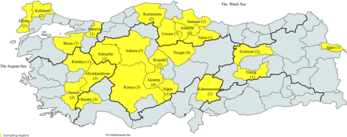
Soil samples were collected from 66 sugar beet cul-tivation regions in 24 provinces of Turkey and dried out at room temperature (Fig. 4). Two different soil sam-ples known to be infested with BNYVV were supplied by the Sugar Research Institute (Ankara, Turkey) were included in the study as positive controls for RNA-1–5 components. The samples were pulverized and sieved through 2 mm-pores, mixed with sand (3:1 ratio of soil and sand) and filled into plastic pots (1.5 kg per each pot). The experiment was performed in 3 replicates. Beta vulgaris L. cv. Ansa was selected as the BNYVV-susceptible cultivar to be deployed in virus infection experiments of bait plants. In each pot, 30 seeds of bait plants were planted and kept at 25 °C and 16/8-hour light/dark period for 10 weeks. Following germination, 15 plantlets were randomly collected from each pot. The roots were detached, washed with distilled water and dried out. The root pieces collected from each soil sample were compiled and ground with porcelain mortar and pestle using liquid nitrogen. After grinding, one gram of crushed root tissue was used in the ELISA test and the remaining amount was stored at −80 °C to be utilized for RNA isolations and RT-PCR assays.Figure 4. Distributions of BNYVV sampling regions within Turkey (highlighted with yellow). A total of 66 soil samples were collected from 24 provinces. The number of soil samples collected from each province were given in brackets (The map is created with the online mapchart.net application version 7.9 available at https:// mapchart.net/turkey.html).
enzyme-linked immunosorbent assays.
The DAS-ELISA was performed to quantify the BNYVV accu-mulation in the samples using the Bioreba AG Kit (Catalog No. # 160175). The DAS-ELISA assay was carried out according to the manufacturer’s recommendations. Bio-Rad Novapath Microplate Reader was used to measure absorbance values of the samples at 405 nm wavelength. The data were analyzed following the Meunier47 pro-cedure. The DAS-ELISA test was carried out separately for each replicate and average values were calculated (Table 1).RnA isolations.
Total RNA was isolated from root tissues using the TRI Reagent™
kit using manufacturer’s recommendations (Bioshop Inc., Canada). RNA concentrations were analyzed by a NanoDrop-1000 spectro-photometer (NanoDrop Technologies, Llc., Wilmington, USA) and the quality was checked by 1% agarose gel electrophoresis and visualizing under UV (GeneGenius Gel Imaging system).primer designs and Rt-pcR assays.
BNYVV nucleotide sequences were retrieved from the GeneBank and highly conserved regions were used to design PCR primers (Fig. 1a). The primer sets specific for BNYVV genomic components (RNA-1–5) were designed using the NCBI Primer Designing tool (Table 3).The NG dART RT kit (Eurex, Poland) was used to perform the first strand cDNA synthesis and RT-PCR assay. Prior to screening, positive controls were utilized to optimize the amplification of the RNA species. cDNA concentrations and quality were assessed by a NanoDrop-1000 Spectrophotometer. PCR amplifications were per-formed in a 25 µL reaction mixture containing 200 ng of cDNA, 10 pmol of each primer, 2.5 mM dNTPs, 0.5 units GoTaq DNA polymerase (Promega, Madison, WI, USA) and 1.5 mM MgCl2. PCR was initiated with a denaturing
step of 3 min at 94 °C followed by 35 cycles including 94 °C for 1 min (denaturation), 60 °C for 2 min (annealing) and 72 °C for 2 min (extension), and the final extension period of 10 min at 72 °C. PCR products were electro-phoresed on 1.5% agarose gel and the GeneGenius Gel Imaging system was used to visualize the amplicons.
Sequence analyses.
Some of the isolates were selected based on ELISA and RT-PCR results to undergo further sequence and phylogenetic analyses. The resulting PCR products of RNA-1–5 species were purified from agarose gels after electrophoresis and double sequenced by the Sanger dideoxy method (BM Labosis Ltd. Co., Ankara, Turkey). Sequence analyses were performed for RNA 1–5 species in comparison with relevant sequences retrieved from the GeneBank. The sequences obtained were initially subjected to the NCBI (National Center for Biotechnology Information) Blast analysis. Sequence comparisons were conducted using the CLC Sequence Viewer 7.6.1 workbench (CLC Bio-Qiagen, Aarhus, Denmark). Phylogenetic trees were constructed to study the evolutionary relationships. CLC Sequence Viewer 7.6.1 program was used to perform the phylogenetic analysis48. Trees were created by the Neighbor Joining method using the Jukes-Cantor distance algorithm and bootstrap support of 100. Pairwise identity and similarity of nucleotide sequences were calculated through matrix analyses performed by a SIAS online tool (Immunomedicine Group, UCM, Spain).Received: 2 July 2019; Accepted: 17 February 2020; Published: xx xx xxxx
References
1. Canova, A. A. di Patologie Della Barbabietola. Inf. Fitopatol. 9, 390–396 (1959).
2. Tamada, T. & Baba, T. Beet Necrotic Yellow Vein Virus from “Rhizomania” Affected Sugar Beet in Japan. Annls Phytopath Soc. Jpn.
39, 325–352 (1973).
3. Mcgrann, G. R., Grimmer, M. K., Mutasa-Gottgens, E. S. & Stevens, M. Progress towards the understanding and control of sugar beet rhizomania disease. Mol. Plant. Pathol. 10, 129–141 (2009).
4. Pavli, O. I., Stevanato, P., Biancardi, E. & Skanracis, G. N. Achievements and prospects in breeding for rhizomania resisstance in sugar beet. Field Crop. Res. 122, 165–172 (2011).
5. Putz, C. Composition and structure of beet necrotic yellow vein virus. J. Gen. Virol. 35, 397–401 (1997).
6. Saito, M., Kiguchi, T., Kusume, T. & Tamada, T. Complete nucleotide sequence of the Japanese isolate S of beet necrotic yellow vein virus RNA and comparison with European isolates. Arch. Virol. 141, 2163–2175 (1996).
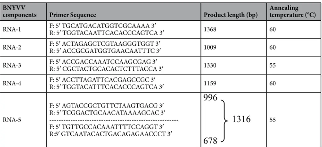
BNYVV
components Primer Sequence Product length (bp) Annealing temperature (°C)
RNA-1 F: 5′ TGCATGACATGGTCGCAAAA 3′R: 5′ TGGTACAATTCACACCCAGTCA 3′ 1368 60 RNA-2 F: 5′ ACTAGAGCTCGTAAGGGTGGT 3′R: 5′ ACCGCGATGGTGAACAATTTC 3′ 1009 60 RNA-3 F: 5′ ACCGACCAAATCCAAGCGAG 3′R: 5′ CGCTACTGCACACTCTTTACCA 3′ 1330 55 RNA-4 F: 5′ ACCTTAGATTCACGAGCCGC 3′R: 5′ TGGTACATTTCACACCCAGTCA 3′ 1159 60
RNA-5 F: 5′ AGTACCGCTGTTCTAAGTGACG 3′ R: 5′ TCGGACTGCAACATAAAAGCAC 3′ ---F: 5′ TGTTGCCACAAATTTTCCAGGT 3′ R:5′ GTCAATACACTGACAGAGAACCCT 3′ 55
7. Koenig, R., Haeberlé, A. M. & Commandeur, U. Detection and characterization of a distinct type of Beet necrotic yellow vein virus RNA 5 in a sugar beet growing area in Europe. Arch. Virol. 142, 1499–1504 (1997).
8. Tamada, T. Benyvirus, in Webster, R.G., Granoff, A. (eds.) Encyclopedia of virology, Academic Press, London, UK pp 154–160 (1999a). 9. Harju, V. A. et al. Occurrence in the United Kingdom of Beet necrotic yellow vein virus isolates which contain RNA 5. Plant. Pathol.
51, 811 (2002).
10. Schirmer, A. et al. Phylogenetic analysis of isolates of Beet necrotic yellow vein virus collected worldwide. J. Gen. Virol. 86, 2897–2911 (2005).
11. Tamada, T. Susceptibility and resistance of Beta vulgaris subsp. maritima to foliar rub-inoculation with Beet necrotic yellow vein virus. J. Gen. Plant. Pathol. 73, 76–80 (2007).
12. Ward, L. et al. Occurrence of two different types of RNA-5-containing beet necrotic yellow vein virus in the UK. Arch. Virol. 152, 59–73 (2007).
13. Richards, K. E. & Tamada, T. Mapping functions on the multipartite genome of beet necrotic yellow vein virus. Annu. Rev.
Phytopathol. 30, 291–313 (1992).
14. Bouzoubaa, S., Quillet, L., Guilley, H., Jonard, G. & Richards, K. Nucleotide sequence of beet necrotic yellow vein virus RNA-1. J.
Gen. Virol. 68, 615–626 (1987).
15. Keskın, B. Polymyxa betae n.sp., ein parasit in den wurzeln von beta vulgaris tournefort, besonders waehrend der jugendentwicklung der zuckerruebe. Arch. Mikrobiol. 49, 348–374 (1964).
16. Tamada, T., Uchino, H., Kusume, T. & Saito, M. RNA 3 deletion mutants of beet necrotic yellow vein virus do not cause rhizomania in sugar beets. Phytopathology 89, 1000–1006 (1999b).
17. Jupin, I., Tamada, Y. & Richards, K. Pathogenesis of beet necrotic yellow vein virus. Sem. Virol. 2, 121–129 (1991).
18. Rush, C. M., Liu, H. Y., Lewellen, R. T. & Acosta-Leal, R. The continuing saga of rhizomania of sugar beets in the United States. Plant.
Dis. 90, 4–15 (2006).
19. Tamada, T. & Abe, H. Evidence that beet necrotic yellow vein virus RNA-4 is essential for efficient transmission by the fungus Polymyxa betae. J. Gen. Virol. 70, 3391–3398 (1989).
20. Tamada, T. et al. Production and pathogenicity of isolates of beet necrotic yellow vein virus with different numbers of RNA components. J. Gen. Virol. 70, 3399–3409 (1989).
21. Heijbroek, W., Musters, P. M. S. & Schoone, A. H. L. Variation in pathogenicity and multiplication of beet necrotic yellow vein virus (BNYVV) in relation to the resistance of sugar-beet cultivars. Eur. J. Plant. Pathol. 105, 397–405 (1999).
22. Arif, M., Khan, W. & Shafi, A. Detection of beet necrotic yellow vein virus in Pakistan using bait-plant bioassay, ELISA and RT-PCR.
Afr. J. Biotechnol. 14, 3206–3215 (2015).
23. Sohi, H. H. & Maleki, M. Evidence for Presence of Types A and B of Beet Necrotic Yellow Vein Virus (BNYVV) in Iran. Virus Genes.
29, 353–358 (2004).
24. Koenig, R., Lüddecke, P. & Haeberle, A. M. Detection of beet necrotic yellow vein virus strains, variants and mixed infections by examining single-strand conformation polymor- phisms of immunocapture RT-PCR products. J. Gen. Virol. 76, 2051–2055 (1995). 25. Miyanishi, M., Kusume, T., Saito, M. & Tamada, T. Evidence for three groups of sequence variants of beet necrotic yellow vein virus
RNA. 5. Arch. Virol. 144, 879–892 (1999).
26. Henry, C. Rhizomania-its effect on sugar beet yield in the UK. Br. Sugar Beet Rev. 64, 24–26 (1996).
27. Tamada, T. General Features of Beet Necrotic Yellow Vein Virus. In Biancardi, E. & Tamada, T. (eds.) Rhizomania, Springer, Cham pp 55-83 (2016).
28. De Biaggi, M. & Biancardi, E. Breeding Methods. In Biancardi, E. & Tamada, T. (eds.) Rhizomania, Springer, Cham pp 233-247 (2016).
29. Nolasco, G., Blas de, C., Torres, V. & Ponz, F. A method combining immunocapture and PCR amplification in a microtiter plate for detection of plant viruses and subviral pathogens. J. Virol. Methods 45, 201–218 (1993).
30. Morris, J., Clover, G. R. G., Harju, V. A., Hugo, S. A. & Henry, C. M. Development of a highly sensitive nested RT-PCR method for Beet Necrotic Yellow Vein Virus detection. J. Virol. Methods 95, 163–169 (2001).
31. Suarez, M. B., Grondona, I., García-Benavides, P., Monte, E. & García-Acha, I. Characterization of beet necrotic yellow vein furovirus from Spanish sugar. Int. Microbiology 2, 87–92 (2010).
32. Yu, C., Wu, J. & Zhou, X. Detection and subgrouping of Cucumber mosaic virus isolates by TAS-ELISA and immunocapture RT-PCR. J. Virol. Methods 123, 155–161 (2005).
33. Zizyte, M., Valkonen, J. & Staniulis, J. Characterization of Beet Necrotic Yellow Veinvirus Infecting Sugar Beet in Lithuania. J Plant
Pathol 95, 211-216 (2013).
34. Ebrahim-Ghomi, M., Mahmoodi, B., Rakhshanderoo, F., Naderpour, M. & Norouzi, P. Widespread distribution of Beet necrotic yellow vein virus in Iranian sugar beet industry. Arch. Phytopathology Plant. Prot. 49, 1–10 (2016).
35. Kutluk Yilmaz, N. D., Uzunbacak, H., Arli-Sokmen, M. & Kaya, R. Distribution of resistance-breaking isolates of beet necrotic yellow vein virus differing in virulence in sugar beet fields in Turkey. Acta Agr. Scand. B-S P 68, 546–554 (2018).
36. Yilmaz, N. D. K., Arli-Sokmen, M. & Kaya, R. p25 pathogenicity factor deletion mutants of beet necrotic yellow vein virus occurring in sugar beet fields in Turkey. J. Plant. Dis. Prot. 125, 89–98 (2018).
37. Fomitcheva, V. W. & Kühne, T. Beet soil-borne mosaic virus: development of virus-specific detection tools. J. Plant. Dis. Prot. 126, 255–260 (2019).
38. Stevanato, P., Biancardi E., & Norouzi P. Assisted Selection. In Biancardi E. & Tamada T. (eds.) Rhizomania, Springer, Cham pp 249–261 (2016).
39. Yılmaz, N. D. K. et al. The widespread occurrences of Beet soil borne virus and RNA-5 containing Beet necrotic yellow vein virus isolates in sugar beet production areas in Turkey. Eur. J. Plant. Pathol. 144, 443–455 (2016).
40. Koch, F. Bericht über eine reise in verschiedene zuckerrübenanbaugebiete der Türkşeker in Anatolien und Thrazien zum studium von wurzelerkrankungen, KWS Kleinwanzlebener Saatzucht AG, Einbeck, Deutschland (1987).
41. Kruse, M. et al. Restriction fragment lenght polymorphism analysis of reverse transcription PCR products reveals the existence of two major strain groups of beet necrotic yellow vein virus. J. Gen. Virol. 75, 1835–1842 (1994).
42. Yılmaz, N. D. K. Identification of strain types of some Beet necrotic yellow vein virus isolates determined in Northern and Central Parts of Turkey. Eurasian J. Soil. Sci. 5, 241–248 (2016).
43. Kutluk Yilmaz, N. D., Meunier, A., Schmit, J. F., Stas, A. & Bragard, C. Partial nucleotide sequence analysis of Turkish isolates of Beet necrotic yellow vein virus (BNYVV) RNA‐3. Plant. Pathol. 56, 311–316 (2007).
44. Li, M. et al. Phylogenetic analysis of Beet necrotic yellow vein virus isolates from China. Virus Genes. 36, 429–432 (2008). 45. Koenig, R. et al. A single U/C nucleotide substitution changing alanine to valine in the beet necrotic yellow vein virus p25 protein
promotes increased virus accumulation in roots of mechanically inoculated, partially resistant sugar beet seedlings. J. Gen. Virol. 90, 759–763 (2009).
46. Ratti, C., Clover, G. R., Autonell, C. R., Harju, V. A. & Henry, C. M. A multiplex RT-PCR assay capable of distinguishing beet necrotic yellow vein virus types A and B. J. Virol. Methods 124, 41–47 (2005).
47. Meunier, A., Schmit, J. F., Stas, A., Kutluk, N. & Bragard, C. Multiplex reverse transcription-PCR for simultaneous detection of Beet necrotic yellow vein virus, Beet soilborne virus, and Beet virus Q and their vector Polymyxa betae Keskin on sugar beet. Appl. Env.
Microbiol. 69, 2356–2360 (2003).
Acknowledgements
The financial support from the Scientific and Technological Research Council of Turkey (TUBITAK), Project No: TOVAG-113O096, is deeply appreciated.
Author contributions
C.Y.Ö. performed the molecular assays (PCR, RT-PCR, ELISA), compiled the data and provided the sequences; S.D.K. and A.D.K. analyzed the sequences, performed the bioinformatics studies, interpreted the data and wrote the paper; S.G., R.K. and E.G. supplied the soil material and performed ELISA; M.Ç.O., F.T., F.R. conducted virus inoculation and bait plant growth; U.K. contributed to bioinformatics analyses and designed primers; A.E. designed, outlined and supervised the whole study.
competing interests
The authors declare no competing interests.
Additional information
Supplementary information is available for this paper at https://doi.org/10.1038/s41598-020-61091-2. Correspondence and requests for materials should be addressed to A.E.
Reprints and permissions information is available at www.nature.com/reprints.
Publisher’s note Springer Nature remains neutral with regard to jurisdictional claims in published maps and institutional affiliations.
Open Access This article is licensed under a Creative Commons Attribution 4.0 International License, which permits use, sharing, adaptation, distribution and reproduction in any medium or format, as long as you give appropriate credit to the original author(s) and the source, provide a link to the Cre-ative Commons license, and indicate if changes were made. The images or other third party material in this article are included in the article’s Creative Commons license, unless indicated otherwise in a credit line to the material. If material is not included in the article’s Creative Commons license and your intended use is not per-mitted by statutory regulation or exceeds the perper-mitted use, you will need to obtain permission directly from the copyright holder. To view a copy of this license, visit http://creativecommons.org/licenses/by/4.0/.