GENETIC AND ENVIRONMENTAL INTERVENTIONS ALTERING THE COURSE OF BRAIN AGING: EVIDENCE FROM THE ZEBRAFISH (DANIO
RERIO) MODEL
A DISSERTATION SUBMITTED TO
THE GRADUATE SCHOOL OF ENGINEERING AND SCIENCE
OF BILKENT UNIVERSITY
IN PARTIAL FULFILLMENT OF THE REQUIREMENTS FOR
THE DEGREE OF
DOCTOR OF PHILOSOPHY
IN
NEUROSCIENCE
By
Elif Tuğce Karoğlu Eravşar
GENETIC AND ENVIRONMENTAL INTERVENTJONS AL TERING THE
COURSE OF BRAIN AGING: EVIDENCE :FROM THE ZEBRAFISH (DAN/O
RER/0) MODEL
By ElifTuğce Karoğlu Eravşar
April, 2021
We certify that we have read this dissertation and that in mır opınıon it is fully adequate, in scope and in quality, asa thesis for the degree of Doctor of Philosophy.
Michelle Marie Adams (Advisor)
Özlen Konu Karakayalı
Erkan Kiriş
Hacı
Hu
~
afalıgönül
Approved for the Graduate School of Engineering and Science
Ezhan Karaşan
Director of the Graduate S.::hool
iii ABSTRACT
GENETIC AND ENVIRONMENTAL INTERVENTIONS ALTERING THE COURSE OF BRAIN AGING: EVIDENCE FROM THE ZEBRAFISH (DANIO
RERIO) MODEL
Elif Tuğce Karoğlu Eravşar PhD in Neuroscience Advisor: Michelle Marie Adams
April, 2021
Age-related cognitive decline occurs during normal aging, although there is no prominent neural loss in the brain. Subtle molecular alterations in synaptic and cellular dynamics are likely underlying these cognitive alterations. One challenge is the widely heterogeneous profile regarding age-related behavioral changes and neurobiological underpinnings. Therefore, it is crucial to characterize how individual factors can contribute to successful or unsuccessful aging and whether these factors can induce shared patterns of alterations in the cellular and synaptic dynamics. Three different intervention approaches were utilized in the current study. The first intervention was a genetic manipulation in the cholinergic system component acetylcholinesterase (AChE), which results in reduced levels of this enzyme in the achesb55/+ mutants. Previous studies have characterized this model as a delayed aging model because of its preserved cognitive abilities at an older age. The current study
iv
was the first study analyzing the neurobiological changes in this mutant model within the context of aging. It was shown that reduced brain AChE activity levels persist in different age groups, including old age in the mutant animals. This reduction was accompanied by subtle decreases in the other elements of the cholinergic system, including acetylcholine and nicotinic acetylcholine receptor subunit alpha-7. Genotype significantly altered key glutamatergic receptor subunits such as N-methyl D-aspartate-type receptor subunit 2B (NR2B) and glutamate receptor subunits 2 and 3 (GluR2/3), with these markers significantly reduced in the achesb55/+ mutants and likely maintaining homeostatic synaptic scaling. At old age, a significant age-related elevation was observed in the synaptophysin levels (SYP) of the old achesb55/+ mutants, and this mutation prevented an age-related decline in the gephyrin (GEP) levels which was evident in the wild-type controls. This mutation also altered the cellular dynamics; an immature neuronal marker, embryonic lethal abnormal-vision (ELAV Drosophila) like-3 (HuC) was significantly upregulated in the achesb55/+ mutants at all ages. In contrast, the levels of inflammation-related markers, glial fibrillary acidic protein (GFAP) and reactive oxygen species (ROS), were downregulated subtly in the mutants. It can be concluded that reduced levels of brain AChE can be associated with altered excitatory homeostasis and preserved levels of GEP and SYP through aging. At the same time, the neuronal marker was upregulated, and inflammation-related markers were downregulated. The second intervention was applying short-term environmental enrichment using the sensory cues to young and old zebrafish to induce successful aging. It was shown that environmental enrichment increases the brain weight in old zebrafish, prevents age-related decrements in the levels of synaptic proteins, including SYP and NR2B, and doublecortin-like kinase
v
(DCAMKL1). Additionally, environmentally enriched old zebrafish had elevated levels of GEP while applying this environmental intervention did not modulate the age-related increases in oxidative stress indicators. The third intervention was also a non-genetic approach. Two short-term opposing dietary treatments, such as caloric restriction (CR) and over-feeding (OF), were applied to young and old zebrafish and an ad-libitum diet. It was demonstrated that a short-term CR regimen upregulated the glutamatergic components of neurotransmission such as GluR2/3 and post-synaptic density 95 (PSD95). Significant age-related decline in GEP levels was observed in old zebrafish in the OF dietary condition. Expression levels of synaptic and regulatory genes were relatively stable, while inflammation-related gene tnfa was altered in an age-dependent manner. Additionally, in the young zebrafish, a significant elevation of trunk cortisol was demonstrated in the OF group compared to CR-fed young zebrafish. Taken together, evaluating different components such as the cholinergic system, diet, and environment can provide us insights into the neurobiological underpinning of successful aging and possible determinants of unhealthy aging.
Keywords: cholinergic system, aging brain, environmental enrichment, synaptic proteins, calorie intake, zebrafish
vi ÖZET
BEYİN YAŞLANMASINI DEĞİŞTİREN GENETİK VE ÇEVRESEL MÜDAHALELER: ZEBRABALIĞI (DANIO RERIO) MODELİNDEN
BULGULAR Elif Tuğce Karoğlu Eravşar Nörobilim Lisansüstü Programı, Doktora
Tez Danışmanı: Michelle Marie Adams Nisan, 2021
Yaşlanan beyinde belirgin bir nöral kayıp olmamasına rağmen, yaşa bağlı bilişsel düşüş normal yaşlanmada ortaya çıkmaktadır. Sinaptik ve hücresel dinamiklerdeki moleküler değişiklikler muhtemelen bu bilişsel değişikliklerin altında yatan nedenlerdir. Yaşa bağlı davranış değişimleri ve nörobiyolojik parametrelerde oldukça heterojen bir profil gözlemlenmektedir, bu durum da karakterizasyon ve tedavi geliştirme amaçlı çalışmaları zorlaştırmaktadır. Bu nedenle, bireysel faktörlerin başarılı veya başarısız yaşlanmaya nasıl katkıda bulunabileceğini ve bu faktörlerin hücresel ve sinaptik dinamiklerde ne gibi değişimlere sebep olabileceğini karakterize etmek oldukça önemlidir. Bu çalışmada yaşlanma sürecini değiştiren üç farklı müdahale yaklaşımı uygulanmıştır. İlk müdahale bir genetik modeli içermektedir, düşük asetilkolinesteraz (AChE) seviyelerine sahip genç ve yaşlı achesb55/+
mutantları kullanılmıştır. Önceki çalışmalar, bu modeli ileri yaşlarda gözlemlenen korunmuş bilişsel yeteneklerinden dolayı gecikmiş yaşlanma modeli olarak nitelendirmiştir ve bu çalışma, yaşlanma bağlamında bu mutant modelindeki nörobiyolojik değişiklikleri
vii
analiz eden ilk çalışmadır. Mutant hayvanlarda farklı yaş gruplarında azalmış beyin AChE aktivitesi seviyelerinin devam ettiği ve bu azalmaya asetilkolin ve nikotinik asetilkolin reseptörü alt birimi alfa-7 dahil olmak üzere kolinerjik sistemin diğer elementlerinde hafif düşüşlerin eşlik ettiği gösterilmiştir. Genotipin etkisi, N-metil D-aspartat tipi reseptör alt birimi 2B (NR2B) ve glutamat reseptör alt birimleri 2 ve 3 (GluR2/3) gibi anahtar glutamaterjik reseptör alt birimlerini önemli ölçüde değiştirmiştir, bu belirteçler achesb55/+
mutantlarında anlamlı olarak azalmıştır. Yaşlanmayla birlikte, achesb55/+
mutantlarının sinaptofizin (SYP) seviyelerinde önemli bir artış gözlemlenmiştir. Yabanıl-tip kontrol grubunda gefirin (GEP) seviyelerinde yaşa bağlı azalmalar meydana gelirken; achesb55/+
mutantlarında GEP seviyelerinde yaşlanmaya bağlı değişikler önlenmiştir. Mutasyonun ayrıca hücresel dinamikler üzerinde de etkileri vardır, nöronal belirteç HuC, achesb55/+
mutantlarında tüm yaş gruplarında önemli ölçüde artmıştır, enflamasyon ile ilgili belirteçlerden olan glial fibril asidik protein (GFAP) ve reaktif oksijen türleri (ROS) seviyeleri mutantlarda azalan bir trende sahiptir. İkinci müdahale, başarılı yaşlanmayı teşvik etmek için genç ve yaşlı zebra balıklarına duyusal elemanlar kullanarak kısa süreli çevresel zenginleştirme uygulamasıdır. Çevresel zenginleştirmenin yaşlı zebra balıklarında beyin ağırlığını artırdığı, SYP, NR2B ve DCAMKL1 gibi proteinlerin düzeylerindeki yaşa bağlı düşüşleri önlendiği gösterilmiştir. Ek olarak, çevresel olarak zenginleştirilmiş yaşlı zebra balıkları yüksek GEP seviyelerine sahipken, bu çevresel müdahalenin uygulanması oksidatif stres göstergelerindeki yaşa bağlı artışları modüle etmemiştir. Üçüncü müdahale de genetik olmayan bir müdahaledir. Kalori kısıtlaması (CR) ve aşırı beslenme (OF) gibi kısa süreli karşıt diyet tedavileri, genç ve yaşlı zebra balıklarına uygulanmıştır. Kısa süreli CR rejiminin, GluR2/3 ve
viii
post sinaptik yoğunluk 95 (PSD95) gibi nörotransmisyonun glutamaterjik bileşenlerini artırabileceği gözlemlenirken; OF diyet grubunda GEP seviyelerinde yaşa bağlı anlamlı bir düşüş gösterilmiştir. Sinaptik ve düzenleyici genlerin ekspresyon seviyeleri nispeten sabitken, enflamasyon ile ilgili gen tnfa yaşa bağlı bir şekilde değişim göstermiştir. Ek olarak, CR ile beslenen genç zebra balıklarına kıyasla OF grubunda önemli bir kortizol yükselmesi bulunmuştur. Birlikte ele alındığında, kolinerjik sistem, diyet ve çevre gibi farklı bileşenlerin değerlendirilmesi, bize başarılı yaşlanmanın nörobiyolojik temelleri ve sağlıksız yaşlanmanın olası belirleyicileri hakkında önemli bilgiler sağlayabilmektedir.
Anahtar Sözcükler: kolinerjik sistem, beyin yaşlanması, çevresel zenginleştirme, sinaptik proteinler, kalori alımı, zebra balığı
ix
x
ACKNOWLEDGEMENTS
Foremost, I would like to express my sincere gratitude to my advisor Prof. Dr. Michelle Adams, for giving me an opportunity to work in such a productive and dynamic research environment and research group which was more like a family to me. I am very grateful for her supporting and encouraging attitude for all these years, and everything that I have learned from her has helped me grow as a young scientist; she has always been inspiring me as a role model and great scientist.
I would like to thank my TIK committee members Assoc. Prof. Dr. Özlen Konu, and Assist. Prof. Dr. Erkan Kiriş, for their feedbacks and constructive discussions. Thanks to their help during this process, my thesis has been improved prominently. I would like to thank Assist. Prof. Dr. Hulusi Kafalıgönül and Assoc. Prof. Dr. Çağdaş Devrim Son for kindly accepting to be on the jury.
I would like to thank my dearest friend Melek Umay Tüz-Şaşik for her support, guidance, help and friendship throughout these years. I would like to thank Assoc. Prof. Dr. Ayça Arslan-Ergül for her support and guidance. I would like to express my sincere gratitude to current and former members of the Adams Lab; Meriç Kınalı, Dilan Ergül Çelebi-Birand, Naz Mengi, Begün Erbaba, Narin Ilgım Ardıç, Beyza Özen, Duygu Mutlu, Serena Aktürk, Füsun Doldur-Ballı, Özge Pelin Burhan, Göksemin Fatma Şengül, Naz Şerifoğlu, Bilge Aşkın, Hande Aydoğan and Duygu Macaroğlu. I would also thank my friends and colleagues at Selcuk University Department of Psychology for their support through this process.
xi
I would like to express my gratitude to my dearest friends Ayşegül Düşündere, Tuba Şahin, Zeynep Çenesiz and Zeynep Yıldırım for their endless support and love. We have learned and grown together and shared lots of great memories; I will always feel blessed for their friendship.
I want to express my greatest gratitude to my mother, Hülya Karoğlu, for her endless support, love, encouragement, and teaching me how to be a good, honest and fair person. I would like to thank my brothers Dursun Emre Karoğlu and Mehmetali İlbey Karoğlu for their support and their help with the figures' preparations. I would like to thank Alaybey Karoğlu, Muallim Eravşar, Rabia Eravşar and Taha Eravşar for being very supportive and wonderful family to me. Last but not least, I would like to thank my spouse Ebubekir for encouraging, supporting, motivating, and loving me through the good times and the bad. I am profoundly grateful and feeling very lucky to have him.
I would like to acknowledge The Scientific and Technological Research Council of Turkey (TÜBİTAK) for financial support as a scholarship holder of 2211-A National Graduate Scholarship Program (BIDEB) and TÜBİTAK 1001 grant to Assist. Prof. Dr. Hulusi Kafalıgönül (Grant number: 215S701) for financially supporting the experiments in Chapter 3. I would like to acknowledge European Molecular Biology Organization (EMBO) Installation Grant with funds provided by TÜBİTAK to Prof. Dr. Michelle M. Adams to support the experiments in Chapters 4 and 5.
xii CONTENTS
ABSTRACT ... iii
ÖZET ... vi
ACKNOWLEDGEMENTS ... x
LIST OF FIGURES ... xvii
LIST OF TABLES ... xxvii
CHAPTER 1 ... 1
INTRODUCTION ... 1
1.1 Aging and cognitive alterations ... 1
1.2 Aging and neurobiological alterations ... 3
1.2.1 Altered synaptic proteome with aging ... 4
1.2.2 Vulnerability of cholinergic system with aging ... 8
1.2.3 Oxidative stress and aging ... 10
1.3 Age-related sex differences ... 11
1.4 Zebrafish as a model organism to study aging ... 12
1.5 Genetic interventions to alter the course of aging ... 13
1.6 Environmental interventions to alter the course of aging ... 14
1.6.1 Environmental enrichment ... 15
1.6.2 Dietary interventions ... 17
xiii CHAPTER 2 ... 21 METHODS ... 21 2.1 Animals ... 21 2.2 Environmental Interventions ... 23 2.2.1 Environmental Enrichment ... 23 2.2.2 Feeding Interventions ... 26
2.3 Euthanization and Dissection of Experimental Subjects ... 27
2.4 Protein Extraction for Western Blot Experiments ... 28
2.5 Protein Extraction for Biochemical Assays ... 29
2.6 Determination of Total Protein Amount ... 30
2.7 DNA Extraction from Tail Samples ... 31
2.8 RNA isolation, DNase treatment, and cDNA Synthesis ... 32
2.9 Cortisol Extraction ... 34 2.10 Cortisol Determination ... 34 2.11 Western Blot ... 35 2.12 Biochemical Assays ... 39 2.12.1 Acetylcholine Assay ... 39 2.12.2 Acetylcholinesterase Assay ... 40
2.12.3 Detection of Reactive Oxygen Species ... 42
xiv
2.13 Genotyping Experiments with Quantitative PCR (qRT-PCR) ... 44
2.14 Quantitative PCR (qRT-PCR) ... 45
2.15 Statistical Analyses ... 47
CHAPTER 3 ... 50
THE EFFECTS OF GENETIC CHOLINERGIC MANIPULATION ON SYNAPTIC, CELLULAR AND INFLAMMATORY PROTEIN LEVELS WITHIN THE CONTEXT OF AGING ... 50
3.1 General body parameters were comparable between achesb55/+ mutants and wild-type zebrafish, while achesb55/+ mutants had lower brain weight at the younger ages ... 50
3.2 Protein markers of cholinergic neurotransmission were significantly altered in the brains of achesb55/+ mutants as compared to age-matched wild-type controls ... 55
3.3 Excitatory, inhibitory, and presynaptic protein levels were affected in achesb55/+ mutants differentially in age and gender-dependent way ... 62
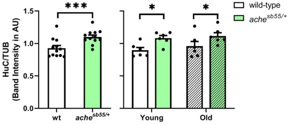
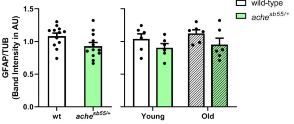
3.4 The brain levels of the immature neuronal marker were promoted while glial marker was downregulated slightly in the achesb55/+ mutants ... 69
3.5 Subtle decreases were observed on the free radical content in the brain of achesb55/+ mutants while no evident alterations in the brain levels of inflammatory protein ... 73
3.6 Multivariate analyses revealed genotype and age-dependent clustering patterns on the synaptic, cellular, and inflammatory proteins ... 76
xv
3.7 Discussion and Conclusions ... 79 CHAPTER 4 ... 90 THE EFFECTS OF SENSORY ENVIRONMENTAL ENRICHMENT ON BODY PARAMETERS, SYNAPTIC AND CELLULAR PROTEINS WITHIN THE
CONTEXT OF AGING AND GENDER ... 90 4.1 Effects of environmental enrichment, aging, and gender on body
parameters including body weight, length, body mass index (BMI),
and wet brain weight ... 90 4.2 Environmental enrichment alters the synaptic protein levels in an
age-dependent manner ... 95 4.3 Environmental enrichment altering post-mitotic neuronal marker in
an age-dependent manner ... 103 4.4 Age is significantly increasing the activity of reactive oxygen
species (ROS) and lipid peroxidation while no environment-dependent
modulations are evident ... 108 4.5 Multivariate analyses showed age and environment-dependent
clustering on synaptic and cellular proteins ... 110 4.6 Discussion and Conclusions ... 117 CHAPTER 5 ... 131 THE EFFECTS OF OPPOSING DIETARY TREATMENTS OF CALORIC
xvi
PROTEIN LEVELS, AND GENE EXPRESSION PATTERNS ON THE BRAIN
WITHIN THE CONTEXT OF AGING ... 131
5.1 Dietary interventions have altered the general body parameters as well as cortisol levels ... 131
5.2 Dietary interventions were altering excitatory synaptic markers while aging changed the inhibitory and presynaptic indicators ... 138
5.3 Genes related to synaptic neurotransmission, neurotrophic factors, and inflammation were not altered significantly by dietary interventions ... 145
5.4 Further correlational and multivariate analyses revealed a diet-dependent clustering in synaptic proteins ... 150
5.5 Correlations and multivariate analyses revealed positive correlations among synaptic and regulatory genes and diet-driven clustering pattern ... 154
5.6 Discussion and Conclusions ... 158
CHAPTER 6 ... 169
OVERALL CONCLUSIONS AND FUTURE PROSPECTS ... 169
xvii LIST OF FIGURES
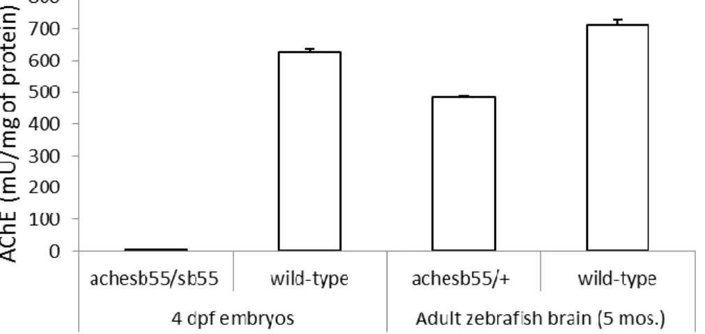
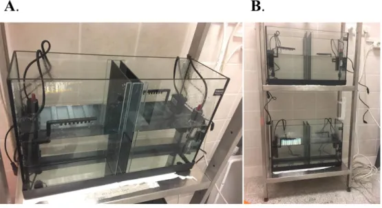
Figure 2.1 Representative pictures of the tank design equipped with air pumps and heaters (A) a view of experimental tanks (B). Adapted from Karoglu-Eravsar et al., 2021 [74]. ... 24 Figure 2.2 A pilot study with white background (A). Used experimental setup for enrichment study with dark blue background (B) Adapted from Karoglu-Eravsar et al., 2021 [74]. ... 25 Figure 2.3 Optimization of AChE assay by using achesb55/sb55 embryos and achesb55/+ brains. ... 41 Figure 3.1 Body weight assessments across genotype and age groups. A significant main effect of age was shown altering body weight. a: Significant main effect of age was observed in a genotype group. ***: p < 0.005. Error bars = +Standard Error Mean. ... 51 Figure 3.2 Body length assessments across genotype and age groups. A significant main effect of age was shown altering body length. a: Significant main effect of age was observed in a genotype group. ***: p < 0.005. Error bars = +Standard Error Mean. ... 52 Figure 3.3 Body mass indices (BMI) across genotype and age groups. A significant main effect of age was shown altering BMIs. a: Significant main effect of age was observed in a genotype group. ***: p < 0.005. Error bars = +Standard Error Mean. . 53 Figure 3.4 Brain weight measures across genotype and age groups. Significant main effects of age and genotype were shown, which altered brain weight. a: Significant main effect of age was observed in a genotype group. *: p < 0.05, ***: p < 0.005. Error bars = +Standard Error Mean. ... 54
xviii
Figure 3.5 Brain AChE levels across genotype and age groups. Significant main effects of genotype and age were shown altering the levels of AChE. *: p < 0.05, ***: p < 0.005. Error bars = +Standard Error Mean. ... 56 Figure 3.6 Brain ACh levels across genotype and age groups. A significant main effect of genotype was shown altering the levels of ACh. *: p < 0.05. Error bars = +Standard Error Mean. ... 58 Figure 3.7 Brain AChE levels across genotype and age groups with very young age was incorporated. Significant main effects of genotype and age were shown altering the levels of AChE. ***: p < 0.005. Error bars = +Standard Error Mean. ... 59 Figure 3.8 Brain ACh levels across genotype and age groups with very young age was incorporated. Significant main effects of genotype and age were shown altering the levels of ACh. *: p < 0.05, **: p < 0.01, ***: p < 0.005. Error bars = +Standard Error Mean. ... 60 Figure 3.9 Representative western blot image was indicated for nAChR-a7 antibody. nAChR-a7 levels across genotype and age groups. Error bars = +Standard Error Mean. F: Female; M: Male. ... 62 Figure 3.10 Representative pictures from one cohort for Western blot experiments of synaptic protein levels examined in the current Chapter for effects of age and genotype. Bands were obtained at the expected molecular weights for all antibodies. F: Female; M: Male. ... 63 Figure 3.11 PSD95 levels across genotype and age groups. Error bars = +Standard Error Mean. ... 63 Figure 3.12 GluR2/3 levels across genotype and age groups. GluR2/3 levels were altered significantly by genotype. *: p < 0.05. Error bars = +Standard Error Mean. .. 64
xix
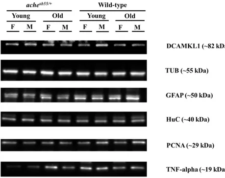
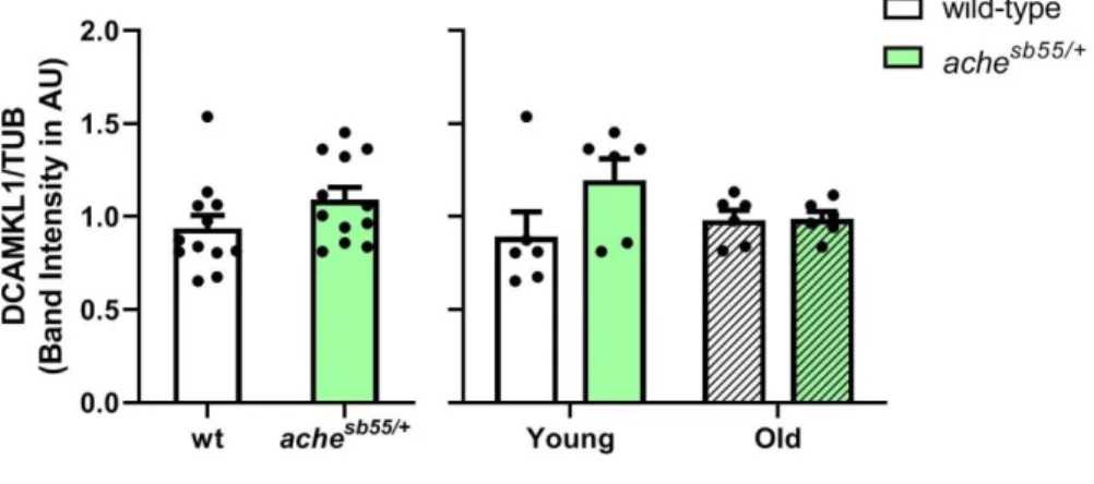
Figure 3.13 NR2B levels across genotype and age groups. NR2B levels were altered significantly by genotype. *: p < 0.05, **: p < 0.01, ***: p < 0.005. Error bars = +Standard Error Mean. ... 65 Figure 3.14 SYP levels across genotype and age groups. SYP levels were altered significantly by age. *: p < 0.05. Error bars = +Standard Error Mean. ... 66 Figure 3.15 GEP levels across genotype and age groups (A). Effect of age and gender by genotype interaction was demonstrated (B). *: p < 0.05; **: p < 0.01; ***: p < 0.005. Error bars = +Standard Error Mean. ... 67 Figure 3.16 GABA-A-a1 levels across genotype and age groups (A). The effect of gender was demonstrated (B). *: p < 0.05. Error bars = +Standard Error Mean. ... 69 Figure 3.17 Representative pictures from one cohort for Western blot experiments of cellular and inflammatory protein levels examined in the current Chapter for effects of age and genotype. Bands were obtained at the expected molecular weights for all antibodies. F: Female; M: Male. ... 69 Figure 3.18 HuC levels across genotype and age groups. Effect of genotype was demonstrated. *: p < 0.05, ***: p < 0.005. Error bars = +Standard Error Mean. ... 70 Figure 3.19 DCAMKL1 levels across genotype and age groups. Error bars = +Standard Error Mean ... 71 Figure 3.20 GFAP levels across genotype and age groups. Error bars = +Standard Error Mean ... 72 Figure 3.21 PCNA levels across genotype and age groups. Error bars = +Standard Error Mean ... 73 Figure 3.22 TNF-alpha levels across genotype and age groups. Error bars = +Standard Error Mean ... 74
xx
Figure 3.23 ROS activity levels across genotype and gender groups. Gender groups collapsed together (A). Age, gender, and genotype groups were separated gender by genotype interaction was revealed (B). *: p < 0.05, **: p < 0.01. Error bars = +Standard Error Mean ... 75 Figure 3.24 Analyses revealed significant correlations among the proteins. *: p<0.05, **: p<0.001. ... 77 Figure 3.25 Factor loading scores for two principal components with respect to proteins. PC1 was contributed by nAChR-a7, ACh, GFAP and TNF-alpha positively (A). PC2 was contributed by GluR2/3 and NR2B positively while negatively controlled by PSD95 and SYP (B). ... 78 Figure 3.26 Clustering profiles of components in which both age groups and genotype groups were separated. Age groups were shown in separate panels while genotype groups were denoted by different colors Wild-type: White, achesb55/+: Black. ... 79 Figure 4.1 Body weight of the zebrafish was altered by the factors of gender and age while no specific effects of environment were revealed. *: p < 0.05, ***: p < 0.005. Error bars = +Standard Error Mean. Adapted from Karoglu-Eravsar et al., 2021 [74]. ... 91 Figure 4.2 Body length of the zebrafish was altered by the factor of age only while no specific effects of environment or gender were revealed. ***: p < 0.005. Error bars = +Standard Error Mean. Adapted from Karoglu-Eravsar et al., 2021 [74]. ... 92 Figure 4.3 Body mass index values were significantly altered by a factor of age; no gender or environment-dependent alterations were observed. ***: p < 0.005. Error bars = +Standard Error Mean. Adapted from Karoglu-Eravsar et al., 2021 [74]. ... 93
xxi
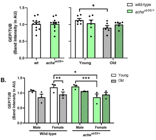
Figure 4.4 Wet brain weight measures were significantly altered by factors of age and gender, and a significant environment by age interaction was revealed. ***: p < 0.005. Error bars = +Standard Error Mean. Adapted from Karoglu-Eravsar et al., 2021 [74]. ... 94 Figure 4.5 Representative pictures from one cohort for Western blot experiments of synaptic protein levels examined in the current Chapter for effects of age, gender and the environmental condition. Bands were obtained at the expected molecular weights for all antibodies. B: Barren environmental condition; E: enriched environmental condition. Adapted from Karoglu-Eravsar et al., 2021 [74]. ... 96 Figure 4.6 PSD95 levels were not altered significantly. Error bars = +Standard Error Mean. Adapted from Karoglu-Eravsar et al., 2021 [74]. ... 97 Figure 4.7 GluR2/3 levels were changed significantly by age and gender-dependent interactions. **: p < 0.01. Error bars = +Standard Error Mean. Adapted from Karoglu-Eravsar et al., 2021 [74]. ... 98 Figure 4.8 NR2B levels were altered by the age-environment interaction and gender. *: p < 0.05, ***: p < 0.005. Error bars = +Standard Error Mean. Adapted from Karoglu-Eravsar et al., 2021 [74]. ... 99 Figure 4.9 SYP levels were altered significantly by age and age by environment interaction. ***: p < 0.005. Error bars = +Standard Error Mean. Adapted from Karoglu-Eravsar et al., 2021 [74]. ... 100 Figure 4.10 GEP levels were elevated in the enriched environment with aging. *: p < 0.05. Error bars = +Standard Error Mean. Adapted from Karoglu-Eravsar et al., 2021 [74]. ... 101
xxii
Figure 4.11 GABA-A-a1 levels tend to be stable across the environment, age, and gender groups. Error bars = +Standard Error Mean. Adapted from Karoglu-Eravsar et al., 2021 [74]. ... 102 Figure 4.12 Representative pictures from one cohort for Western blot experiments of cellular protein levels examined in the current Chapter for effects of age, gender and the environmental condition. Bands were obtained at the expected molecular weights for all antibodies. B: Barren environmental condition; E: enriched environmental condition. Adapted from Karoglu-Eravsar et al., 2021 [74]. ... 103 Figure 4.13 HuC levels tend to be stable across the environment, age, and gender groups. Error bars = +Standard Error Mean. Adapted from Karoglu-Eravsar et al., 2021 [74]. ... 104 Figure 4.14 DCAMKL1 levels decreased with aging in the barren environmental condition. *: p < 0.05. Error bars = +Standard Error Mean. Adapted from Karoglu-Eravsar et al., 2021 [74]. ... 105 Figure 4.15 GFAP levels tend to be stable across the environment, age, and gender groups. Error bars = +Standard Error Mean. Adapted from Karoglu-Eravsar et al., 2021 [74]. ... 106 Figure 4.16 PCNA levels tend to be stable across the environment, age, and gender groups. Error bars = +Standard Error Mean. Adapted from Karoglu-Eravsar et al., 2021 [74]. ... 107 Figure 4.17 AChE activity levels tend to be stable across the environment, age, and gender groups. Error bars = +Standard Error Mean. Adapted from Karoglu-Eravsar et al., 2021 [74]. ... 108
xxiii
Figure 4.18 ROS activity levels were significantly altered by the factors of age and gender. *: p<0.05. Error bars = +Standard Error Mean. Adapted from Karoglu-Eravsar et al., 2021 [74]. ... 109 Figure 4.19 MDA levels were significantly altered by the factor of age. *: p<0.05. Error bars = +Standard Error Mean. Adapted from Karoglu-Eravsar et al., 2021 [74]. ... 110 Figure 4.20 Analyses revealed significant correlations among the synaptic and cellular proteins. *: p<0.05. ... 111 Figure 4.21 Factor loading scores for three principal components with respect to proteins. PC1 is contributed by DCAMKL1 and NR2B positively and PSD95 negatively (A). PC2 was contributed by GFAP positively (B). PC3 was altered by GEP positively and PCNA negatively (C). ... 113 Figure 4.22 Three components were visualized by separating the age groups; PC1 showed an age-specific clustering profile. Age groups were denoted by different colors. Young: Blue, Old: Green. ... 114 Figure 4.23 Three components were visualized by separating the environmental conditions, which were denoted by different colors. Barren: Black, Enriched: Red.116 Figure 4.24 Clustering profiles of components in which both age groups and environmental conditions were separated. Age groups were shown in separate panels while environmental conditions were denoted by different colors Barren: Black, Enriched: Red. ... 117 Figure 5.1 Total body weight measurements across age and diet groups. Significant effects of age and diet were demonstrated on the body weight measures. a: Significant main effect of age was observed in a diet group. *: p < 0.05, ***: p < 0.005. Error
xxiv
bars = +Standard Error Mean. Adapted from Karoglu-Eravsar et al., 2021 (under-review) [128]. ... 132 Figure 5.2 Total body length measurements across age and diet groups. Significant effects of age and diet were demonstrated on the body length measures. a: Significant main effect of age was observed in a diet group. ***: p < 0.005. Error bars = +Standard Error Mean. Adapted from Karoglu-Eravsar et al., 2021 (under-review) [128]. ... 133 Figure 5.3 Total body mass index (BMI) measurements across age and diet groups. Significant effects of age and diet were demonstrated on the BMI. a: Significant main effect of age was observed in a diet group. *: p < 0.05, ***: p < 0.005. Error bars = +Standard Error Mean. Adapted from Karoglu-Eravsar et al., 2021 (under-review) [128]. ... 135 Figure 5.4 Wet brain weight measurements across age and diet groups. Significant effects of age and diet were demonstrated on brain weight. a: Significant main effect of age was observed in a diet group. **: p < 0.01, ***: p < 0.005. Error bars = +Standard Error Mean. Adapted from Karoglu-Eravsar et al., 2021 (under-review) [128]. ... 136 Figure 5.5 Trunk cortisol levels across age and diet groups. A significant effect of diet was demonstrated on trunk cortisol levels. *: p < 0.05. Error bars = +Standard Error Mean. Adapted from Karoglu-Eravsar et al., 2021 (under-review) [128]. ... 137 Figure 5.6 Representative pictures from one cohort for Western blot experiments of synaptic protein levels examined in the current Chapter for effects of age and dietary interventions. Bands were obtained at the expected molecular weights for all
xxv
antibodies. AL: Ad-libitum; OF: overfeeding; CR: Caloric restriction; M: Male; F: Female. Adapted from Karoglu-Eravsar et al., 2021 (under-review) [128]. ... 139 Figure 5.7 PSD95 levels across age and diet groups. A significant effect of diet was demonstrated on PSD95 levels. *: p < 0.05. Error bars = +Standard Error Mean. Adapted from Karoglu-Eravsar et al., 2021 (under-review) [128]. ... 140 Figure 5.8 GluR2/3 levels across age and diet groups. Significant main effects of diet and age were demonstrated on GluR2/3 levels. *: p < 0.05, **: p < 0.01. Error bars = +Standard Error Mean. Adapted from Karoglu-Eravsar et al., 2021 (under-review) [128]. ... 141 Figure 5.9 NR2B levels across age and diet groups. Error bars = +Standard Error Mean. Adapted from Karoglu-Eravsar et al., 2021 (under-review) [128]. ... 142 Figure 5.10 GEP levels across age and diet groups. A significant effect of age was demonstrated on the GEP. a: Significant main effect of age was observed in a diet group. *: p < 0.05. Error bars = +Standard Error Mean. Adapted from Karoglu-Eravsar et al., 2021 (under-review) [128]. ... 143 Figure 5.11 GABA-A-a1 levels across age and diet groups. Error bars = +Standard Error Mean. Adapted from Karoglu-Eravsar et al., 2021 (under-review) [128]. ... 144 Figure 5.12 SYP levels across age and diet groups. A significant effect of age was demonstrated on the SYP levels. **: p < 0.01. Error bars = +Standard Error Mean. Adapted from Karoglu-Eravsar et al., 2021 (under-review) [128]. ... 145 Figure 5.13 Relative expression levels of the synaptic genes across age and diet groups. (A) dlg4, (B) dlg4b, (C) sypa and (D) gepb. Error bars = +Standard Error Mean. FC: Fold Change ... 147
xxvi
Figure 5.14 Relative expression levels of the regulatory genes across age and diet groups. (A) igf1, (B) bdnf, (C) tnfa and (D) il10. ***: p < 0.005. a: Significant main effect of age was observed in a diet group. Error bars = +Standard Error Mean. FC: Fold Change ... 149 Figure 5.15 Analyses revealed significant correlations among the synaptic proteins. *: p<0.05, **: p<0.001. ... 151 Figure 5.16 Factor loading scores for two principal components for synaptic proteins. PC1 is contributed by PSD95, GluR2/3, NR2B, and SYP positively (A). PC2 was contributed by GEP and GABA-A-a1 positively (B). ... 152 Figure 5.17 Clustering profiles of components extracted from protein expression data in which both age groups and dietary conditions were separated. Diet groups were shown in separate panels, while age groups were denoted by black and white. Young: Black, Old: White. ... 153 Figure 5.18 Analyses revealed significant correlations among synaptic and regulatory genes. *: p<0.05, **: p<0.001. ... 155 Figure 5.19 Factor loading scores for two principal components for synaptic proteins. PC1 is contributed by bdnf, sypa, gepb, dlg4 and dlg4b positively (A). PC2 was contributed by tnfa and il10 positively (B). ... 156 Figure 5.20 Clustering profiles of components extracted from gene expression data in which both age groups and dietary conditions were separated. Diet groups were shown in separate panels, while age groups were denoted by black and white. Young: Black, Old: White. ... 157
xxvii LIST OF TABLES
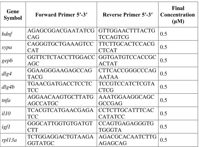
Table 2. 1 Dietary regimens for feeding interventions ... 27 Table 2. 2 Reactions for Bradford assay ... 31 Table 2.3 List of the antibodies utilized in the current study ... 37 Table 2.4 Reaction volumes for controls and unknown samples ... 43 Table 2.5 Primer sequences utilized in genotyping experiments ... 45 Table 2.6 Sequences and concentrations of primers used in the gene expression analyses ... 46
1
CHAPTER 1
INTRODUCTION 1.1 Aging and cognitive alterations
Normal aging without the presence of neuropathological conditions such as Alzheimer's disease can lead to alterations in the cognitive abilities of older adults. These alterations are associated with improvements in specific cognitive ability categories such as language skills. In contrast, in a group of cognitive domains, aging can lead to deteriorations which can be referred to as aging-related cognitive decline. One domain that stays relatively stable and even improved with aging is language skills. The specific categories of this cognitive ability, such as vocabulary knowledge and reserve, show improvements with aging. On the other hand, even within the same cognitive ability, age-related changes can occur differentially, and certain categories can present more vulnerability against age-related changes. To illustrate, language-related processes, which were mainly modulated by the processing speed, such as word generation and verbal fluency, can demonstrate significant declines and deteriorations with aging [1], [2].
Another cognitive ability is a speed of processing, and dysregulation in this domain can impair multiple related cognitive skills. Deteriorations in the speed of processing can alter multiple cognitive categories such as speed of motor activities, response latency, and perception. Processing speed is vulnerable against aging, and at older ages, people tend to have disrupted overall processing speed compared to younger people [3]. Furthermore, it was proposed that deteriorations in processing
2
speed can underlie cognitive decline in different cognitive skills and have an explanatory role in these changes [3]. Age-related changes can also be observed in attention and its categories like selective attention and divided attention. Old adults tend to perform drastically slower in conditions demanding selective attention, focusing on the particular features of the task while ignoring the others [4]. Additionally, another category of attention, divided attention which refers to directing the attentional sources to multiple cues or tasks, is significantly impaired with aging [5].
Memory-related deficits are pronounced within the context of aging. It has been shown that encoding and retrieval steps were generally jeopardized with aging while no evident changes were reported in retention processes [6],[7]. Additionally, the different categories of memory are selectively impaired while the other categories remain relatively stable. Studies have reported that semantic and implicit memory components were stable through aging, while episodic memory raising from an individual's collection of experiences was impaired significantly at older ages [2],[8].
Overall, cognitive decline can occur in normal aging, but this decline is not a unitary process indicating impairments in all measures. Certain cognitive skills are selectively affected with normal aging, while others tend to remain stable and spared. However, these declining components can impair the self-sufficiency, well-being, and mind-span of older adults. Therefore, characterization of the neurobiological underpinning of these cognitive changes is very crucial to understand the mechanism of age-related changes and develop intervention strategies.
3 1.2 Aging and neurobiological alterations
Normal aging does not promote global loss of neurons and synapses in the cortex and subcortical regions, unlike age-related neuropathological conditions such as Alzheimer's disease characterized by progressive neuronal death and synaptic loss as well as the existence of neuropathological profile [9]. Despite the fairly maintained number of neurons and synapses, age-related cognitive decline still occurs in normal aging. It affects the cognitive domains, including episodic memory, executive functions, and processing speed [2]. Therefore, it is crucial to dissect the elements contributing to normal aging-related cognitive decline and target these elements for further interventions to ameliorate the aging phenotype.
In normal aging, there are no apparent histopathological alterations; yet changes occur in more subtle levels. These changes can include disturbances in synaptic integrity and content, decreased neurogenesis, increased inflammatory markers, altered metabolic activity, and transcriptional changes [10]. It is crucial to note that these elements are not totally disconnected from each other. For example, it was shown that with normal aging, the transcriptional profile could be skewed on genes regulating synaptic plasticity, inflammation, and metabolic activity [11], which in turn can alter all the elements. Therefore, there is no single contributing factor; instead, there is extensive cross-talk between multiple systems. Complexity is not limited to the cross-talk between different components; each system, such as different neurotransmitter systems, is not equally vulnerable against age-related alterations. Therefore, it can be promising to focus on the more vulnerable systems to have effective interventions.
4 1.2.1 Altered synaptic proteome with aging
Age-related disturbances in synaptic integrity and plasticity can be considered as a more direct cause of age-related cognitive decline. Analysis of age-related changes in the synaptic proteome has crucial contributions to age-related cognitive decline. The levels of some synaptic proteins can give information about the age-related impairments on neurotransmission. Post-synaptic 95 (PSD95) is a clustering and scaffolding protein, and it is a member of the membrane-associated guanylate kinase family; with its PDZ domain, it can cluster glutamate receptors; α-amino-3-hydroxy-5-methyl-4-isoxazole propionic acid receptors (AMPARs), and N-methyl-D-aspartate receptors (NMDARs) on the post-synaptic site. It also clusters cytoskeletal elements, ion channels, and molecules responsible for signal transduction depending on the synaptic activity [12]. It has roles in the maturation of the synaptic elements, including terminals and spines. Additionally, PSD95 has a crucial role in the maintenance of excitatory-inhibitory balance. Overexpression studies showed that while glutamatergic excitatory clusters increased in terms of their size, numbers of gamma-Aminobutyric acid (GABA)ergic synapses become reduced. Parallel to that, knock-down studies with PSD95 revealed a decreased number of glutamatergic synaptic innervations, whereas the number of GABAergic innervations was increased [13]. In the aging literature, it was shown that hippocampal levels of PSD95 were reduced with aging, and this decrease was more prominent in the old groups with impaired cognitive performance [14].
PSD95 can cluster different glutamate receptors such as AMPA and NMDA; it indirectly clusters the AMPA type receptors with additional proteins such as TARP
5
and Stargazin while directly binds to the NMDA receptors. Glutamate receptor subunit 2 (GluR2) is a subunit in AMPA receptors expressed abundantly in most AMPA receptors [13]. The presence or absence of this subunit can alter the calcium regulation, to illustrate if GluR2 was in lower levels or lacking cell becomes more permeable to calcium [15]. Similarly, if GluR2 was expressed in high levels, cells become less calcium-permeable. Additionally, GluR2 was exposed to RNA editing unedited glutamine (Q) transformed to arginine (R) in an edited form; and this RNA editing can also regulate the calcium homeostasis while unedited form was permeable to calcium and edited form renders low calcium permeability [15]. However, this editing occurs in almost all the cases when GluR2 was expressed. Declines in GluR2 levels were reported with aging; reduced levels of GluR2 make the cell more vulnerable to neurotoxic damage because of the elevated permeability to calcium [16]. Glutamate receptor subunit 3 (GluR3) is another AMPA type receptor whose functions are relatively enigmatic compared to the GluR2 subunit. GluR3 is involved in the insertion and trafficking of AMPA receptors and the induction of fast currents of glutamatergic transmission and responsible for regulating short-term plasticity dynamics [17].
NMDA-type receptor subunit 2B (NR2B) is one of the receptor subunits that contribute to channel formations, and also, this subunit can prolong and sustain the response of the receptor. NR2B subunit expression can be altered developmentally, while its expression levels are very high in the post-natal brain, decrements in NR2B subunit levels are evident at the adult ages [18]. Additionally, over expression studies targeting NR2B showed that elevations in NR2B levels are associated with promoted
6
synaptic plasticity as well as memory and learning performances, and age-related declines were previously reported with regards to this subunit [19][20]. There are different views concerning this age-related decline in NR2B levels. The first view suggests that this reduction in NR2B manifests protection at older ages against the excitotoxicity since NR2B is associated with high calcium influx. The other opinion indicates that higher expression of NR2B is beneficial in all age groups, including old and young ages, and NR2B enhancement at an older age is inducing promoted plasticity and improved behavioral performance [21].
Gephyrin (GEP) can be considered as an inhibitory counterpart of the PSD95. GEP is found in the GABAergic and glycinergic synapses; it anchors and clusters GABAA, the ionotropic GABA receptors, and glycine receptors. Knock-down studies
indicated that reduced GEP expression is associated with larger glutamatergic terminals and PSD95-NMDA clusters while GABAergic innervations decrease. Overexpression of GEP induces smaller glutamatergic terminals, PSD95-NMDA clusters, while GABAA and GEP clusters become larger [22]. It was shown that GEP
levels could be altered depending on the age and cognitive status of the animal; it was reported that in the old and cognitively impaired groups, GEP levels were significantly increased in the parietal cortex, whereas no change was observed in the prefrontal cortex [23]. Additionally, evidence suggested that GEP levels were downregulated in neuropathological conditions, while maintained and elevated levels of GEP were protective against harmful effects of these insults [24]. GABAA
receptors are the clustering partners of GEP, and these receptors have several subunits. One of these subunits is alpha 1 (GABA-A-a1). GABA-A-a1 is one of the
7
predominant subunits widely expressed in the central nervous system and obtaining binding sites for psychiatric drugs like benzodiazepines [25]. In terms of aging literature, it was shown that GABA-A-a1 subunit levels are relatively stable at different age groups [26].
Synaptophysin (SYP) is a transmembrane glycoprotein, and SYP can be found in the presynaptic region and on synaptic vesicles. It was suggested that SYP could have potential roles in the trafficking of the synaptic vesicles and endocytosis [27]. In aging literature, it was demonstrated that increased SYP levels were significantly correlated with intact cognitive performance at older ages [28], and age-related decreases at SYP levels in the hippocampus of old animals were reported [20].
To conclude, aging is associated with subtle alterations in synaptic integrity and plasticity associated with age-related cognitive decline. When synaptic integrity and plasticity elements were evaluated together, one of the most intriguing shared features of PSD95 and GEP is their heterotypic effects. Their effects are not limited to their clustering targets, but they can alter the strength of synaptic innervations of their inhibitory or excitatory counterparts. PSD95 and GEP can have combinatory contributions to excitatory/inhibitory balance, which can be disturbed with aging. Taken together, the protein levels of PSD95, GEP, and their clustering partners GluR2/3, NR2B, and GABA-A-a1, as well as SYP, can give general information regarding excitatory/inhibitory balance and presynaptic integrity, which can be disturbed with aging.
8
1.2.2 Vulnerability of cholinergic system with aging
One of the most studied neurotransmitter systems in the scope of aging research is the cholinergic system. Bartus and his colleagues suggested that dysregulation in cholinergic neurotransmission contributed to age-related cognitive decline and Alzheimer's disease [28]. Acetylcholine is the neurotransmitter of cholinergic neurotransmission; it is composed of two elements; choline and acetate. Acetate is carried by coenzyme A, and this complex consisting of acetate and coenzyme A is called acetyl coenzyme A. The choline acetyltransferase enzyme (ChAT) transfers the acetate from the complex of acetyl coenzyme A to the choline, and at the end of this synthesis, acetylcholine is obtained. After acetylcholine is released from the cholinergic terminals, termination of this neurotransmitter is conducted with the enzyme acetylcholinesterase. Acetylcholinesterase breaks acetylcholine in the synaptic cleft into choline and acetate, and after this enzymatic degradation, choline molecules are taken up by choline transporters back to the cell. This reuptake process has high efficiency, and it is a rate-limiting factor for the acetylcholine synthesis; because the amount of choline in the cell body which is transported by axonal transport to the terminals is not adequate when it is compared with the amount of released choline; that is why uptake of choline is a crucial step [29].
After the release, acetylcholine can stimulate its receptors; there are two types of acetylcholine receptors. The first class is the nicotinic acetylcholine receptors (nAChRs); they are ionotropic receptors and coupled with ligand-gated ion channels. There are two subunits of nAChRs, α and β, coded by different genes in the central nervous system. In mammalian brain 12 subunits of nAChRs were identified; α-2, 3,
9
4, 5, 6, 7, 8, 9, 10; and β-2, 3, 4. These ionotropic receptors in the central nervous system can be permeable to calcium, potassium, and sodium. Additionally, it was suggested that nAChRs were anchored and clustered at the pre-synaptic region with rapsyn, a cytoskeletal protein [30]. The second class is the muscarinic acetylcholine receptors (mAChRs); they are metabotropic receptors coupled with class A G proteins. Different subtypes of mAChRs were reported; M1, M2, M3, M4, and M5; and their activation associated with the second messenger system and protein kinase activity. Knock-out studies revealed their various functions: regulation of dopaminergic release, learning, locomotion, activating MAPK signaling, and regulating stress response [31]. With all this information, it could be said that cholinergic neurotransmission has multiple components, and age-related hypothesized dysfunctions can alter these various elements.
Deficiencies in acetylcholine biosynthesis were reported in old mice with a 75% reduction in the biosynthesis of acetylcholine compared to the younger ages; additionally, a parallel decrease was reported in the release of acetylcholine with increasing age following the stimulation [32]. Besides the synthesis and release of the neurotransmitter of acetylcholine, dysfunctions can occur in the other components, including; the high-affinity choline uptake, which was considered a rate-limiting process for the acetylcholine synthesis and alterations in the expressions of the critical subunits in the nAChRs and mAChRs. Interestingly, it was reported that different subunits of the nAChRs could have selective vulnerabilities depending on aging and age-related diseases. For example, in Alzheimer's disease, the α4 subunit of
10
nAChRs was severely reduced, whereas the affected subunit is α7 in Lewy body dementia [33].
1.2.3 Oxidative stress and aging
Besides alterations in the main neurotransmitter systems and synaptic dynamics, aging is also associated with low-grade inflammation, which persists through aging and becomes chronic. Aging-related changes in oxidative stress mediating mechanisms can also contribute to this low-grade inflammation [34]. Accumulation of these chronic detrimental processes can lead to dysregulated cellular and synaptic dynamics, as well as neuropathological conditions. It was reported that aging disrupts the glial dynamics and can lead to aberrant changes and reactivity in the regulation of microglial cells and astrocytes [10]. Astrocytes are also closely related to the glutamatergic neurotransmission system. When glutamate was released to the synapse, astrocytes were involved in the uptake of glutamate. Deficiencies in this system might lead to the accumulation of glutamate and elevated excitation, which can be detrimental in the long term [35]. Aberrant glial reactivity can be accompanied by elevation on the levels of inflammatory cytokine tumor necrosis factor-alpha (TNF-alpha) in the aging brain.
Aging triggers the accumulation of oxidative materials such as reactive oxygen species (ROS) and induces oxidative alterations and peroxidation in certain cellular elements such as lipids. The brain can have selective vulnerabilities against these age-related changes in oxidative stress mechanisms because of its higher metabolic demands and oxygen consumption compared to other organs [36]. Elevated ROS levels can increase the incidences of lipid peroxidation. The brain is vulnerable to
11
lipid peroxidation due to the larger surface area of the membranes contributed by dendrites and extended axons [37]. Increases in these measures can lead to membrane instability and elevated ROS damage, altering the cellular and synaptic dynamics in the aging brain [38].
1.3 Age-related sex differences
Another crucial and generally overlooked factor is gender in the aging literature. Gender can alter the course of aging at many levels, molecular to behavioral, and the course of age-related neurodegenerative diseases as well. For example, male subjects tend to experience steeper declines in cognitive performance than females [40]. The alterations associated with aging in glucose metabolism and blood flow rate are sex-specific [39]. The levels of circulating sex hormones such as estrogen are associated with lower peroxide production, regulating cellular aging [38]. It was reported that age-related gene expression changes could occur in a sexually dimorphic pattern. In humans, it was shown that critical time windows in which more prominent changes were observed in the gene expression were different between males and females; more drastic changes occur in females at 80-90 years of age, whereas this window was 60-70 years of age for males. Interestingly altered genes were skewed in different categories between sexes; in females, gene groups associated with immune activation were selectively increased, whereas, in males, gene groups related with protein transport-synthesis and energy production were decreased with aging [11].
Sexual dimorphism can also be observed in the dynamics of brain mitochondria. Mammalian studies indicated that in adulthood, females tend to have significantly
12
lower peroxide production than males, but after the operation of ovariectomy, peroxide levels of brain mitochondria have become similar to males, and interestingly with the estradiol treatment, this effect was reversed in the females. Additionally, when males were treated with estradiol, a similar improvement, reduction in the peroxide production, was also observed [38].
The effects of the circulating sex hormones are not limited only to mitochondrial integrity; they can exert their effects on synaptic integrity. It was shown that estradiol treatment on ovariectomized rats increased the NMDR1 subunit of NMDA receptors at protein and mRNA levels in the hippocampus and increased dendrites in the CA1 region [40]. It could be said that circulating sex hormones can profoundly affect mitochondrial exhaustion, synaptic integrity, and gene expression patterns associated with age-related symptoms.
1.4 Zebrafish as a model organism to study aging
Zebrafish have high physiological and genetic homology to humans. The developmental and mutagenesis studies are benefited robustly from this model; one pair can produce approximately 200 embryos; since their development occurs ex Utero, transparent embryos can be observed and manipulated easily [41]. Various genetic strains are available in the zebrafish, and some strains can be very effective compared to the other models. In terms of aging research, zebrafish is a valuable model. A gradual cognitive decline was observed in the aging zebrafish, which is very similar to humans [42]. Beyond that, aging markers, including reactive oxygen species, senescence-associated beta-galactosidase (SABG) activity, lipofuscin
13
deposits, and telomere attrition, were demonstrated parallel changes in response to aging as in humans [43]. Moreover, sexual dimorphism in terms of age-related synaptic protein alterations, gene expression changes, and neurogenesis was observed in the zebrafish brain [44]–[46].
1.5 Genetic interventions to alter the course of aging
Genetic interventions can give information about the importance of the interfered system and present more causative explanations. Due to the fact that the cholinergic system has a selective vulnerability against age-related changes, it might be promising to focus on the cholinergic system as an intervention strategy. Generally, acetylcholinesterase inhibitors were used widely to reduce Alzheimer's disease-related cognitive deficits and pathology, although this treatment's efficiency is still questionable [47]. Reducing acetylcholinesterase levels could be promising. Conceptually, it will increase acetylcholine levels in the synapse and lead to its stimulation. These changes might have the potential to prevent age-related cholinergic hypofunction. Knock-out studies with mice targeting acetylcholinesterase did not reveal prominent impacts. Homozygous mutants had severe developmental muscle abnormalities, while heterozygous mutants were viable. However, other compensatory cholinesterase activities were observed in the heterozygous mutants, despite the reduced acetylcholine levels [48]. In mammals, acetylcholinesterase is not the only cholinesterase responsible for the termination of acetylcholine; butyrylcholinesterase can also cleave acetylcholine at lower rates. Therefore, when mutation reduces acetylcholinesterase levels, compensatory increases occur at the butyrylcholinesterase levels [49].
14
In that case, to see more noticeable changes, vertebrate models could be a better option. To illustrate, in zebrafish, there is no functional butyrylcholinesterase, and acetylcholinesterase is the only cholinesterase responsible for cleaving the acetylcholine. Homozygous mutation in the acetylcholinesterase coding gene (achesb55/sb55 line) results in the complete abolishment of acetylcholinesterase functions, yet it is associated with abnormalities in the development of musculature; sensory neurons, and more importantly, lethal [50]. On the other hand, heterozygous mutants (achesb55/+ line) show no developmental abnormality, characterized by decreased brain levels of acetylcholinesterase and increased acetylcholine levels [51]. More importantly, these heterozygous mutants were behaviorally characterized as a delayed aging model. After 24 months of age, wild-type zebrafish demonstrated a significant cognitive decline in the domains of entrainment to temporal-spatial cues, learning performance in conditioned place preference tests, and flexibility of the learning strategies. On the other hand, 24 months old achesb55/+ mutants showed comparable performance with the young (6 months of age) groups, with intact behavioral performance [42]. Analysis of the neurobiological basis of this improvement can have explanatory roles in the contribution of altered cholinergic neurotransmission to age-related cognitive decline.
1.6 Environmental interventions to alter the course of aging
Genetic models of interventions are powerful in terms of the establishment of a more focused relationship between the gene of interest and its targets, but since they were induced at very early stages of the development; they can alter the developmental dynamics; change the microenvironment of the brain and lead to
15
different base-line compared to the wild-type controls. At this point, along with the genetic interventions, environmental interventions can create a more holistic view.
1.6.1 Environmental enrichment
Environmental enrichment is one of the environmental interventions, and it is referring to exposure to perceptually, socially, physically, and cognitively stimulating environments. It could be thought that the level of stimulation may not be so comparable between humans and laboratory animals; an enriched environment would be associated with high-level tasks such as increased intellectual activities, participating in group activities in humans. On the other hand, in laboratory animals, the setup is generally achieved by introducing large cages, new toys, mazes, wheels, and group housing [52]. To understand the underlying mechanisms and distinguish neurobiological contributors of the ameliorative effects of environmental enrichment, animal models are still required. Environmental enrichment was carried out with success in the previous works using different model organisms, including zebrafish.
In the aging literature, it was shown that environmental enrichment could reduce cognitive decline in the old groups in terms of their performances in spatial memory acquisition [53], radial arm maze, and Morris water maze tasks [54]. Neurobiological changes have accompanied these improvements. To illustrate, enriched and old animals had higher neurogenesis, higher cortical thickness, increased spine density, increased branching of dendrites than old control groups raised in a barren environment [52]. Additionally, an increased expression of neurotrophins, including BDNF and NGF, was reported in the rats exposed to an enriched environment [55].
16
Also, it was indicated that environmental enrichment could increase the levels of the synaptophysin in the hippocampus and frontoparietal cortex in old rats [53]. Moreover, in the literature, pioneering studies indicated that environmental enrichment could modulate the activity of particular neurotransmitter systems. It was noted that brain acetylcholine and acetylcholinesterase levels were increased in the enriched animals [56].
Evidence suggested that the application of environmental enrichment can be associated with better cognitive performance and altered neurobiological dynamics. However, the literature is still inadequate to point out certain factors that might alter or potentiate the impacts of environmental enrichment. The first factor is the ideal time to initiate this treatment. Environmental enrichment can be started at very young, adult, or old ages, and the obtained effects may not be so comparable. For example, it was shown that old subjects could be benefitted from the environmental enrichment, while behavioral and neurobiological measures were more drastically affected at very young ages [57][58]. Another critical factor is the gender, effectiveness and neurobiological outcomes of the environmental enrichment can be differentially affecting the males and females, to illustrate in the rat models, it was shown that old female animals with enrichment could have higher levels of SYP, which is an overall synaptic protein; but another study demonstrated no profound effects of enrichment in male animals [53][54]. Finally, the components through which the EE is working need to be determined. Traditionally these interventions were applied in a mixed fashion, and the individual components of the EE such as sensory, exercise, and socialization were blended. A recent study indicated that the
17
magnitude of the effects of EE differed depending on these individual factors. For example, environmental (sensory) enrichment and physical exercise have prevented amyloid-beta infusion-related memory deficits and decreased lipid peroxidation in the brain, while social enrichment merely altered social recognition performance antioxidant capacity [59]. Therefore, dissecting out the components of EE in terms of their specific molecular mechanisms within the context of aging is essential for translational applications.
1.6.2 Dietary interventions
Another environmental intervention that can alter the course of aging is dietary interventions. Dietary interventions can have opposing actions on achieving successful aging. For example, one strategy applied as a dietary intervention is restricting the total calorie intake, and the application of the caloric restriction (CR) can delay age-related impairments. Effects of caloric restriction can alter many different pathways, including; nutrient-sensing signaling, autophagy, survival, growth, and so on [60]. Evidence suggested that the CR diet could improve old rats' behavioral performance and prevented declining levels of glutamatergic proteins such as NR2B and GluR2 and general synaptic protein SYP [61]. Besides its effects on the synaptic proteins, the application of CR can reduce the ROS activity and the lipid peroxidation in the aging brain, which can manifest the further protective effects of the CR on brain metabolism [62].
While CR has possible ameliorating impacts on the aging brain, other dietary interventions can accelerate brain aging. These dietary interventions generally consist
18
of increased fat content in the regular diet by inducing higher calorie intake. Evidence suggested that elevated intake in the total calorie amount could cause cognitive deteriorations at adult age and the older ages [63]–[66]. Additionally, it was indicated in the human studies that people with high BMI scores in their middle age had an increased risk of the incidence of dementia and neuropathological diseases at older ages than people having normal BMI scores [67].
Higher calorie intake leads to detrimental changes in neurobiological components. Evidence suggested that elevated calorie consumption inducing obesity led to significant reductions in the brain levels of vesicular glutamate and GABA transporters, altering the syntheses of these key neurotransmitters [68]. It was demonstrated that the levels of PSD95 and Activity Regulated Cytoskeleton-associated protein (Arc) declined with the overfeeding regimen in the mammalian models [64]. Previous studies showed that a high-calorie diet with elevated fat intake could increase the protein levels of inflammatory markers, microglial markers and decrease Brain-derived neurotrophic factor (BDNF) on the brain [63], [69].
1.7 Aims and Hypothesis
The main aim of this thesis is to characterize the synaptic proteome and cellular markers with aging. In this respect, genetic and environmental interventions were utilized to decelerate or accelerate the aging profile in the zebrafish brain. Possible sexually dimorphic patterns were also taken into account by incorporating females and males in equal ratios to all designated experimental designs. A genetic intervention mentioned in Chapter 3 was targeting the cholinergic system, and the
![Figure 2.2 A pilot study with white background (A). Used experimental setup for enrichment study with dark blue background (B) Adapted from Karoglu-Eravsar et al., 2021 [74]](https://thumb-eu.123doks.com/thumbv2/9libnet/5765120.116747/52.918.213.847.122.387/figure-background-experimental-enrichment-background-adapted-karoglu-eravsar.webp)