Clin Rheumatol (2006) 25: 889–890 DOI 10.1007/s10067-006-0284-4
B R I E F R E P O RT
Ali Kemal Oğuz . Levent Özçakar . Bayram Kaymak
Rare abdominal findings in Behçet
’s disease
Received: 10 February 2006 / Revised: 6 March 2006 / Accepted: 6 March 2006 / Published online: 22 March 2006 # Clinical Rheumatology 2006
To the editor,
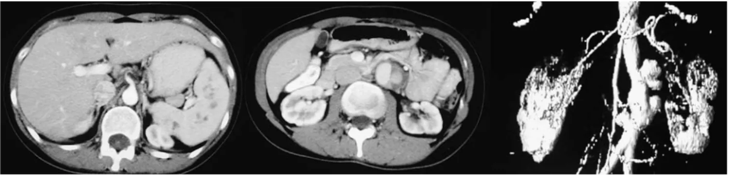
A 30-year-old woman was examined for her complaints of abdominal pain. She had a history of Behçet’s disease (BD) for 7 years. She is Turkish and a Caucasian. No HLA typing was performed. Ultrasonography results revealed splenomegaly and diffuse widening in distal portions of abdominal aorta. An abdominal computed tomography (CT) image demonstrated multiple splenic lesions (Fig. 1a), irregularities and scarring in the renal parenchyma bilaterally (Fig. 1b), and an infrarenal aneurysm in the abdominal aorta (Fig. 1c). She refused a secondary immunosuppressive agent (azathioprine and
cyclophosphamide) to be added to her treatment protocol and also refused splenectomy. She has been closely followed-up with a maintenance steroid and colchicine therapy and is still in remission. She currently has a depressive mood mostly related to medial advice against a pregnancy; otherwise, she is living a normal life (she is a biologist and very busy working as a consultant for a famous microscope company).
Splenic involvement is very seldom in BD [1] and is usually a slight splenomegaly [2]. In our patient, irregular multiple lesions, which are quite rare in BD, were detected in the spleen. The pattern of renal involvement was the other atypical finding in our patient. Renal involvement in Behcet’s disease is more frequent than has previously been recognized (0–55%) [3]. As a vasculitic syndrome affecting all types and sizes of blood vessels and also complicated by vascular throm-bosis in one third of cases, the renal lesions in Behcet’s disease display a wide spectrum. It characteristically involves secondary amyloidosis (with nephrotic syn-drome and renal failure), glomerulonephritis (with several different types of glomerular lesions), renal vascular disease (renal artery thrombosis and aneurysms, and renal vein thrombosis), and interstitial nephritis [3]. In our patient with normal urinalysis, bilateral irregular scars were detected in renal parenchyma—something not routinely observed in BD. The infrarenal multilobular abdominal aortic aneurysm was the last but perhaps the most potentially devastating lesion.
A. K. Oğuz
Department of Internal Medicine, Ufuk University Medical School, Ankara, Turkey
L. Özçakar (*) . B. Kaymak
Department of Physical Medicine and Rehabilitation, Hacettepe University Medical School,
Ankara, Turkey e-mail: [email protected] Tel.: +90-312-3094142 Fax: +90-312-3105769 Present address: L. Özçakar .
Department of Physical Medicine and Rehabilitation, Hacettepe University Medical School,
References
1. Gürler A, Boyvat A, Tursen U (1997) Clinical manifestations of Behcet’s disease: analysis of 2147 patients. Yonsei Med J 38: 423–427
2. Bayraktar Y, Özaslan E, Van Thiel DH (2000) Gastrointestinal manifestations of Behçet’s disease. J Clin Gastroenterol 30:144–154
3. Akpolat T, Akkoyunlu M, Akpolat I, Dilek M, Odabas AR, Ozen S (2002) Renal Behçet’s disease: a cumulative analysis. Semin Arthritis Rheum 31:317–337
Fig. 1 a Abdominal CT; multiple irregular hypodense lesions in the spleen with a maximum size of 2 cm. b Abdominal CT demonstrating bilateral renal cortical scars. c Three-dimensional CT angiography depicting multisaccular infrarenal abdominal aortic
aneurysm, extending from an infrarenal localization—2 cm inferior to the origin of the renal arteries—to a place proximal to the aortic bifurcation, with a maximum diameter of 4.5×3 cm