©Copyright 2017 by The Medical Bulletin of University of Health Sciences Haseki Training and Research Hospital The Medical Bulletin of Haseki published by Galenos Yayınevi. ©Telif Hakkı 2017 Sağlık Bilimleri Üniversitesi Haseki Eğitim ve Araştırma Hastanesi Haseki Tıp Bülteni, Galenos Yayınevi tarafından basılmıştır. AddressforCorrespondence/Yaz›flmaAdresi:Cansu Benli Işık
University of Health Sciences Haseki Training and Research Hospital, Clinic of Medical Pathology, İstanbul, Turkey
Phone: +90 505 798 14 89 E-mail: [email protected] ORCID ID: orcid.org/0000-0003-3805-3143 Received/GeliflTarihi:09 January 2017 Accepted/KabulTarihi:11 February 2017 Med Bull Haseki 2017;55:216-20
Tiroid İnce İğne Aspirasyonda Hangi Sitolojik Özellikler Papiller Karsinom için
Daha Değerlidir?
Aim:Fine-needle aspiration plays a significant role in the evaluation
of solitary thyroid nodules. Several cytological criteria are defined and these primarily aim to distinguish papillary carcinoma (PC), the most common thyroid malignancy, from other benign diseases. In our study, we aimed to determine which cytologic criteria are more valuable in diagnosing PC.
Methods: Patients, who had undergone thyroidectomy operation
between January 2013 and December 2014 and had preoperative fine-needle aspiration cytology, were screened. Among cases that were postoperatively diagnosed as benign, 62 cases and among those diagnosed as PC and papillary microcarcinoma, 24 cases were included in this study. The cytologic criteria were reassessed. In the linear regression analysis, the variables associated with the diagnosis of malignancy at the level of p<0.05 were considered significant for predicting the diagnosis of PC.
Results: The variables that remained as predictive for the diagnosis of
cancer as the result of the Backward analysis following the multivariate analysis performed with the variables with a p value of less than 0.1 were cellular pleomorphism, nuclear atypia, papillary structures with fibrovascular cores, and intranuclear pseudoinclusion.
Conclusion:Although every cytological feature is characteristic for PC,
none of them alone is specific; they should be evaluated together with all nuclear and structural features during the diagnostic approach.
Keywords: Thyroid, fine-needle aspiration, papillary carcinoma,
Bethesda system
Amaç:Tiroidin soliter nodül değerlendirmesinde ince iğne aspirasyonu
önemli bir rol oynar. Bir takım sitolojik kriterler tanımlanmış olup, bunlar öncelikle tiroid malignitesi içinde en sık rastlanılan papiller karsinomu (PK) diğer benign hastalıklardan ayırmayı amaçlar. Çalışmamızda hangi sitolojik kriterlerin PK tanısı koydurmada daha değerli olduğunu saptamayı amaçladık.
Yöntemler: Hastanemizde Ocak 2013 ve Aralık 2014 tarihleri
arasında tiroidektomi operasyonu gerçekleşen ve preoperatif dönemde ince iğne aspirasyon sitolojisi uygulanan olgular tarandı. Postoperatif tanısı benign olanlardan 62’si, PK ve papiller mikrokarsinomlardan 24’ü çalışmaya dahil edildi. Sitolojik değerlendirme kriterleri yeniden değerlendirildi. Lineer regresyon analizinde malignite tanısıyla p<0,05 düzeyinde ilişkili bulunan değişkenler PK tanısını öngörmede anlamlı kabul edildi.
Bulgular: Çalışmamızda p<0,1 olan değişkenlerle çok değişkenli analiz
yapılıp Backward analiz sonucunda kanser tanısını öngörmede anlamlı kalan değişkenler; hücresel pleomorfizm, nükleer atipi, fibrovasküler korlu papiller yapılar ve intranükleer psödoinklüzyondur.
Sonuç: Her bir sitolojik özellik PK için karakteristik olmakla birlikte hiç
birinin tek başına varlığı spesifik değildir. Tanı yaklaşımında tüm nükleer ve yapısal özelliklerle birlikte değerlendirilmelidir.
Anahtar Sözcükler: Tiroid, ince iğne aspirasyonu, papiller karsinom,
Bethesda sistemi
Öz Abs tract
Cansu Benli Işık, Halide Nur Ürer*, Kamile Altundal*, Mehmet Zeki Günlüoğlu**, Pınar Fırat***
University of Health Sciences Haseki Training and Research Hospital, Clinic of Medical Pathology, İstanbul, Turkey
*University of Health Sciences Yedikule Chest Diseases and Thoracic Surgery Training and Research Hospital, Clinic of Medical Pathology, İstanbul, Turkey
**Medipol University Faculty of Medicine, Department of Chest Surgery , İstanbul, Turkey
***İstanbul University İstanbul Faculty of Medicine, Department of Medical Pathology, İstanbul, Turkey
Which Cytological Characteristics are More Valuable for
Papillary Carcinoma in Thyroid Fine-needle Aspiration?
Introduction
Fine-needle aspiration plays a significant role in the evaluation of solitary thyroid nodules. Fine-needle aspiration cytology (FNAC) is a simple, minimally invasive, inexpensive, and easily performed procedure. It is considered a valuable diagnostic method due to its high specificity and sensitivity. The number of thyroidectomies has gradually decreased with the routine use of FNAC in benign diseases (1). Another role of FNAC is to differentiate patients in regarding the options of surgical and conservative treatment (1,2). The Bethesda system is used for standard reporting of thyroid cytology specimens. The defined criteria primarily aim to distinguish papillary carcinoma (PC), the most common thyroid malignancy, from the other benign diseases. However, while the cytomorphological changes are typical, they are not lesion-specific.
In our study, we aimed to determine which cytologic criteria are more valuable in diagnosing PC by investigating the cytologic features defined by FNAC.
Methods
Patients, who had undergone thyroidectomy operation between January 2013 and December 2014 and had preoperative FNAC, were screened. A total of 444 cases were detected. The postoperative histopathological diagnoses were benign, PC, and papillary microcarcinoma, and follicular neoplasm with indeterminate malignancy potential was found in 303 (68.2%), 62 (13,9%), 39 (8.7%), and 40 (9%) patients, respectively. The cytological samples of the cases were examined. The cases with inadequate cytologic preparation, in which the microscope slides were inaccessible due to consultation and the preoperatively biopsied nodule was suspected not to be the nodule that pathology was identified in the postoperative period, were excluded. Among patients who were postoperatively diagnosed with nodular or adenomatous hyperplasia, 62 cases were randomly sampled and were assigned to benign thyroid hyperplasia (BTH) group. PC and papillary microcarcinomas with or above 0.8 cm diameter were selected among the cases postoperatively diagnosed as malignant. A total of 24 cases were included in this group of PC.
The microscope slides belonging to resection and routine cytologic specimens of all cases were brought from the archive. Initially, the cytology preparations were evaluated by the pathologists. Conventional smear samples were stained using the Papanicolaou technique. Light-microscopic examination was performed again by three pathologists together (H.N.U., K.A., and C.B.I.). The Bethesda system for reporting thyroid cytopathology was taken into consideration in the cytological examination of the fine-needle aspiration specimens. The cytologic evaluation criteria were reassessed and graded with
the joint decision of pathologists. Cytomorphological criteria, such as cellularity level and presence of colloid were rated as “low” or “high”; honeycomb appearance, lymphocytic ground, hemosiderin-containing histiocytes, three-dimensional cell clusters, micro-follicles, squamous metaplastic cells, Hurthle cells, cellular pleomorphism, nuclear atypia, papillary cell clusters with/without fibrovascular cores, swirling cell layers, large nuclei, nuclear overlapping, long nuclear clefting, intranuclear
pseudoinclusion, pale nucleus, perimembranous
micronucleolus, psammoma body, and multinuclear giant cells were rated as “present” or “absent”. After completion of the cytological examinations of needle aspiration specimens, resection specimens of all cases were examined with the same methodology and the final diagnoses were confirmed.
StatisticalAnalysis
Frequency differences between the malignancy and BTH groups of cytology criteria were initially analyzed by using chi-square (pearson) test or Fisher’s exact test. Multivariate analysis was performed with the variables, the frequencies of which were found to be different with the probability ratio of p<0.1 between the two groups in this univariate analysis. Linear regression analysis was used for this purpose. In the linear regression analysis performed with the backward method, the variables associated with the diagnosis of malignancy at the level of p<0.05 were considered significant for predicting the diagnosis of PC.
Results
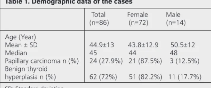
The demographic data of 86 cases included in the study are shown in Table 1.
All cytomorphological criteria were evaluated in PC and BTH. Resection results were found to be compatible with those of fine-needle biopsy aspiration.
While the most common alterations in PC were found to be long nuclear clefting, nuclear overlapping, large nucleus, nuclear pleomorphism, and insufficient colloid; psammoma bodies, Hurthle cells, and lymphocytic background were not observed at all. All cytologic features were determined in benign thyroid hyperplasia, although with variable rates.
Table 1. Demographic data of the cases
Total Female Male (n=86) (n=72) (n=14) Age (Year) Mean ± SD Median Papillary carcinoma n (%) Benign thyroid hyperplasia n (%) 44.9±13 43.8±12.9 50.5±12 45 44 48 24 (27.9%) 21 (87.5%) 3 (12.5%) 62 (72%) 51 (82.2%) 11 (17.7%) SD: Standard deviation
The intranuclear pseudoinclusion was determined with the rates of 58.3%, and 1.6% in the PC and BTH, respectively. Interestingly, the feature was common and was detected very easily in the specimens containing pseudoinclusion; in other cases, it was never seen despite prolonged scanning.
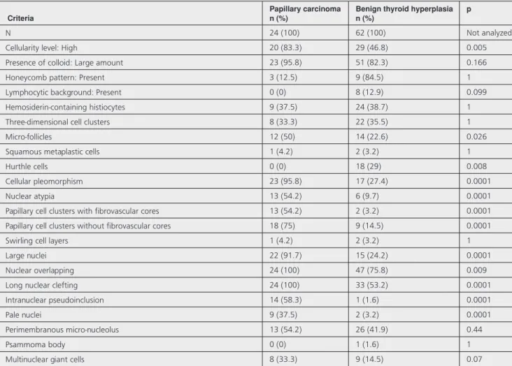
Each criterion was compared by univariate analysis. The results obtained are shown in Table 2.
The variables that remained predictive for the diagnosis of cancer as the result of the backward analysis with the linear regression method following the multivariate analysis performed with the variables with p values <0.1 were cellular pleomorphism, nuclear atypia, papillary structures with fibrovascular cores and intranuclear pseudoinclusion. The cytological changes are shown in Figures 1 to 4. In Table 3, the B coefficients and p values of these variables are given.
Discussion
Differentiation between benign and malignant thyroid nodules is important for avoiding unnecessary surgical
procedures and complications as well as determining the treatment protocol. Currently, the initial step in approaching the thyroid nodule is indisputably the FNAC method (2-4).
Based on the defined structural and cytomorphological characteristics, the success rate of detection of PC by FNAC is over 95% (2). Various cytomorphological features have been determined for this purpose. These features might not be possible to co-exist in every sample.
Table 2. Cytological alterations in papillary carcinoma and benign thyroid hyperplasia together with the significance levels of the frequency differences of these cytological alterations between the two groups
Criteria Papillary carcinoman (%) Benign thyroid hyperplasian (%) p
N 24 (100) 62 (100) Not analyzed
Cellularity level: High 20 (83.3) 29 (46.8) 0.005
Presence of colloid: Large amount 23 (95.8) 51 (82.3) 0.166
Honeycomb pattern: Present 3 (12.5) 9 (84.5) 1
Lymphocytic background: Present 0 (0) 8 (12.9) 0.099
Hemosiderin-containing histiocytes 9 (37.5) 24 (38.7) 1
Three-dimensional cell clusters 8 (33.3) 22 (35.5) 1
Micro-follicles 12 (50) 14 (22.6) 0.026
Squamous metaplastic cells 1 (4.2) 2 (3.2) 1
Hurthle cells 0 (0) 18 (29) 0.008
Cellular pleomorphism 23 (95.8) 17 (27.4) 0.0001
Nuclear atypia 13 (54.2) 6 (9.7) 0.0001
Papillary cell clusters with fibrovascular cores 13 (54.2) 2 (3.2) 0.0001
Papillary cell clusters without fibrovascular cores 18 (75) 9 (14.5) 0.0001
Swirling cell layers 1 (4.2) 2 (3.2) 1
Large nuclei 22 (91.7) 15 (24.2) 0.0001
Nuclear overlapping 24 (100) 47 (75.8) 0.009
Long nuclear clefting 24 (100) 33 (53.2) 0.0001
Intranuclear pseudoinclusion 14 (58.3) 1 (1.6) 0.0001
Pale nuclei 9 (37.5) 2 (3.2) 0.0001
Perimembranous micro-nucleolus 13 (54.2) 26 (41.9) 0.44
Psammoma body 0 (0) 1 (1.6) 1
Multinuclear giant cells 8 (33.3) 9 (14.5) 0.07
Table 3. The cytological variables that were found to be significant for predicting the diagnosis of papillary cancer in multivariate analysis
Criterion B coefficient p
Cellular pleomorphism 271 <0.0001
Nuclear atypia 266 <0.0001
Papillary cell clusters with
fibrovascular cores 342 <0.0001
Moreover, similar cytomorphological features can also be partially present in BTH. In this study, we investigated which feature (s), and their co-existence might be valuable in the diagnosis of carcinoma by comparing the cytology of PC and BTH. We demonstrated the combination of cellular pleomorphism, atypia, and papillae with cores and
intranuclear pseudoinclusion could be useful in diagnosing carcinoma.
In papillary thyroid carcinoma, the sizes and shapes of the cells are variable. The cells may be medium-sized or large; their shape may be cuboidal, columnar, polygonal, fusiform, or even “histiocytoid” (5,6). The last described cellular alteration may lead to a false-positive diagnosis due to the confusion created by the histiocyte clusters showing nuclear atypia in cystic nodular hyperplasia (7). Moreover, radioactive iodine treatment also causes similar changes (8). Cellular pleomorphism and atypia, having high diagnostic value, can be easily identified, however, the evaluation might be rather difficult.
It has been asserted that well-formed papillary structures with fibrovascular cores in BTH characterized by florid hyperplasia can frequently be confused with PC (9,10). On the other hand, in a colloid-rich background, the diagnosis of malignancy can come to the forefront only if the short, nonbranching papillae are co-existent with the typical nuclear alterations (11). The nature of the papillary structures and associated changes might be the factors to consider in the cytological evaluation.
Intranuclear cytoplasmic pseudoinclusions are present in 50-100% of PC aspirations (5). In our study, the rate was determined as 58.3% in PC. It is known that they can also be observed in BTH, although rare. It can be suggested that intranuclear cytoplasmic pseudoinclusions accompany malignancies, although it is not a validated feature if present alone. It has been suggested that a nuclear clefting of 20% or more can be diagnostic for thyroid neoplasm in FNAC in cases that this feature is not available (12). The probability of PC could be eliminated when the clefting rate was below 10%, whereas it was emphasized that the probability of malignant lesions
Figure1aand1b. Papillary structure in papillary carcinoma on
fine needle aspiration cytology (Papanicolaou stain)
Figure2.This sheet of follicular cells displays some features of
papillary carcinoma, including nuclear enlargement, powdery ch-romatin, prominent intranuclear pseudoinclusions (arrow) and nuclear molding (Papanicolaou stain x 400)
Figure3.These follicular cells demonstrate marked
anisonucle-osis, pale chromatin and overlapping (Papanicolaou stain x 400)
Figure4. Multinuclear giant cells may be accompanied to
could occur when the clefting rate is 10-19%. The results of our study showed that nuclear clefting was valuable in diagnosing PC, whereas it did not take part in the differential diagnosis.
It has been reported that nuclear crowding and overlapping were frequently at striking levels in PC (5). However, the striking point that we identified in this regard was that the criterion was rather nonspecific due to the 75.8% rate that we found in BTH.
Psammoma body can rarely be found in benign thyroid lesions (4,5,13). LiVolsi (13) reported that the psammoma body was below 1% in benign hyperplasia. For PC, the positive predictive value of psammoma body alone was 50%, whereas it was 100% when taken into account with the other cytologic features (5,14). On the other hand, it is also the fact that psammoma bodies may not be present in every PC.
StudyLimitations
In this study, there were various limitations. Firstly, it had the general limitations of the retrospective method. In addition, the bias can occur as a result of the sampling process.
Conclusion
Although every cytological feature is characteristic for PC, none of them alone is specific; they should be evaluated together with all nuclear and structural features during the diagnostic approach. However, when it is not possible to determine all of them in practice, the togetherness of cellular pleomorphism, nuclear atypia, papillary structures with fibrovascular cores and the intranuclear pseudoinclusion can gain importance.
Ethics
EthicsCommitteeApproval: It was not taken. InformedConsent: It was not taken.
Peer-review: Externally and internally peer-reviewed. AuthorshipContributions
Concept: H.N.Ü. Design: H.N.Ü., C.B.I. Data Collection or Processing: H.N.Ü., C.B.I., K.A. Analysis or Interpretation: H.N.Ü., K.A., P.F., C.B.I., M.Z.G. Literature Search: C.B.I., H.N.Ü. Writing: C.B.I., H.N.Ü.
Conflict of Interest: No conflict of interest was
declared by the authors.
Financial Disclosure: The authors declared that this
study received no financial support.
References
1. Handa U, Garg S, Mohan H, Nagarkar N. Role of fine needle aspiration cytology in diagnosis and management of thyroid lesions: A study on 434 patients. J Cytol 2008;25:13-7. 2. Karataş A, Giray S, Peker Ö, et al. Tiroid Nodüllerinin
Değerlendirilmesinde Bethesda 2007 Sınıflamasının Klinik Sonuçları. Ulusal Cerrahi Dergisi 2009;25:92-6.
3. İmamoğlu Ç, İmamoğlu FG, Dizen H, et al. Ultrasound guided fine needle aspiration cytology in thyroid nodules: Cytohistologic correlation. Medical journal of Mugla Sitki Kocman University 2015;2:7-11.
4. Pandey P, Dixit A, Mahajan NC. Fine-needle aspiration of the thyroid: A cytohistologic correlation with critical evaluation of discordant cases. Thyroid Res Pract 2012;9:32-9.
5. Ali SZ, Cibas ES. The Bethesda System for Reporting Thyroid Cytopathology. Definitions, Criteria and Explanatory Notes. New York, NY, USA: Springer; 2010.
6. Renshaw AA, Gould EW. Why there is the tendency to “overdiagnose” the follicular variant of papillary thyroid carcinoma. Am J Clin Pathol 2002;117:19-21.
7. Nassar A, Gupta P, LiVolsi VA Baloch Z. Histiocytic aggregates in benign nodular goiters mimicking cytologic features of papillary thyroid carcinoma(PTC). Diagn Cytopathol 2003;29:243-5.
8. Anderson SR, Mandel S, LiVolsi VA, Gupta PK, Baloch ZW. Can cytomorphology differentiate between benign nodules and tumors arising in Graves’ disease? Diagn Cytopathol 2004;31:64-7.
9. Pusztaszeri MP, Krane JF, Cibas ES, Daniels G, Faquin WC. FNAB of benign thyroid nodules with papillary hyperplasia: a cytological and histological evaluation. Cancer Cytopathol 2014;122:666-77.
10. Perez-Montiel MD, Suster S. The spectrum of histologic changes in thyroid hyperplasia: a clinicopathologic study of 300 cases. Hum Pathol 2008;39:1080-7.
11. Khayyata S, Barroeta JE, LiVolsi VA, Baloch ZW. Papillary hyperplastic nodule: pitfall in the cytopathologic diagnosis of papillary thyroid carcinoma. Endocr Pract 2008;14:863-8. 12. Yang YJ, Demirci SS. Evaluating diagnostic significance
of nuclear grooves in thyroid fine needle aspirates with a semiquantitative approach. Acta Cytol 2003;47:563-70. 13. LiVolsi VA. Papillary thyroid carcinoma: an update. Modern
Pathol 2011;24(Suppl 2):1-9.
14. Ellison E, Lapuerta P, Martin SE. Psammoma bodies in fine-needle aspirates of the thyroid: predictive value for papillary carcinoma. Cancer 1998;84:169-75.