experimental renal ischemia-reperfusion in rats
Aynur Koç
1, Neyhan Ergene
2, Abdülkerim Kasim Baltaci
3and Rasim Mogulkoc
3 1Necmettin Erbakan University, Meram Medical Faculty, Deparment of Physiology, Konya, Turkey 2
KTO Karatay University, Medical Faculty, Deparment of Physiology, Konya, Turkey 3
Selcuk University, Medical Faculty, Deparment of Physiology, Konya, Turkey
Abstract:This study aimed to examine the affects of 3’-4’-dihydroxyflavonol (DiOHF) on lipid peroxidation in
experimental renal ischemia-reperfusion. The research was conducted on Wistar-albino type male rat. The experimental groups were formed as 1.Control; 2.Sham; 3.Ischemia; 4.Ischemia+reperfusion; 5.DiOHF+Ischemia; 6.Ischemia+ DiOHF + reperfusion. The highest tissue glutathione levels were found in groups 5 and 6. Groups 1 and 2, which were control and sham groups respectively, had the lowest tissue GSH values. Ischemia group was found to have the highest tissue malondialdehyde (MDA) level. Tissue MDA levels in group 4 were lower than those in group 3, however, higher than the levels in all other groups. Erythrocyte GSH levels in groups 5 and 6 were higher than the levels in all other groups. Group 4 has highest plasma MDA values. Plasma MDA levels in group 3 were lower than the levels in Group 4, but higher than those in other groups. The results of the study indicate that intraperitoneal DiOHF administration inhibits lipid per oxidation that intensifies in the case of renal ischemia-reperfusion injury in rats.
Keywords: Renal ischemia-reperfusion, 3’-4’-Dihydroxyflavonol, lipid per oxidation, rat.
INTRODUCTION
Reduction in or interruption of blood supply to an organ due to various reasons (particularly during vascular surgical procedures and organ transplantation) is called ischemia. Ischemia causes the tissue to become hypoxic and hypoxic tissue injury follows. Extended hypoxia impairs the cellular integrity and may even lead to cell death. Reperfusion, on the other hand, is the restoration of blood supply to the tissue. In the course of reperfusion, free oxygen radicals (ROS) released by polymorpho nuclear leukocytes (PMNL) that increase the destruction of the cell. This condition is called tissue injury associated with reperfusion (Duman et al., 2015). Renal ischemia-reperfusion (I/R) injury is a common outcome of clinical procedures such as vascular surgery, organ removal and a cause of renal failure (Koga et al., 2012). Renal ischemia is a frequent complication that can lead to high morbidity and mortality in the perioperative period (Hutchens et al., 2008). Lipid peroxidation is an autocatalytic pathway causing oxidative stress in cell membranes and resulting in the release of reactive lipid aldehydes. Lipid per oxidation is commonly used as a marker of stress in the tissues (Walker et al., 2001). Lipid peroxidation products like malondialdehyde (MDA) determine the severity of per oxidation (Bicer et al., 2012). Of the peroxidation products, two have particularly more marked mutagenic effects: MDA and hydroxynonenal (HNE). MDA is a product that results from the breakdown of three or more double-bond fatty acids (Mogulkoc et al., 2006). I/R injury contribute to lipid peroxidation associated with
ROS (Koga et al., 2012). Glutathione (GSH) is the major intracellular antioxidant against oxidative stress. It is produced in many tissues, particularly those of the liver, from glutamate, cysteine and glycine. Glutathione keeps sulfhydryl groups in reduced form and provides protection against oxidation. Thus, it prevents the inactivation of functional proteins (Ross 1988; Baltaci et al., 2014). Flavonoids with antioxidant characteristics have the ability of scavenging free radicals. Flavonoid radicals that are formed after the separation of hydrogens become stabilized by forming chelate rings with trace metals in the medium (Bors et al., 1990). Flavonoids prevent the formation of ROS by discarding reactive species as much as possible, and thus, restrict the prolongation of oxidative reactions (Akhlaghi and Bandy 2009). The present study investigated the effect of 3’-4’-dihydroxyflavonol (DiOHF), a synthetic flavonoid, which was shown in previous studies to have protective effects in heart and brain ischemia, on experimental renal I/R.
MATERIALS AND METHODS
The present study was carried out at the 56 Wistar-albino type male rats supplied by the Experimental Medicine Research and Application Center of Necmettin Erbakan University. Research was approved by the local ethics committee of the same center. The experimental animals weighed between 200 and 250 g. The rats were randomly divided into 6 groups, of which two included 8 and the other four included 10 rats. The feed and water were given as ad libitum.
Experimental animals were given 10 g/100 g body weight of feed daily. They were kept in a room having a standard
room temperature (of 21±1oC) in 12 hour dark/ 12 hour light cycle.
Surgical procedures were performed after the animals were anesthetized using intraperitoneal ketamine hydrochloride (50 mg/kg), Xysaline (rompun) (5 mg/kg) injection. After the induction of renal ischemia-reperfusion, midline laparotomy was performed. A vascular clamp was placed at the hilus level in the left kidney to interrupt the blood circulation in the left renal artery for one hour to induce ischemia.
Control (n=8): In this group, animals were not subjected to any procedure. Blood samples were collected by cardiac puncture from the animals by general anesthesia. After the animals were sacrificed by cervical dislocation, their kidney tissues were taken.
Sham Group (n=8): After the animals were anesthetized (with ketamine + rompun), 1 ml of the 3’,4’-dihydroxyflavonol (DiOHF) solvent solution was injected into the peritoneum and the kidney site was opened and closed surgically. The animals were then sacrificed under general anesthesia to collect blood and kidney tissue samples.
Renal-Ischemia Group (n=10): The animals were put under general anesthesia and their left kidneys were subjected to ischemia for one hour. At the end of this period, the animals were sacrificed and their blood and kidney tissue samples were taken.
Renal Ischemia + Reperfusion Group (n=10): After the animals were put under general anesthesia, their renal tissues were subjected to ischemia for one hour and then to reperfusion for another hour. The animals were sacrificed after I/R and their blood and kidney tissue samples were collected.
DiOHF + Renal Ischemia Group (n=10): Before renal ischemia, DiOHF was injected as 30 mg/kg intraperitoneal and ischemia was induced in their left kidney. At the end of the ischemia period, the animals were sacrificed to collect blood and renal tissue samples. Renal Ischemia + DiOHF + Reperfusion Group (I/R) (n=10): Following anesthesia, ischemia was induced in their left kidney for 1 hour. Ischemia induction was followed by intraperitoneal DiOHF (30 mg/kg) injection. Then reperfusion was induced for another hour. At the end of this period, blood and kidney tissue were taken. Dihydroxyflavonol administration
DiOHF of 100ml (Indofine Chemical Co., USA) was dissolved in a mixture of dimethyl sulfoxide (2 ml) + polyethylene glycol (11ml) + distilled water (7 ml). The animals were injected with intraperitoneal DiOHF (Wang
et al., 2009).
Collection of blood and renal tissue samples
Blood samples of 3-4 ml were taken from the anesthetized animals and put into EDTA tubes. The blood in EDTA tubes were centrifuged at 3000 rpm for 10 min in a cooling centrifuge device adjusted to +4oC. The plasma samples separated as such were kept at -80oC by analysis. The renal tissue samples were kept as dry tissue at -80oC until the time of analysis.
Blood and tissue analyses Tissue protein analysis
Tissue protein was tested using total protein test kit PRO-10300 according to spectrophotometric method.
Determination of tissue malondialdehyde (MDA) levels MDA levels in tissue were determined by Uchiyama and Mihara (1977) method. The resulst were given as mg/g protein.
Tissue glutathione analysis
The GSH quantities in the samples were measured according to Ellman’s method. The values were presented as nmol/g protein (Ellman 1959).
Plasma malondialdehyde analysis
Plasma levels of MDA were determined as previously defined (Draper andHadley, 1990).
Erythrocyte glutathione analysis
Erythrocyte glutathione analysis was performed according to previously mentioned (Atroshi and Sandholm, 1982).
STATISTICAL ANALYSES
SPSS statistical software was used in the statistical analysis. The results were described as mean ± SD. Kruskall Wallis variance analysis was used in the comparisons among the groups and Mann Whitney U test was employed to identify the p<0.05 level. Results p<0.05 were evaluated as significant.
RESULTS
Tissue MDA and GSH values of the groups are presented in table 1. An examination of the tissue GSH values of the study groups reveal that Groups 5 and 6 had the highest GSH values (p<0.005). GSH values of the ischemia and ischemia + reperfusion groups, which were Groups 3 and 4, were found higher than those in Groups 1 and 2. There was no difference between the GSH values of other groups.
The highest tissue MDA levels were found in Group 3, which was the ischemia group (p<0.005). Tissue MDA values in the ischemia+ reperfusion group were lower than the levels in Group 3 (p<0.005), but higher than those in all other groups (p<0.005). There was not any difference between the tissue MDA levels of Groups 1, 2, 5, and 6 (table 1).
Erythrocyte GSH and plasma MDA levels were given at the table 2. In terms of erythrocyte GSH values, Groups 5 and 6, which were administered DiOHF, were found to have the highest values. Erythrocyte GSH levels in Groups 1, 2, 3, and 4 did not show any statistically significant difference (table 2).
When plasma MDA values were examined, group 4, which was I/R group, was seen to have the highest plasma MDA values. Plasma MDA level was lower than those in Group 4 (p<0.005), but higher than the levels in all other groups (p<0.005). Differences between the plasma MDA values of other groups were not statistically significant.
DISCUSSION
This study shows that the changes that occur in lipid peroxidation have actually taken place in renal ischemia-reperfusion injury experimentally induced in animals. The changes in the levels of MDA, which can be considered a marker of injury, in particular, indicate that the ischemia targeted in the study was indeed induced in renal tissue. Ischemia-reperfusion leads to acute renal damage associated with high mortality and morbidity. In addition to the direct damage caused by reduced blood flow, I/R injury is related with large-scale production of ROS, cytokines and chemokines (Kim et al., 2010). Despite being necessary for the tissue to sustain its viability, reperfusion causes further cellular damage. It initiates a
series of events involving the damaging of renal cells and finally cell death through apoptosis and necrosis (Sharples
et al., 2004). Levels of ROS are elevated especially
during reperfusion leading to the lipid peroxidation. Mitochondrial dysfunction, energy shortage, and impaired cellular calcium homeostasis resulting from oxidative stress lead to further ROS formation, which in turn causes further damage to cellular proteins, DNA, and membrane lipids (Aruna Devi et al., 2010; Kunduzova et al., 2010). As it contains a plentitude of long-chain, multi-unsaturated fatty acids in its lipid composition, the kidney is particularly vulnerable to the damage caused by ROS (Ozbek, 2012).
DiOHF has antioxidant activity (Leo et al., 2011). This study aimed to explore how DiOHF affects the lipid peroxidation to result from in vivo unilateral ischemia and I/R induced in the kidney of experimental animals for one hour and the antioxidant defense mechanism. Experiment groups were formed to find out whether effects of DiOHF on the I/R injury resulting from oxidative stress occurred in the ischemia period or I/R period. In order to identify the oxidative stress that may occur as a result of renal ischemia and I/R, MDA was determined. GSH and total protein levels were studied to show the antioxidant defense that could develop against oxidative stress. In the experimental model where Woodman and Chan (2009) induced I/R in the back feet of rats, DiOHF produced its protective effect during reperfusion. In
Table 1: Renal tissue GSH and MDA levels
Groups GSH (nmol/g protein) MDA (mg/g protein)
1-Control (n=8) 0.41±0.06 c 0.03±0.00 c
2-Sham-Control (n=8): 0.35±0.06 c 0.05±0.02 c
3-Renal Ischaemia (n=10) 0.45±0.10 b 0.33±0.03 a
4- Renal Ischaemia-Reperfusion (I/R) (n=10) 0.44±0.09 b 0.29±0.09 b
5- 3',4'-Dihydroxyflavonol (DiOHF) + Renal Ischaemia 0.50±0.07 a 0.04±0.00 c
6-Renal Ischaemia + 3',4'-Dihydroxyflavonol (DiOHF) +
Reperfusion (I/R) (n=10) 0.54±0.17 a 0.05±0.02 c
*Different letters in same column indicate differences among the groups. DiOHF supplementation increased renal tissue GSH levels and reduced tissue MDA levels after the renal ischemia. a>b>c (p<0.005)
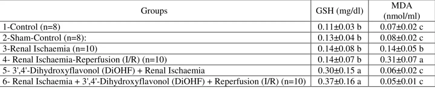
Table 2: Erythrocytes GSH and plasma MDA levels
Groups GSH (mg/dl) MDA
(nmol/ml)
1-Control (n=8) 0.11±0.03 b 0.07±0.02 c
2-Sham-Control (n=8): 0.13±0.04 b 0.08±0.02 c
3-Renal Ischaemia (n=10) 0.14±0.08 b 0.14±0.05 b
4- Renal Ischaemia-Reperfusion (I/R) (n=10) 0.14±0.07 b 0.31±0.07 a
5- 3',4'-Dihydroxyflavonol (DiOHF) + Renal Ischaemia 0.30±0.15 a 0.06±0.02 c
6- Renal Ischaemia + 3',4'-Dihydroxyflavonol (DiOHF) + Reperfusion (I/R) (n=10) 0.37±0.16 a 0.05±0.01 c *Different letters in same column indicate differences among the groups. DiOHF supplementation increased erythrocytes GSH levels and reduced plasma MDA levels after the renal ischemia. a>b>c (p<0.005)
contrast, in this study where we induced unilateral kidney I/R in rats the protective effect of I/R was observed in both ischemia and I/R groups. The reduction of tissue and plasma MDA levels which were elevated in ischemia and I/R groups back to control levels after DiOHF supplementation and the increase in the tissue and GSH levels in the DiOHF-supplemented groups were taken as the indicators of the protective effect of DiOHF in ischemia and reperfusion.
Ischemia reduces the activity of cellular defense enzymes against ROS and reperfusion or oxygen supply causes further impairments in the oxidant/antioxidant balance which has already been rendered delicate by increased ROS production (Malakul et al., 2011). Excessive ROS production impacts membrane lipids, inactivation of antioxidant enzymes, impairment of cellular skeleton, cell integrity, DNA degeneration, leukocyte activation, endothelial cell injury, and cytokine production. Antioxidant agents were used in experimental models (Kim et al., 2010). In a study where the effect of resveratrol, an antioxidant polyphenolic compound, on renal injury associated with I/R was examined, it was established on the basis of MDA and GSH parameters that resveratrol alleviated renal injury resulting from I/R (Sener et al., 2006). Edaravone is a strong scavenger of hydroxyl and peroxyl radicals and shows antioxidant activity to prevent lipid peroxidation. It was demonstrated that edaravone supplementation after the induction of ischemic acute renal failure by clamping the renal artery for 45 minutes mitigated I/R injury by cleaning intracellular ROS in renal tubule cells and inhibiting lipid peroxidation in cell membranes (Doi et al. 2004). Supplementation of vitamin E and C antioxidants to experimental ischemic kidneys increased the renal hemodynamics and reduced oxidative stress, inflammation and fibrosis by chronically blocking oxidative stress pathways (Lerman et al., 2009). Similar to the previous I/R studies (Chan et al., 2003; Singh et al., 2000), at our research aimed to determine the antioxidant activity of DiOHF, a synthetic flavonoid with well-established antioxidant action, on renal I/R injury. GSH is a critical element in the cellular protection mechanism against various harmful stimuli including oxidative stress. Low-molecular weight GSH (reduced glutathione) is a significant scavenger of free radicals in the cytoplasm (Hensley et al., 2000; Sener et al., 2006). Glutathione is a crucial intracellular antioxidant, both because it directly cleans ROS and as a substrate for glutathione peroxidase that catalyzes ROS destruction (Nitescu et al., 2009). It plays a remarkable part in nitroxide bioreduction at the cell and organelle level (Hirayama et al., 2005). High concentrations of intracellular glutathione and other antioxidant compounds build a strong capacity for free radical scavenging (Dröge, 2002).
As opposed to early studies (Nitescu et al., 2006; Scaduto
et al., 1988; Slusser et al., 1990), it was found in our
study that in vivo renal I/R injury elevated renal GSH amount. It is speculated that this difference can be attributed to the difference in the length of I/R and that the elevated renal tissue GSH levels found in ischemic and ischemic-reperfused groups in this study can be explained by an increased defense response to lipid peroxidation. Besides ischemia and I/R groups, ischemia and I/R groups given with DiOHF were seen to have higher GSH levels in the renal tissue. Increased tissue GSH levels found in DiOHF-supplemented groups can be considered an indicator of the further strengthening of the renal tissue defense mechanism against lipid peroxidation.
In a study where rat kidneys were subjected to 60 minutes of ischemia and 24 hours of reperfusion to examine the effects of N-acetylcysteine on renal I/R injury, GSH levels were established to drop in the I/R group (Aydoğdu et al., 2005). This result is not consistent with the results of our study where we found elevated tissue GSH levels in the I/R group. When this and some other I/R studies are considered, it is seen that the duration of reperfusion in the concerned studies is longer than the reperfusion duration in our study (Rhodenet al., 2002; Woodman and Chan 2004). It is believed that the level of free radicals that formed over the course of the one-hour reperfusion period has elevated GSH levels by increasing the antioxidant defense response. The longer reperfusion durations and the higher levels of free radical formation as a result might have weakened the antioxidant mechanism and led to a fall in the GSH levels in other studies.
Erythrocytes are continuously exposed to intra- and extra-cellular free radicals and consequently, erythrocyte membranes that are rich in multi-unsaturated fatty acids are inflicted by lipid peroxidation. In our study, although the tissue GSH parameter increased in I/R and DiOHF-supplemented groups, erythrocyte GSH levels were elevated in DiOHF-supplemented groups only. This can be seen as proof that DiOHF supplementation strengthens the antioxidant defense mechanism.
I/R injury has been established to cause accumulation of oxidation products like MDA, changes in antioxidant enzymes, and induction of apoptosis (Aruna Devi et al., 2010). Elevated MDA levels may produce a negative effect through the increase in peroxynitrite free radical activity (Rhoden et al., 2002). DiOHF effectively cleans super oxide and thus, increases basal and stimulated NO bioavailability. Consequently, it may offer protection against the vascular impairment stemming from I/R (Woodman and Chan2004). In this research, renal tissue MDA values were examined to determine the lipid peroxidation resulting from I/R. The highest MDA values were obtained in the ischemia group and this provided
evidence of the full-fledged development of ischemia in the kidney. Nonetheless, the increase found in the MDA levels of I/R group indicates that the oxidant damage continues during reperfusion. Tissue MDA levels in the DiOHF-supplemented groups dropped almost to the levels found in the control groups. The post-DiOHF decrease found in tissue MDA levels that had elevated in I/R demonstrates that DiOHF alleviated oxidant stress to a large extent.
When the results of the study are evaluated, the full development of ischemia and reperfusion injury can be seen from the increase in oxidants and inhibition of the antioxidant system. The increase in MDA levels as an indicator of the oxidant stress in the tissue during ischemia-reperfusion injury was shown. Additionally, GSH values, which have a protective effect against lipid per oxidation, were demonstrated to increase as a result of increased antioxidant activity in the tissue. Besides, it is seen in the study that DiOHF produced a protective effect against lipid per oxidation in the experimental ischemia-reperfusion injury both by inhibiting oxidants and increasing the effectiveness of antioxidants.
CONCLUSION
The clinical importance of this study that present used DiOHF doses may be useful for renal ischemia as drug. However, the limitation of performed study that experimental procedure and different doses of DiOHF may be in consider in future.
ACKNOWLEDGEMENT
This study was supported by a grant Necmettin Erbakan University, Scientific Research Council (Grant number is NEU-BAP-131318001). The authors declare that there are no conflicts of interest.
REFERENCES
Akhlaghi M and Bandy B (2009). Mechanisms of flavonoid protection against myocardial ischemia-reperfusion injury. J. Mol. Cell Cardiol.,46(3): 309-317.
ArunaDevi R, Lata S, Bhadoria BK, Ramteke VD, Kumar S, Sankar P, Kumar D and Tandan SK (2010). Neuroprotective effect of 5,7,3',4',5'-pentahydroxy
dihydroflavanol-3-O-(2''-O-galloyl)-beta-D-glucopyranoside, a polyphenolic compound in focal cerebral ischemia in rat.Eur. J. Pharmacol.,626(2-3): 205-212.
Atroshi F and Sandholm M (1982). Red blood cell glutathione as a marker of milk production in finn sheep. Res. Vet. Sci.,33 (2): 256-259.
Aydogdu N, Kaymak K and Yalçın O (2005). The effect of N-asetilsistein on renal ischemia-reperfusion damage in rat. Fırat Med. J.,10(4): 151-155.
Baltaci AK, Mogulkoc R, Ayyildiz M, Kafali E and Koyuncuoglu T (2014).Lipid peroxidation in kidney and testis tissues in experimental hypothyroidism: the role of zinc.Bratisl. Lek. Listy.,115(8): 498-501. Bicer M, Gunay M, Baltaci AK, Uney K, Mogulkoc R
and Akil M (2012). Effect of zinc supplementation on lipid peroxidation and lactate levels in rats with diabetes induced by streptozotocin and subjected to acute swimming exercise.Bratisl. Lek. Listy.,113(4): 199-205.
Bors W, Heler W, Michel C and Saran M (1990). Flavonoids as antioxidants: Determination of radical-scavenging efficiencies. Methods Enzymol.,186(1): 343-355.
Chan ECH, Drummorol DR and Woodman OL (2003). 3’,4’-Dihydroxyflavonol enhances nitric oxide bioavailability and improves vascular function after ischemia and reperfusion injury in the rat. J.
Cardiovasc. Pharmacol.,42(6): 727-735.
Doi K, Suzuki Y, Nakao A, Fujita T and Noiri E (2004). Radical scavenger edaravone developed for clinical use ameliorates ischemia/reperfusion injury in rat kidney.
Kidney Int.,65(5): 1714-1723.
Draper HH and Hadley M (1990). Malondialdehyde determination as index of lipid peroxidation.Methods
Enzymol.,186(1): 421-431.
Dröge W (2002). Free radicals in the physiological control of cell function.Physiol. Rev.,82(1): 47-95. Duman A, Mogulkoc R, Baltaci AK and Menevse E
(2015). 3',4'-dihydroxyflavonol attenuates tissue damage in unilateral testis ischemia-reperfusion in rats.
Bratisl. Lek. List.,116(12): 735-740
Ellman GL (1959). Tissue sulfhydryl groups. Arch.
Biochem. Biophys.,82(1): 70-77.
Hensley K, Robınson KA, Gabbıta SP, Salsman S and Floyd AR (2000). Reactive oxygen species, cell signaling, and cell injury. Free Radical. Bio.
Med.,28(10): 1456-1462.
Hirayama A, Nagase S, Ueda A, Oteki T, Takada K, Obara M, Inoue M, Yoh K, Hirayama K and Koyama A (2005). In vivo imaging of oxidative stress in ischemia-reperfusion renal injury using electron paramagnetic resonance. Am. J. Physiol. Renal. Physiol., 288(3): 597-603.
Hutchens PM, Dunlap J, Hurn DP and Jarnberg PO (2008). Renal ischemia: Does sex matter. Int. Anest.
Res. Soc.,107(1): 239-249.
Kim J, Jang HSand Park KM (2010). Reactive oxygen species generated by renal ischemia and reperfusion trigger protection against subsequent renal ischemia and reperfusion injury in mice.Am. J. Physiol. Renal
Physiol.,298(1): 158-166.
Koga H, Hagiwara S, Kusaka J, Goto K, Uchino T, Shingu C, Kai S and Noguchi T (2012). New α-lipoic acid derivative, DHL-HisZn, ameliorates renal ischemia-reperfusion injury in rats.J. Surg. Res.,174(2): 352-358.
Kunduzova OR, Escourrou G, De La Farge F, Salvayre R, Séguélas MH, Leducq N, Bono F, Herbert JM and Parini A (2004). Involvement of peripheral benzodiazepine receptor in the oxidative stress, death-signaling pathways, and renal injury induced by ischemia-reperfusion.J. Am. Soc. Nephrol.,15(8): 2152-2160.
Leo CH, Hart JL and Woodman OL (2011). 3′,4′-Dihydroxyflavonol reduces superoxide and improves nitric oxide function in diabetic rat mesenteric arteries.
PLoS One.,6(6): e20813.
Lerman LO, Textor SC and Grande JP (2009). Mechanisms of tissue injury in renal artery stenosis: ischemia and beyond.Prog. Cardiovasc. Dis.,52(3): 196-203.
Malakul W, Ingkaninan K, Sawasdee P and Woodman OL (2011). The ethanolic extract of Kaempferia parviflora reduces ischaemic injury in rat isolated hearts.J.
Ethnopharmacol.,137(1): 184-191.
Mogulkoc R, Baltaci AK, Oztekin E, Aydin L and Sivrikaya A (2006). Melatonin prevents oxidant damage in various tissues of rats with hyperthyroidism.
Life Sci.,79(3): 311-315.
Nitescu N, Ricksten SE, Marcussen N, Haraldsson B, Nilsson U, Basu S and Guron G (2006). N-acetylcysteine attenuates kidney injury in rats subjected to renal ischaemia-reperfusion. Nephrol. Dial.
Transplant.,21(5): 1240-1247.
Ozbek E (2012). Induction of oxidative stress in kidney.
Int. J. Nephrology., 2012(1):465897.
Rhoden EL, Rhoden CR, Lucas ML, Lima LP, Zettler C and Klein AB (2002). The role of nitric oxide pathway in the renal ischemia-reperfusion injury in rats.
Transpl. Immunol.,10(4): 277-284.
Ross D (1988). Glutathione, free radicals and chemotherapeutic agents. Pharmacol. Ther., 37(2): 231-249.
Scaduto RC, Gattone VH, Grotyohann LW, Wertz J and Martin LF (1988). Effect of an altered glutathione content on renal ischemic injury.Am. J. Physiol.,255(5-2): 911-921.
Sharples EJ, Patel N, Brown P, Stewart K, Mota-Philipe H, Sheaff M, Kieswich J, Allen D, Harwood S, Raftery M, Thiemermann C and Yaqoob MM (2004). Erythropoietin protects the kidney against the injury and dysfunction caused by ischemia-reperfusion. J.
Am. Soc. Nephrol.,15(8): 2115-2124.
Singh D, Chander V and Chopra K (2000). Protective effect of catechin on ischemia-reperfusion-induced renal injury in rats. Pharmacol Rep.,57(1): 70-76. Slusser SO, Grotyohann LW, Martin LF and Scaduto RC
(1990). Glutathione catabolism by the ischemic rat kidney. Am. J. Physiol.,258(6): 1546-1553.
Şener G, Tusğtepe H, Yüksel M, Cetinel S, Gedik N and Yeğen BC (2006). Resveratrol improves ischemia/ reperfusion-induced oxidative renal injury in rats. Arch.
Med. Res., 37(7): 822-829.
Uchiyama M and Mihara M (1977). Determination of malondyaldehyde precurser in tissues by thiobarbituric acid test. Anal. Biochem.,86(1): 271-278.
Walker LM, York JL, Imam SZ, Ali SF, Muldrew KL and Mayeux PR (2001). Oxidative stress and reactive nitrogen species generation during renal ischemia.
Toxicol. Sci..63(1): 143-148.
Wang S, Thomas CJ, Dusting GJ, Woodman OLand May CN (2009). 3',4'-Dihydroxyflavonol improves post-ischaemic coronary endothelial function following 7 days reperfusion in sheep.Eur. J. Pharmacol.,624(1-3): 31-37.
Woodman OL and Chan ECH (2004). Vascular and anti-oxidant actions of flavonols and flavones. Annual Scientific Meeting of ASCEPT 2003. Clin. Exp.