Rethinking isolated cleft lip and palate as a syndrome
Mine Koruyucu, DDS, PhD,a Yelda Kasimog˘lu, DDS, PhD,a Figen Seymen, DDS, PhD,a Merve Bayram, DDS, PhD,b Asli Patir, DDS, PhD,b Nihan Ergöz, DDS, PhD,a Elif B. Tuna, DDS, PhD,a Koray Gencay, DDS, PhD,a Kathleen Deeley, MS,c Diego Bussaneli, DDS, PhD,c,d Adriana Modesto, DDS, MS, PhD, DMD,c,eand Alexandre R. Vieira, DDS, MS, PhDc,e
Objective. The goal of the present work was to use dental conditions that have been independently associated with cleft lip and palate (CL/P) as a tool to identify a broader collection of individuals to be used for gene identification that lead to clefts. Study design. We studied 1573 DNA samples combining individuals that were born with CL/P or had tooth agenesis, supernu-merary teeth, molar incisor hypomineralization, or dental caries with the goal to identify genetic associations. We tested 2 single-nucleotide polymorphisms that were located in the vicinity of regions suggested to contribute to supernumerary teeth. Overrepresentation of alleles were determined for combinations of individuals as well as for each individual phenotypic group with anα of .05.
Results. We determined that the allele C of rs622260 was overrepresented in all individuals studied compared with a group of unrelated individuals who did not present any of the conditions described earlier. When subgroups were tested, associations were found for individuals with hypomineralization.
Conclusions. Although we did not test this hypothesis directly in the present study, based on associations reported previously, we believe that CL/P is actually a syndrome of alterations of the dentition, and considering it that way may allow for the iden-tification of genotype-phenotype correlations that may be useful for clinical care. (Oral Surg Oral Med Oral Pathol Oral Radiol 2018;125:307–312)
The endeavor of identifying genes responsible for the etiology of common complex oral and craniofacial con-ditions has been the focus of many research groups for the last 3 decades. For isolated forms of cleft lip and palate, it started with TGFA in 19891and continued more recently with hypothesis-free work scanning the whole genome continues to suggest new associations. A concept our group has proposed is that isolated forms of cleft lip and palate are often accompanied by other minor anoma-lies of the dentition. Individuals born with cleft lip and palate are at least 4 times more likely to present dental anomalies, such as tooth agenesis and supernumerary teeth.2
We also know that individuals born with clefts have more enamel defects.3
Individuals born with cleft lip and palate are also historically suggested to have more dental caries,4,5
even though our own data from populations that have no or very little access to dental care suggest otherwise.6,7
Furthermore, tooth agenesis on occasion is associated with supernumerary teeth,8,9
and individuals
with enamel defects such as molar incisor
hypomineralization are suggested to have more dental
caries10or tooth agenesis of second premolars11or co-occurrence of tooth agenesis and supernumerary teeth.12 Based on these correlations (Figure 1), we designed an experiment that included individuals born with clefts or who have tooth agenesis, supernumerary teeth, molar incisor hypomineralization, or dental caries, and com-pared them with individuals ascertained as having none of these conditions, to test for association with markers in the vicinity of 2 genes that have been suggested as possibly contributing to supernumerary teeth (HMCN1 and IGSF9 B)12
with the assumption that isolated forms of clefts are actually syndromes that combine 1 or more alterations of the dentition that share genetic contribu-tors. The goal of the present work was to use dental conditions that have been independently associated with cleft lip and palate as a tool to identify a broader col-lection of individuals to be used for gene identification that lead to clefts. The combined analysis would yield the identification of genes that contribute to the occur-rence of multiple signs (syndrome) or a particular one. Here we report these analyses and continue to provide evidence that isolated forms of cleft lip and palate are likely syndromes that potentially include multiple al-terations of the dentition.
aDepartment of Pedodontics, School of Dentistry, Istanbul University,
Istanbul, Turkey.
bDepartment of Pedodontics, School of Dentistry, Istanbul Medipol
University, Istanbul, Turkey.
cDepartment of Oral Biology, University of Pittsburgh School of Dental
Medicine, Pittsburgh, PA, USA.
dDepartment of Pediatric Dentistry, UNESP, Araraquara, Brazil. eDepartment of Pediatric Dentistry, University of Pittsburgh School of
Dental Medicine, Pittsburgh, PA, USA.
Received for publication Sep 21, 2017; returned for revision Jan 2, 2018; accepted for publication Jan 8, 2018.
© 2018 Elsevier Inc. All rights reserved. 2212-4403/$ - see front matter
https://doi.org/10.1016/j.oooo.2018.01.007
Statement of Clinical Relevance
We believe that isolated cleft lip and palate is actu-ally a syndrome of alterations of the dentition and considering it that way may allow for the identifica-tion of genotype-phenotype correlaidentifica-tions that may be useful for clinical care.
PATIENTS AND METHODS
We used DNA samples from 1573 individuals who were ascertained to have distinct oral and craniofacial altera-tions as part of our decade-long studies in oral clefts and other dental abnormalities. These individuals are sum-marized in Table I and described in this section. All participants signed an informed consent document before entering into this study. Parents consented for their off-spring, and age-appropriate consent was obtained from all children older than 7 years. This protocol was ap-proved by both the Istanbul University and University of Pittsburgh Institutional Review Boards. Genomic DNA was obtained from whole saliva.
The first study group14was composed of 573 indi-viduals, 158 with clefts (59 with bilateral cleft lip and cleft palate, 10 with cleft palate only, 3 with left cleft lip only, 67 with left cleft lip and cleft palate, and 23 with right cleft lip and cleft palate), 254 unaffected family members, and 161 nonrelated individuals with no history of syndromic clefting. Seventy-three individuals were the product of consanguineous marriages (41 with clefts, 32 from the nonrelated individual pool).14All participants were recruited at the Department of Pedodontics clinics, Istanbul University, Turkey. Individuals born with
isolated forms of cleft of the lip with or without cleft palate or cleft palate only and all of their available first-degree relatives were invited to participate in this study between October 2007 and October 2009. We also invited at least 1 unrelated individual for each cleft case re-cruited of the same age and sex during the same period. All study participants were examined in a dental office by the same professional (M.K.), and panoramic radio-graphs were available for all participants. Dental anomalies outside the cleft area (tooth agenesis, supernumerary teeth, macrodontia, microdontia, malocclusion, and enamel hy-poplasia) were recorded. Tooth agenesis was defined based on the age of the individual and when initial tooth for-mation would be visible radiographically. As expected,2 participants with clefts had more dental anomalies outside the cleft area than controls (tooth agenesis in 31 cases vs in 7 controls, supernumerary teeth in 34 cases vs in 1 control, macrodontia and microdontia in 40 cases vs in 9 controls, enamel hypoplasia in 30 cases vs in 21 controls).14
The second study group15
consisted of 52 unrelated pa-tients with sporadic tooth agenesis (29 participants had missing incisors, 44 had missing premolars, 8 had missing molars, and 10 had missing canines; 7 participants were missing just 1 tooth, 17 were missing 2 teeth, and 27 were missing 3 teeth or more) and their parents and siblings (total of 170 individuals) who were recruited in the met-ropolitan area of Istanbul. No one was the result of a consanguineous marriage. No one reported to have another relative affected by tooth agenesis, oral clefts, or anosmia. Probands had at least 1 developmentally missing tooth, excluding third molars. Twenty-seven participants in this group were female and 25 were male. All cases were of sporadic origin. Tooth agenesis was the sole disorder af-fecting these patients, and second premolars were the teeth most commonly absent, followed by lateral incisors. None of the families reported a history of clefts.15
The third study group included unrelated individuals who were enrolled in the Pedodontics Clinics of Istan-bul University and in daycare facilities in IstanIstan-bul, Turkey (n= 245) and were ascertained to have molar incisor hypomineralization (MIH).16
No one was the result of
Fig. 1. Schematic representation of oral and craniofacial phe-notypes that occur in association.
Table I. Summary of the individuals studied (N= 1573)
Condition originally ascertained
Phenotype
Consanguinity (N) Total (N) Affected (N) Unaffected (N)
Cleft lip and palate 158 415 73 573
Tooth agenesis 52 118 0 170
Supernumerary teeth 83 129 21 212
Molar-incisor hypomineralization 163 82 0 245
Dental caries 170 203 Unknown 373
Total individuals included in analyses 508* 88† — 596
*Individuals with associated asthma from Ergöz et al.13were excluded from further analyses.
a consanguineous marriage. The exclusion criteria in-cluded having evidence of a syndrome, fluorosis, or use of a fixed appliance. Calibrated examiners carried out the clinical examination (E.B.T. calibrated M.B.). Exami-nation calibrations were performed according to the following protocol: First, the calibrator presented to the examiner the criteria for MIH detection, showing pic-tures of several situations to be noted in the examination and discussing each of these situations in a session that lasted 1 to 2 hours. Next, the calibrator and examin-er(s) examined 10 to 20 patients and discussed each case. E.B.T. and M.K. prescreened patients, and M.B. per-formed the full examination, with aκ of 1.0. The MIH diagnosis was performed according to the European As-sociation of Paediatric Dentistry criteria.17 The 245 individuals were defined as either a participant with the MIH phenotype or as patients with no evidence of MIH (including no evidence of fluorosis).
The fourth study group was composed of 373 indi-viduals with or without caries experience.13,18,19
These patients were recruited at the Pedodontics Clinics of Is-tanbul University and daycare facilities in the city of Istanbul. One of the authors (A.P., N.E., M.B.) carried out the clinical examination after being calibrated by an experienced specialist (F.S.). The intraexaminer agree-ment was assessed by a second clinical examination in 10% of the sample after 2 weeks, with aκ of 1.0. Pres-ence of caries was defined as individuals having decayed or filled tooth surfaces or teeth extracted due to caries, and individuals with no evidence of caries (including no evidence of white-spot lesions) and no history of caries were called caries free. Examinations were done with the use of a flashlight and mouth mirror. Caries experience was scored by the decayed, missing, and filled teeth (dmft/ DMFT) and decayed, missing, and filled surfaces (dmfs/ DMFS) indexes according to World Health Organization guidelines. We had no information about consanguinity in this group.
The final study group consisted of 212 individuals, in which 83 were ascertained as having supernumerary teeth and their parents and siblings were included in the study when possible. Supernumerary teeth were confirmed by radiographic examination. Twenty-one individuals were the result of a consanguineous marriage. Most of cases had mesiodens (N= 74), with 5 cases having supernu-merary maxillary premolars or molars and 4 cases having supernumerary mandibular premolars or molars.
Because we had both families (parents with between 1 and 5 offspring) and unrelated unaffected individu-als, we could perform family-based and case-control analyses. Genomic DNA samples were obtained from saliva. Genotyping was performed by the TaqMan method,20 with a QuantStudio 6 Flex instrument and predesigned probes (Applied Biosystems, Foster City, CA, USA). The markers rs10798049 and rs622260 were
genotyped (supplementary file). For all the compari-sons, we used Fisher exact test for both the allelic transmission and logistic regression to determine the ge-notypic association between case (as defined by presence of a dental or craniofacial alteration) and the single-nucleotide polymorphism genotypes, as implemented in PLINK.21The primary comparison group consisted of 88 individuals that we could confirm were not born with oral clefts, did not have dental anomalies, and were caries free. This group includes everyone in the sample who were ascertained for all phenotypes we included in this studied and confirmed as being unaffected. No one was the result of consanguineous marriage. This was a rigorously defined group to allow for effectively testing our hypothesis. In the first pass, we compared these 88 unaffected indi-viduals with all indiindi-viduals affected by at least 1 of the previously described conditions (N= 508). We per-formed just 2 tests, 1 for each genetic marker. In the second pass, we compared unaffected individuals with subsets of affected conditions and to just 1 condition at a time (or composite of conditions). We did that for both markers, for the one that had an association to see if a particular phenotype or certain combinations of these phe-notypes were associated with the genetic variants, and for the other that did not have an association with the total sample to determine that a lack of evidence for as-sociation would remain when the subsets were tested. We decided not to test for additional subgroups within each phenotype (ie, clefts based on laterality or anatomic struc-ture affected, dental anomalies based on teeth affected) to not increase further the number of tests to be performed.
RESULTS
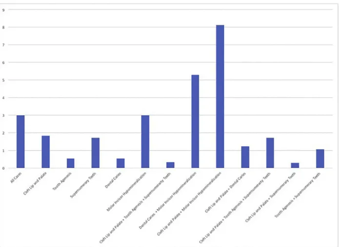
In the analysis of the group that combined individuals affected by isolated cleft lip and palate, tooth agenesis, supernumerary teeth, molar incisor hypomineralization, or dental caries, we found an association with rs622260 but not with the rs10798049 marker. We determined that the allele C of rs622260 was overrepresented in all in-dividuals studied compared with a group of unrelated individuals who did not present any of these conditions (P= .05). To determine if this association was related to a particular subgroup, we then tested each phenotype in-dividually. We found evidence that the association with the marker rs622260 was driven by the subset of samples with molar incisor hypomineralization (Table II;Figure 2). When subgroups were tested, associations were identi-fied for individuals with molar incisor hypomineralization (P= .05), isolated cleft lip and palate or molar incisor hypomineralization (P= .0003), and dental caries or molar incisor hypomineralization (P= .005). No other combi-nations had suggestive association. No further analyses for the marker rs10798049 had an association. Family-based analysis did not reveal any associations (data not shown).
DISCUSSION
Our group has approached the challenge of identifying genes contributing to isolated cleft lip and palate by ex-ploring the presence of concomitant dental anomalies and the occurrence of cancer in the families that had chil-dren born with oral clefts (summarized by Vieira22,23). We have proposed the concept that isolated forms of cleft lip and palate are quite rare and that the majority of cases actually have accompanying signs, particularly in the den-tition. Evidence indicates that incisor development is intrinsically linked with that of the upper lip and primary palate, such that cleft lip and hypodontia or supernumerarism of the lateral incisor are likely in some cases to arise from shared pathogenesis.24
In fact, “su-pernumerary” lateral incisors may actually reflect a failure of the normal fusion of 2 independent dental epithelial thickenings to form a single incisor—the same basic mechanism that results in a typical cleft lip. This exem-plifies that the coincidence of phenotypes occurring in the same individual or these clinical presentations oc-curring individually but segregating in the same family, at least in some cases, have a common anatomic devel-opment component. Descriptions such as “isolated” vs
“syndromic” may hinder gene discovery, and more com-plete clinical descriptions, such as “right cleft lip with cleft palate and bilateral mandibular second premolar and left maxillary lateral incisor agenesis with family history of breast cancer” may be more suited to provide genotype-phenotype correlations. With that in mind, we designed an experiment in which we combined individuals with conditions that we know are reported to be associated with each other (Figure 1). This criterion fits the defi-nition of “syndrome,” which is a term that defines a group of signs and symptoms that consistently occur together (or a condition characterized by a set of associated signs or symptoms). This is a clinically defined term that does not necessarily relate to the etiology of the signs and symptoms but rather how consistently they associate with each other. One limitation of our work is that we did not test for subtypes of clefts (bilateral, unilateral left or right, cleft lip only, cleft palate only) or types of teeth affect-ed by agenesis or having a supernumerary in the vicinity, and this should be done in future analyses with more sub-stantial sampling.
For our experiment, we purposely chose markers that have not been previously associated directly with isolated
Table II. Summary of the association results
Gene marker Phenotype
Minor allele frequency in affected individuals
Minor allele frequency in unaffected individuals
(N= 88) P
IGSF9 B rs10798049 All cases combined (N= 508) 0.27 0.31 .37
Isolated cleft lip and palate (N= 195) 0.25 0.31 .21
Tooth agenesis (N= 52) 0.24 0.31 .35
Supernumerary teeth (N= 122) 0.27 0.31 .47
Dental caries (N= 134) 0.29 0.31 .77
Molar incisor hypomineralization (N-140) 0.28 0.31 .52
Isolated cleft lip and palate+ tooth agenesis + supernumerary teeth (N= 369)
0.26 0.31 .22
Dental caries+ molar incisor hypomineralization (N = 274) 0.28 0.31 .69
Isolated cleft lip and palate+ molar incisor hypomineralization (N= 335)
0.26 0.31 .31
Isolated cleft lip and palate+ dental caries (N = 229) 0.27 0.31 .41
Isolated cleft lip and palate+ tooth agenesis (N = 247) 0.25 0.31 .25
Isolated cleft lip and palate+ supernumerary teeth (N = 317) 0.26 0.31 .25
Tooth agenesis+ supernumerary teeth (N = 174) 0.26 0.31 .35
HCMN1 rs622260 All cases combined (N= 508) 0.41 0.32 .05
Isolated cleft lip and palate (N= 195) 0.39 0.32 .16
Tooth agenesis (N= 52) 0.35 0.32 .58
Supernumerary teeth (N= 122) 0.25 0.32 .18
Dental caries (N= 134) 0.35 0.32 .58
Molar incisor hypomineralization (N-140) 0.41 0.32 .05
Isolated cleft lip and palate+ tooth agenesis + supernumerary teeth (N= 369)
0.34 0.32 .72
Dental caries+ molar incisor hypomineralization (N = 274) 0.45 0.32 .005
Isolated cleft lip and palate+ molar incisor hypomineralization (N= 335)
0.5 0.32 .0003
Isolated cleft lip and palate+ dental caries (N = 229) 0.37 0.32 .29
Isolated cleft lip and palate+ tooth agenesis (N = 247) 0.39 0.32 .18
Isolated cleft lip and palate+ supernumerary teeth (N = 317) 0.34 0.32 .75
cleft lip and palate but are in the vicinity of genes re-cently suggested to contribute to supernumerary teeth. The single-nucleotide polymorphism rs10798049 is located in 1 q31.1 and rs622260 is located in 11 q25. A duplication of 1 q31.1 was reported to lead to hemifa-cial macrosomia, anophthalmia, anotia, macrostomia, and cleft lip and palate in a 2-year old boy.25
The analysis yielded results that suggest rs622260 is associated with molar incisor hypomineralization, and when this condition was studied in combination with in-dividuals born with isolated cleft lip and palate. The single-nucleotide polymorphism rs622260 is located in the intron of NCAPD3, which is a gene that forms a complex that establishes mitotic chromosome architecture. Interest-ingly, 30 individuals (15.4%) in the present study group of 195 born with clefts had associated enamel hypopla-sia, and 17 of them (57%) carried 1 copy of the associated allele, a higher frequency than found for the overall group of cases with molar incisor hypomineralization or any combinations (Table I). The association was also iden-tified when the analysis combined individuals with caries experience and individuals with molar incisor
hypomineralization. We have proposed that minor al-terations of the developing enamel may lead to an enamel surface more susceptible to demineralization and hence to a higher caries experience.19,26
Being concerned about multiple testing, we avoided applying the strict Bonferroni correction and increas-ing the type II error. If we had used Bonferroni correction, we would have lowered theα to .00027 (.05/180). We have reported before27
that known true associations are missed when correction for multiple testing is imple-mented. The results of our work should be considered with caution and serve to generate hypotheses to be di-rectly tested in larger and more homogeneous samples. On the other hand, simply disregarding the nominal as-sociations presented here may delay discovery by misleading the field to believe that no true biological re-lationships exist. We believe that the approach presented here, if implemented in a larger scale, can lead to the de-termination of more homogeneous study groups and the identification of genotype-phenotype correlations that may aid in the future to personalized clinical management of individuals born with cleft lip and palate.
Fig. 2. Summary of P values of the association between the phenotypes tested and rs622260. P values were transformed in the negative of their logarithm of base 10; 3 in the x-axis corresponds to P= .05; 5 in the x-axis corresponds to P = .0067.
REFERENCES
1. Ardinger HH, Buetow KH, Bell GI, VanDemark DR, Murray JC.
Association of genetic variation of the transforming growth factor-alpha gene with cleft lip and palate. Am J Hum Genet. 1989;45: 348-353.
2. Letra A, Menezes R, Granjeiro JM, Vieira AR. Defining cleft
subphenotypes based on dental development. J Dent Res. 2007; 86:986-991.
3. Malanczuk T, Optiz C, Retzlaff R. Strcutural changes of dental
enamel in both dentitions of cleft lip and palate patients. J Orofac
Orthop. 1999;60:259-268.
4. Antonarakis GS, Palaska PK, Herzog G. Caries prevalence in
non-syndromic patients with cleft lip and/or palate: a meta-analysis.
Caries Res. 2013;47:406-413.
5. Worth V, Perry R, Ireland T, Wills AK, Sandy J, Ness A. Are people
with orofacial cleft at higher risk of dental caries? A systematic review and meta-analysis. Br Dent J. 2017;223:37-47.
6. Jindal A, McMeans M, Narayanan S, et al. Women are more
sus-ceptible to caries but individuals born with clefts are not. Int J Dent. 2011;454532:2011.
7. Howe BJ, Cooper ME, Wehby GL, et al. Dental decay
pheno-type in nonsyndromic orofacial clefting. J Dent Res. 2017;96: 1106-1114.
8. Vargas RAP, Castillo Tauscher S, Vieira AR. Genetic studies of
a Chilean family with three different dental anomalies. Rev Med
Chile. 2006;134:1550-1557.
9. Küchler EC, Costa MC, Vieira AR. Concomitant tooth agenesis
and supernumerary teeth: report of a family. Ped Dent J. 2009; 19:154-158.
10. Americano GC, Jacobsen PE, Soviero VM, Haubek D. A
system-atic review on the association between molar incisor hypomineralization and dental caries. Int J Paediatr Dent. 2017; 21:11-21.
11.Baccetti T. A controlled study of associated dental anomalies. Angle
Orthod. 1998;68:267-274.
12. Suprabha BS, Sumanth KN, Boaz K, George T. An unusual case
of non-syndromic occurrence of multiple dental anomalies. Indian
J Dent Res. 2009;20:385-387.
13. Ergöz N, Seymen F, Gencay K, et al. Genetic variation in
ameloblastin is associated with caries in asthmatic children. Eur
Arch Paediatr Dent. 2014;15:211-216.
14. Yildirim M, Seymen F, Deeley K, Cooper ME, Vieira AR.
De-fining predictors of cleft lip and palate risk. J Dent Res. 2012;91: 556-561.
15. Vieira AR, Seymen F, Patir A, Menezes R. Evidence of linkage
disequilibrium between polymorphisms at the IRF6 locus and isolate tooth agenesis, in a Turkish population. Arch Oral Biol. 2008;53: 780-784.
16. Jeremias F, Koruyucu M, Küchler EC, et al. Genes expressed in
dental development are associated with molar-incisor hypomineralization. Arch Oral Biol. 2013;58:1434-1442.
17. Leppaniemi A, Lukinmaa PL, Alaluusua S. Nonfluoride
hypomineralizations in the permanent first molars and their impact on the treatment need. Caries Res. 2001;35:36-40.
18. Patir A, Seymen F, Yildirim M, et al. Enamel formation genes are
associated with high caries experience in Turkish children. Caries
Res. 2008;42:394-400.
19. Bayram M, Deeley K, Reis MF, et al. Genetic influences on dental
enamel that impact caries differ between the primary and perma-nent dentitions. Eur J Oral Sci. 2015;123:327-334.
20. Ranade K, Chang MS, Ting CT, et al. High-throughput genotyping
with single nucleotide polymorphisms. Genome Res. 2001;11: 1262-1268.
21. Purcell S, Neale B, Todd-Brown K, et al. PLINK: A tool set for
whole-genome association and population-based linkage analy-ses. Am J Hum Genet. 2007;81:559-575.
22. Vieira AR. Unraveling human cleft lip and palate research. J Dent
Res. 2008;87:119-125.
23. Vieira AR. Genetic and environmental factors in human cleft lip
and palate. In: Cobourne MT, ed. Cleft Lip and Palate.
Epidemi-ology, Aetiology and Treatment. Frontiers of Oral BiEpidemi-ology, Vol.
16. Basel, Switzerland: Karger; 2012:19-31.
24. Hovorakova M, Lesot H, Peterkova R, Peterka M. Origin of the
deciduous upper lateral incisor and its clinical aspects. J Dent Res. 2006;85:167-171.
25. Huang X, Zhu B, Jiang H, et al. A de novo 1.38 Mb duplication
of 1 q31.1 in a boy with hemifacial macrosomia, anophthalmia, anotia, macrostomia, and cleft lip and palate. Int J Pediatr
Otorhinolaryngol. 2013;77:560-564.
26. Shimizu T, Ho B, Deeley K, et al. Enamel formation genes
in-fluence enamel microhardness before and after cariogenic challenge.
PLoS ONE. 2012;7:e45022.
27. Vieira AR, McHenry TG, Daack-Hisrch S, et al. Candidate gene/
loci studies in cleft lip/palate and dental anomalies finds novel susceptibility genes for clefts. Genet Med. 2008;10:668-674.
Reprint requests:
Alexandre R. Vieira, DDS, MS, PhD University of Pittsburgh
Department of Oral Biology School of Dental Medicine 412 Salk Pavilion Pittsburgh, PA 15261